

 PDF
PDF ePub
ePub Citation
 Print
Print


Fenestration refers to the localized duplication of a vessel. Vascular abnormalities are divided into two main configurations: (1) duplications, which have a relatively large arterial window, and (2) arterial slits, which are assumed to be intraluminal defects.1 Duplications can be subdivided into either partial or segmental types and either true or complete types.
The basilar artery is formed by the fusion of the plexiform, primitive, longitudinal neural arteries in the craniocaudal direction by approximately the 5th fetal week.2 Duplication of the basilar artery can result from a failure of the fusion of these embryonic precursors. Both lumens of the fenestrated segments are lined by endothelial tissue and are separated by a tunica media, but they can share a common tunica adventitia.
Compared with the partial or segmental types, true or complete duplication-type fenestrations are reported to be extremely rare.3,4 Only one case report of autopsy findings of duplication-type basilar artery fenestration and associated brainstem infarction has appeared in the literature.5 In the present article, we present a case of complete duplication-type fenestration of the basilar artery found in a patient with pontine infarction. Both proximal and distal communications of the fenestration were demonstrated with virtual arterial endoscopy, which has never been reported previously.
CASE REPORT
A 55-year-old woman suddenly developed motor weakness of the right side and dysarthria 1 day prior to admission. On admission she had mild dysarthria, especially of the lingual components, hemiparesis of the right side (motor grade III), and right facial palsy. The vascular risk profile included hypertension and type-2 diabetes mellitus. She was a nonsmoker and did not drink alcohol. Electrocardiograms and both hematological and biochemical laboratory findings were all within normal limits.
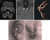
Diffusion- and T2-weighted MR images obtained the same day revealed high signal intensities in the left pontine regions, consistent with an ischemic lesion (Fig. 1-A). The source images for both contrast-enhanced MR imaging and three-dimensional (3-D) MR angiography displayed narrow segments of the basilar artery and double-lumen signs (Fig. 1-B and C). These MR findings were consistent with a long intimal dissection or fenestration of the basilar artery. Using virtual endoscopy (Rapidia; Infinitt, Seoul, Republic of Korea), a diagnosis of an extreme fenestration was confirmed (Fig. 1-D and E). Because of the complete proximal and distal fusion shown by virtual endoscopy, we speculated that this finding was a basilar arterial fenestration of the true duplication type.
The patient was treated with heparin and later was treated with clopidogrel. Two weeks after the onset of symptoms, hemiparesis of the right side improved (motor grade V-) and she was discharged.
DISCUSSION
The reported incidence of fenestration of the basilar artery has varied from 0.3% in angiographic examinations to 5.26% in autopsy series.6,7 Basilar artery fenestration is the most commonly observed form of fenestration of the cerebral arteries.7 The fenestration is usually short and occurs primarily in the proximal segment of the artery,8 with its clinical interest attributable to the possible association with aneurysms localized at the junctions of the fenestrated segments.9 There is known to be a defect in the media of the arterial wall that has been thought to be the predisposing factor for the prevalence of saccular aneurysms together with turbulent blood flow at the joining points.10
In the present case, the lesion was a true duplicationt-type fenestration, with each lumen sharing the medial wall. This finding was consistently observed from the joining portion of the two vertebral arteries to the top of the basilar artery, and each of the posterior cerebral arteries was branched from the adjoined portion of the basilar artery. The pontine infarction was located in the portion of the proximal openings.
There has been speculation about associations between either vertebrobasilar artery fenestrations and brainstem ischemia,10 or infarctions,5,11,12 although their relationships are controversial. Among these reports, some authors showed basilar artery fenestration and associated brainstem infarctions.5,11 One study showed an arterial slit-type fenestration that was confirmed by virtual arterial endoscopy,11 and another study showed a true duplication-type that was confirmed by autopsy.5 The latter case was a true duplication-type, nonseparated form of fenestration that shared the medial wall that was confined to the caudal half portion of the basilar artery. The autopsy findings showed a partially organized thrombus that occluded both halves of the duplicated portion, suggesting possible hemodynamic disturbances and turbulent blood flow at the site of fenestration.
Another case of extreme fenestration of the basilar artery was reported in a retrospective MR angiographic study.4 In that study, 10 basilar artery fenestrations were observed from 600 brain reviews (1.7%) and one case showed a total duplication. The shape of the fenestration was different in the proximal and distal parts of the basilar artery; in the proximal two-thirds of the artery, a true duplication-type fenestration that shared the medial wall was noted, but in the distal one-third of the artery, the artery was fully separated, with each basilar artery terminating in a posterior cerebral artery. However, it was not mentioned whether this finding was combined with a brainstem infarction.
Because of the 'double-lumen sign' in the present case, a basilar artery fenestration might be misinterpreted because it is known as a pathognomonic sign of arterial dissection.13 Even though neuroimaging has improved in both resolution and 3-D capabilities, it remains difficult to diagnose fenestrations of intracranial arteries. This is consistent with the large discrepancy in the reported incidence of fenestration of the basilar artery between angiographic examination and autopsies.6,7
Virtual endoscopy is a new diagnostic method that uses computer processing of 3-D image datasets, such as CT or MR imaging scans, to provide simulated visualization of specific organs.14 The ability to obtain an internal view of a vessel facilitates qualitative analyses and could improve the pretherapeutic visualization of vascular variants, stenosis, and obstruction of cerebral vessels.15 Virtual endoscopy in patients with fenestration, as in the present case, has both improved the anatomical identification of vessels and prevented misdiagnoses of fenestration as either dissection or thrombus.
In summary, we have reported a case of a patient with a duplication-type, nonseparated fenestration of the basilar artery and associated brainstem infarction. Even though a causative linkage between arterial fenestration and cerebral ischemia was not established, a reliable diagnosis of the vascular condition in the affected lesion was important clinically. From this point of view, virtual arterial endoscopy may enhance diagnostic capabilities, especially in certain cases.
 XML Download