

 PDF
PDF ePub
ePub Citation
 Print
Print


Cluster headache (CH) consists mainly of two basic clinical features: unilateral frontalo-periorbital pain and ipsilateral dysautonomic symptoms. Moreover, periodic occurrence is a characteristic but unexplained feature of the disorder. Although little is known about the etiology of CH and the possible anatomical features underlying this headache syndrome, it has been presumed that CH develops from pathophysiological events that activate the trigeminovascular system.1
The cavernous sinus has been suggested as a pathophysiological locus to explain the complex symptomology of CH. A study using orbital phlebograms of CH patients demonstrated vasculitic evidence in the ipsilateral superior ophthalmic vein and cavernous sinus during periods of CH.2 Furthermore, several symptomatic cases have been described in association with cavernous sinus pathology.3,4 The hypothalamus is also a putative pathophysiological locus as it is involved in circadian rhythms and sleep-awake cycles. Both neuroendocrine and positron-emission tomography (PET) studies have suggested the involvement of hypothalamic mechanisms in CH.5,6
However, cluster-like headaches associated with cerebral venous thrombosis (CVT) have not been reported. We describe a case in which CVT presented with periodic painful ophthalmoparesis with autonomic dysfunction that resembles a CH.
CASE REPORT
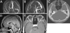
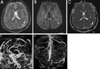
A 26-year-old man visited our hospital because of recurrent headaches since the age of 20 years. He insidiously developed a right-sided headache, especially in the forehead region, accompanied by intermittent eyelid swelling and visual blurring. These attacks occurred every evening over a period of 2 months, and each attack lasted for approximately 10 hours. After this 2-month period, his symptoms resolved completely. At the age of 21 years, he reported more severe headaches with newly developed symptoms of right facial numbness and slow progressive visual disturbances of both eyes. He was treated for presumed optic neuritis at a local ophthalmology clinic, and although the visual disturbances were attenuated after 2 weeks of medication, these headaches persisted for 2 months. At the age of 25 years, similar headache attacks occurred again for a few weeks. Another episode of headache developed 1 month before the present admission. He described the headache involving the right forehead region as "pulsating or stabbing". Lacrimation and nasal congestion also appeared with increasing pain, and he noted symptoms of facial numbness and double vision that were aggravated after attempts to gaze laterally to either side. His past medical history and family history were unremarkable, and his general physical examination was normal. A neurological examination performed when he was free of headache revealed that he was alert and oriented. Adduction and elevation of the left eye and adduction of the right eye were slightly limited. A funduscopic examination showed bilateral papilledema but perimetry revealed no defects in the visual field. No abnormalities were detected in motor and sensory examinations, and deep-tendon reflexes were normoactive. Cerebrospinal fluid examination was normal except for elevated pressure (280 mm Hg) and immunoglobulin G index (1.17). Investigations for collagen vascular diseases were all negative, and there was no evidence of other coagulopathies. Magnetic resonance (MR) venography showed no luminal flow in the right transverse sinus (Fig. 1-A) and partial occlusion in the straight sinus (Fig. 1-B), which were consistent with CVT. Brain MR imaging showed a slightly increased signal in the straight and right transverse sinuses (Fig. 1-C) on a T1-weighted image. There were neither enlargements nor signal changes in the cavernous sinuses or intraorbital structures (Fig. 1-D). In addition, temporal bone computed tomography revealed the presence of bilateral mastoiditis (Fig. 1-E), but an audiogram was normal. Oxygen inhalation during the attack with maximal intensity alleviated his headache completely within 1 minute, but the headache reappeared after the cessation of oxygen administration. Anticoagulation with heparin and antibiotics for CVT and mastoiditis were started, and acetazolamide, corticosteroid, and verapamil were also prescribed. His headache was mildly attenuated by those treatments, but his visual dimness persisted. He continued taking these medications for 2 months, when he decided to stop. Eighteen months after discharge, he developed two generalized tonic-clonic seizures, preceded by scintillations in his right-side visual field and clonic movements of his right arm. A T2-weighted image showed hyperintense lesions in the left temporoparietal area (Fig. 2-A), which were consistent with vasogenic and cytotoxic edema that was suggested by the findings of the diffusion-weighted image (DWI) (Fig. 2-B) and the apparent diffusion coefficient map (Fig. 2-C). MR venography revealed persistent venous thrombosis involving the straight sinus, transverse sinus, and the superior sagittal sinus (data not shown). Anticoagulation therapy, together with antiepileptic medication (valproate: 1350 mg/day) was started, and his headache subsequently disappeared.
DISCUSSION
CH is one of the well-known primary headache syndromes, but its pathophysiological mechanisms are a matter of debate. Associated intracranial lesions have been found in rare cases. Several cases of secondary cluster-like headaches have been reported, and include arteriovenous malformations, aneurysms, sinusitis, and tumors.7 While the present case showed recurrent bouts of cluster-like headaches, the relatively longer durations of pain, papilledema, and ophthalmoparesis were unusual features of a typical CH.
CVT is a rare disease, although the exact frequency of CVT in the general population is not known. Headache, papilledema, and seizure are frequent symptoms of CVT and often are present at diagnosis.8 One of the main mechanisms of CVT is the spread of infection from the mastoid air cells through emissary veins or directly into the adjacent dural sinus.
The exact mechanism of CVT pathology remains unknown. However, it has been suggested that venous stasis of the cavernous sinus contributes to the development of CH.9 This theory is based on observations of subjects suffering from CH who showed a constitutional narrowness of the cavernous sinus. Such a narrowness could predispose individuals to this disorder and render them more prone to develop venous stasis within the cavernous sinus, which could induce pain. Furthermore, based on the abnormal findings in the orbital phlebography of patients with CH, it has been suggested that inflammation would obliterate venous outflow from the cavernous sinus on one side,2 which supports the venous stasis hypothesis.
The periodic nature of CH implicates the involvement of the hypothalamus has been suggested that the circadian periodicity of CH is due to circulatory changes in the hypothalamus and is associated with hypothalamic dysfunction.1 Evidence of hypothalamic activation in a PET study supports this hypothesis.5 It is notable that venous flow from the hypothalamus drains into the basal vein of Rosenthal that passes through the straight sinus, and hence thrombosis in the straight sinus in our patient might affect the periodicity of headaches observed in this case.
The cavernous sinus drains chiefly into the transverse sinuses via the superior petrosal sinus; thus, our patient's symptoms would be the result of a transverse sinus thrombosis that obliterates this outflow. Taken together, we suggest that impaired local venous drainage from the cavernous sinus triggered the ipsilateral periorbital pain and cholinergic symptoms. These symptoms, such as rhinorrhea, nasal congestion, and lacrimation, are related to the nociceptive fibers of the ophthalmic division of the trigeminal nerve and the subsequent activation of the trigeminovascular system and parasympathetic vasomotor efferent fibers via the brainstem.9,10
Nevertheless, in the present case, the coexistence of primary CH and CVT is possible, since venous occlusion was not improved on subsequent MR venography, although his symptoms improved after drug treatment. In this regard, venous stasis in the cavernous sinus might simply aggravate the pre-existing primary CH. However, the dramatic oxygen response and the fact that headaches developed simultaneously with progressive visual disturbances at the initial presentation support our belief that CVT plays a causative or secondary role, triggering cluster-like headaches in a more vulnerable patient.
In conclusion, we believe that a patient with cluster-like headaches can present with CVT and that CVT may also aggravate cluster-like headaches by a hemodynamic mechanism.
 XML Download