

 PDF
PDF ePub
ePub Citation
 Print
Print


Lateral medullary infarction (Wallenberg syndrome) is a relatively common vertebrobasilar vascular syndrome. However, ipsilateral hemiparesis as part of lateral medullary infarction is rare, and is known as Opalski's syndrome. Some pathologic and neuroradiologic reports have shown that the lesion is located lower than in Wallenberg syndrome, and the ipsilateral hemiparesis seen in this syndrome is attributed to the involvement of corticospinal fibers caudal to the pyramidal decussation.1 However, Opalski's syndrome with cerebellar lesion is rare.
CASE REPORT
A 64-year-old male was admitted with sudden onset of right-sided hemiparesis, headache, gait disturbance, and recurrent vomiting. The patient regularly took amlodipine besylate (5 mg once a day) for his blood pressure, but nothing for his diabetes. On examination, his pulse was regular, his blood pressure was 160/100 mmHg, and his temperature and respiratory rate were normal.
A neurological examination revealed right-sided hemiparesis (MRC grade: upper/lower, IV/IV), right Horner syndrome, and ataxia of his right limbs. On sensory examination, pinprick and temperature sensations were decreased on the right side of his face and the left side of his body. Position and vibration senses were also decreased on the right side of his body.
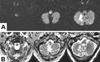
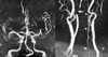
On the second day in hospital, his right side became hemiplegic (upper/lower, II/II) with flaccid tone and hyporeflexia. During the next 7 days, his tone returned, the deep tendon reflex increased, and he developed a right-side Babinski response. Diffusion-weighted and T2-weighted brain MRI performed 24 hours after the onset of his condition revealed a high-intensity area in the right lateral medulla extending from the rostral medulla to the upper cervical cord, and in the right cerebellum in the territory of the medial branch of the posterior inferior cerebellar artery (PICA) (Fig. 1). MR angiography disclosed suspicious narrowing of the proximal and distal portions of the right vertebral artery and hypoplasia of the left vertebral artery (Fig. 2).
The patient was treated with intravenous heparin for 5 days. Low-dose aspirin (100 mg once a day) and clopidogrel (75 mg once a day) were initiated for secondary prevention. On the basis of the MR angiography findings, we recommended digital subtraction angiography and angioplasty, but the patient refused any other treatment modalities. The patient was discharged 36 days after the ischemic accident with hemiparesis (upper/lower, III/III), ataxia of his right side, and residual sensory impairment.
DISCUSSION
Ipsilateral hemiparesis with symptoms and signs of lateral medullary infarction were first described by Opalski in 1946.1 He reported two patients with ipsilateral hemiplegia, ataxia, Horner syndrome, diminished facial sensation, and diminished superficial sensation of the contralateral side. Pathologic and neuroradiologic findings have identified the causal lesion of Opalski's syndrome.2,3 In the present case, the lesion causing ipsilateral hemiparesis was located in the upper cervical cord, also involving the corticospinal tract below the decussation.
The arterial supply of the medulla arises from the vertebral artery, PICA, and anterior and posterior spinal arteries. The PICA and vertebral artery supply the lateral medullary area, and branches of the vertebral artery are distributed to practically the entire lateral medullary region between the medullary pyramids and the fasciculus cuneatus at the caudal medullary level. Norrving and Cronqvist examined the pattern of vascular occlusion in lateral medullary infarctions, and found that the most common vascular lesions involved the vertebral arteries.5 The relative sizes of the vertebral arteries vary considerably, and in approximately 10% of cases one vessel is so small that the other is essentially the only artery supplying the brainstem and cerebellum. When the one vertebral artery responsible for supplying the major source of the blood flow is occluded, the resulting infarction is more severe than in the case of bilaterally competent vertebral arteries.6 Therefore, in the present case we may consider that the right vertebral artery was responsible for supplying the major source of blood flow and that an artery-to-artery embolism with right vertebral arterial atherosclerosis resulted in coexisting cerebellar and medullary lesions extending to the rostral cervical cord.
Several previous reports of Opalski's syndrome2-4 attributed these clinical observations to observed focal ischemic lesions of the medulla. Dhamoon et al. reported one autopsy case with severe atherosclerosis and thrombosis in the proximal and distal sections of the right vertebral artery in Opalski's syndrome.2 In the present case, acute ischemic lesions extended to the rostral medulla and encompassed the medial PICA territory of the cerebellum, and MR angiography showed suspicious severe stenosis or near occlusion of the right vertebral artery, and hypoplasia of the left vertebral artery.
This studies was subject to some limitations. Conventional angiography and angioplasty could not be performed due to patient's refusal. In addition, considering that intrinsic PICA (in situ branch artery) disease can also cause lateral medullary lesion with multiple cerebellar involvement,7 we cannot exclude the possibility of in situ PICA occlusion.
 XML Download