Abstract
Optic aphasia is a rare syndrome in which patients are unable to name visually presented objects but have no difficulty in naming those objects on tactile or verbal presentation. We report a 79-year-old man who exhibited anomic aphasia after a left posterior cerebral artery territory infarction. His naming ability was intact on tactile and verbal semantic presentation. The results of the systematic assessment of visual processing of objects and letters indicated that he had optic aphasia with mixed features of visual associative agnosia. Interestingly, although he had difficulty reading Hanja (an ideogram), he could point to Hanja letters on verbal description of their meaning, suggesting that the processes of recognizing objects and Hanja share a common mechanism.
The disturbance of naming objects is a common symptom in patients with brain damage. However, naming deficits can be modality specific. Optic aphasia is the inability to name objects only when they are presented in a visual mode - the patient can name the objects on tactile or verbal presentation. Since the syndrome was first described by Freund in 1889, patients with optic aphasia have occasionally been reported.1-6 However, the precise level of impairment remains unspecified. Here we describe a patient with optic aphasia. Interestingly, he demonstrated preserved semantic knowledge of Hanja, which is not observed in Hangul. This suggests that the processes of recognizing an ideogram in the Korean language and an object share a common mechanism.
K.D., a 79-year-old, hypertensive, right-handed man who had received 11 years of education, visited our hospital for further management of the stroke that had experienced occurred 9 years previously. We could not obtain the medical records from the previous hospital, and detailed information about his neurological problems at the time of the onset was unavailable. His neurological examination was not significant except for a right-superior-quadrantanopsia. A brain magnetic resonance imaging scan performed 1year after the onset showed an infarct in the left inferior temporooccipital lobe and part of the splenium (Fig. 1). Electrocardiography, chest X-ray, and routine laboratory results were within normal limits.
K.D.'s chief complaints were his language and memory impairments. Neuropsychological tests7 and speech-language assessments were performed for the detailed evaluation of his language and cognitive abilities. The neuropsychological tests revealed severe visual and verbal memory disturbances (<5th percentile for both), visuospatial dysfunction (<1st percentile), and some frontal executive dysfunctions. The result of the Korean version of the Western Aphasia Battery8 showed the profile of an anomic type of aphasia of mild to moderate severity (aphasia quotient=77.3/100). His speech was fluent (9/10) and without dysarthria. He had a prominent naming deficit (5.1/10), a mildly reduced ability for auditory comprehension (7.7/10), and his reading ability was severely impaired even at the word level, showing the characteristics of letter-by-letter reading.
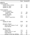
Interestingly, K.D.'s naming deficit was restricted only to stimuli presented visually. His performance on the Korean version of the Boston Naming Test9 was less than the 1st percentile (0/60), whereas it was at the 48th percentile (35/60) on verbal semantic presentation. Object naming by touch was also intact (19/20), with the only error being mistaking a ruler for a thermometer. Thus, his deficits of visual processing of objects and letters were systematically assessed (Table 1).
Tasks of matching objects to pictures, matching for shapes, and matching for colors were used to evaluate K.D.'s visual perceptual ability for objects. K.D. had no difficulties in accomplishing any of the tasks. Structural recognition and semantic recognition tasks were administered for visual recognition tests. For the structural recognition test, six line-drawn pictures were prepared: three pictures for two overlapping shapes and three pictures for objects taken from an unusual viewpoint. He could name all of the overlapped shapes; however, when he was asked to point to the object matching the picture drawn from the unusual view, he missed one item. For the semantic recognition tasks, pantomime and semantic categorization tests were used. Ten objects from everyday life were presented one by one, and he was asked to mime the use of each object. On this test, he kept saying "I don't know" after he missed the first two items. On the test of semantic categorization, he was shown pictures with three objects (e.g., flute, frying pan, and violin) and was asked to select the two that were closely related. He missed 6 out of 30 items (80% accuracy).
The task of matching syllables was used to evaluate his visual perception of letters. K.D. did not experience any problems in this test. The Korean orthographic system consists of both phonograms (Hangul) and ideograms (Hanja), and a double dissociation between the processing of the orthographies have been proposed.10 Thus, the tests for both Hangul and Hanja reading were performed. On the test of reading Hangul, he determined the meaning of words by letter-by-letter reading. Once he converted the graphemes to phonemes, he appeared to understand them via the auditory route, which was intact. On the reading test of Hanja, he demonstrated significant difficulties (1/10) even with the basic level of words (e.g., 耳 [ear] and 雨 [rain]). He showed the same difficulty in matching pictures to Hanja letters. Surprisingly, however, he showed a relatively preserved ability (8/10) when he was asked to point to the Hanja letter associated with the definition of the word presented verbally (e.g., "what you write with" for 鉛筆
[pencil]). The lexical decision-making tests for both Hangul and Hanja were not completed because K.D. refused to finish them after he failed on the first several items.
We report here a patient who presented with selective naming deficits only for visually presented objects, without disturbance in tactile or verbal semantic presentations. This phenomenon can be described as either visual agnosia or optic aphasia. However, the results of systematic assessments of visual processing revealed that K.D.'s symptoms were not fully compatible with any type of visual agnosia.11 Visual naming deficits in optic aphasia are not attributable to the disturbance of visual perception or recognition. His visual perceptual ability was intact, and can thus be differentiated from visual apperceptive agnosia.
Nevertheless, K.D.'s differentiation between visual associative agnosia and optic aphasia was not clear. He had no problems with object use in daily life. He was able to point to objects according to their name. These findings support the diagnosis of optic aphasia and contradict that of associative visual agnosia. However, he could not mime the use of objects at all, which is not compatible with optic aphasia. It may be argued that miming is not a good indication of a preserved semantic system, and many reported cases of optic aphasia have actually shown severe impairments in pantomime.1 He also showed a mildly reduced ability in visual semantic categorization, indicating incomplete semantic specification. Therefore, we concluded that optic aphasia was predominant but that there were some mixed features of visual associative agnosia in this patient.
Although patients with optic aphasia are typically considered to have an unimparied ability to recognize visual objects, some authors have indicated that optic aphasia and visual agnosia may represent different degrees of visual recognition impairment based on the variability of agnostic signs found in patients with optic aphasia.1-4 There is one report of a patient whose condition evolved from visual associative agnosia into optic aphasia, suggesting that both share a common underlying mechanism.2 Alternatively, that patient may have presented with visual agnosia at the time of the onset, the condition evolving into optic aphasia later on, presenting a mild form of visual associative agnosia.
K.D.'s lesions, in the left inferior temporooccipital lobe and a part of the splenium (Fig. 1), are in accordance with the previous case reports. Lesions in the left occipital and posterior temporal area tend to be related to visual associative agnosia, whereas the additional involvement of the splenium is observed only in patients with optic aphasia.5 Based on an analysis of the correlation between lesions and deficits, it was proposed that the right hemisphere also has a semantic system, and optic aphasia showed the variable semantic competence of the right hemisphere without refining the processing of the left hemisphere.4-6 In light of this hypothesis, the incomplete semantic specification of K.D. may reflect a low degree of semantic competence of his right hemisphere.
Interestingly, K.D. exhibited a relatively preserved ability with regard to the task of pointing out Hanja words (ideogram) in accordance with the verbal definition, even though he failed to read those words. This did not occur in Hangul reading because he exhibited the characteristics of letter-by-letter reading for Hangul (phonogram). It has been proposed that the deficit of visual objects and letters share a common mechanism.4 In our patient, the competence of semantic knowledge of ideogram is quite similar to that of visual objects, suggesting that a common neural mechanism underlies the semantic recognition process for both ideogram and objects. Moreover, it may reflect the role of the right hemisphere in reading ideogram12 and supports the different processing of phonograms and ideograms in the Korean orthographic system.10,12
Figures and Tables
References
1. De Renzi E, Saetti MC. Associative agnosia and optic aphasia: qualitative or quantitative difference? Cortex. 1997. 33:115–130.

2. De Renzi E, Zambolin A, Crisi G. The pattern of neuropsychological impairment associated with left posterior cerebral artery infarcts. Brain. 1987. 110:1099–1116.

3. Iorio L, Falanga A, Fragassi NA, Grossi D. Visual associative agnosia and optic aphasia: a single case study and a review of the syndromes. Cortex. 1992. 28:23–37.

4. Chanoine V, Ferreira CT, Demonet JF, Nespoulous JL, Poncet M. Optic aphasia with pure alexia: a mild form of visual associative agnosia? A case study. Cortex. 1998. 34:437–448.

5. Schnider A, Benson DF, Scharre DW. Visual agnosia and optic aphasia; are they anatomically distinct? Cortex. 1994. 30:445–457.

6. Coslett HB, Saffran EM. Optic aphasia and the right hemisphere: a replication and extension. Brain and Language. 1992. 43:148–161.

7. Kang Y, Na DL. Seoul Neuropsychological Screening Battery. 2003. Incheon: Human Brain Research & Consulting Co..
8. Kim H, Na DL. Korean Version - The Western Aphasia Battery. 2001. Seoul: Paradise Institute for Children with Disabilities.
9. Kim H, Na DL. Korean version - Boston Naming Test. 1997. Seoul: Hakjisa Publishers.
10. Kwon M, Kim JS, Lee JH, Sim H, Nam K, Park H. Double dissociation of Hangul and Hanja reading in Korean patients with stroke. European Neurology. 2005. 54:199–203.

11. Farah MJ, Feinberg TE. Feinberg TE, Farah MJ, editors. Visual object agnosia. Behavioral Neurology and Neuropsychology. 1997. New York: McGraw-Hill;239–244.


 PDF
PDF ePub
ePub Citation
 Print
Print


 XML Download