

 PDF
PDF ePub
ePub Citation
 Print
Print


Primary infection with Varicella zoster virus (VZV), a human herpes virus,1 causes chicken pox (varicella), after which the virus lies dormant in the cranial nerve and dorsal root ganglia. The virus frequently reactivates to produce shingles (zoster) and postherpetic neuralgia.1,2 Neurological complications can follow either primary VZV infection or reactivation of the virus.3 Complications associated with herpes zoster are postherpetic neuralgia, vasculopathy (encephalitis), aseptic meningitis, myelitis, optic neuritis, retrobulbar neuritis, cranial nerve palsy, focal motor weakness, neurologic bladder, and Guillain-Barré syndrome.3 Acute cerebellar ataxia is the most common neurological complication of varicella infection in children.3 Cerebellitis associated with herpes zoster is rare in adults. Ramsay Hunt syndrome comprises peripheral facial palsy accompanied by an erythematous vesicular rash on the ear (zoster oticus) or in the mouth.4 Here, we describe the case of an immunocompromised patient who presented with Ramsay Hunt syndrome complicated by cerebellitis, which, to our knowledge, has not been reported previously in the English-language literature.
CASE REPORT
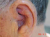
Our 76-year-old male subject had a 10-year history of hypertension and had undergone surgery for sigmoid colon cancer 2 months before this admission. He had been treated with chemotherapy (doxifluridine, 900 mg/day; polysaccharide-K, 3000 mg/day) for 1 month. The patient was admitted for unsteadiness and wide-based gait. One week before admission, he felt a chilling sensation and found vesicles on his left ear (Fig. 1) and neuralgic pain radiating from the left postauricular area to the frontotemporal area. Two days later he developed a wide-based, unsteady gait. He did not complain of dizziness, nausea, or vomiting. On the following day he experienced one episode of vomiting, after which his anticancer medication was stopped. On his visit to the outpatient clinic 2 days thereafter he showed no focal neurologic deficit other than an unsteady gait. On the evening of the same day, he noticed left peripheral facial palsy. A brain magnetic resonance imaging (MRI) scan was performed on the next day on his visit to the emergency room, and was interpreted to be normal (Fig. 2-A and B). On admission, the patient developed fever and worsening unsteady gait. He had no known past history of chicken pox.
Neurologic examination revealed left peripheral facial palsy and a moderate ataxia on bilateral heel-to-shin test. No nystagmus was found in all planes of gaze, and finger-to-nose test revealed no sign of ataxia. He showed truncal ataxia and wide-based unsteady gait. He was unable to perform the tandem walk test. Examinations of all cranial nerves except for the facial nerve were normal. Motor and sensory examinations were unremarkable and posterior column functions were all preserved. Deep tendon reflexes were normal. The patient had erythematous-based vesicular lesions on the left ear.
The patient's vital signs were normal except for a fever of up to 37.7℃. Complete blood counts were normal. The results of cerebrospinal fluid (CSF) examination are listed in Table 1. The CSF exhibited moderate pleocytosis and a modest elevation of protein content; CSF glucose content was normal. We suspected that the patient had Ramsay Hunt syndrome complicated by cerebellitis; he was therefore given intravenous acyclovir (10 mg/kg) and prednisone (60 mg). Electroneurography revealed a 90% degeneration of the left facial nerve. Pure-tone audiometry was normal.
After 1 week, the radiating pain had subsided, but left head tilting developed. Skew deviation also was not noted. Analysis of his CSF revealed mild pleocytosis that was mainly attributable to an increase in the number of lymphocytes, and a mild elevation of protein content (Table 1). The glucose content was normal. His condition was thought to be improving, so we maintained acyclovir while tapering the corticosteroid. On the 13th day of treatment he complained of severe nausea and watery diarrhea. The medications were stopped in consideration of the possibility of side effects from both acyclovir and corticosteroid. On the 15th day, the patient began to experience dizziness in addition to the already existing and persistent nausea, vomiting, diarrhea, and ataxic gait. Hypertropia of the left eye was observed, but diplopia and nystagmus were absent. Extraocular movement was fully intact. The diarrhea had resolved by day 17; however, because of persistent dizziness, nausea, and vomiting, a second brain MRI was performed for further investigation. This follow-up MRI showed gadolinium enhancement of the left facial nerve within the internal acoustic canal (Fig. 2-C). Gastrofiberscopy was also performed for evaluation of the severe nausea and vomiting, demonstrating the presence of flat erosive gastritis. On the 19th day, the patient showed right-beating horizontal nystagmus in all directions of gaze. He had mild limb ataxia on the left finger-to-nose and heel-to-shin tests, and complained of persistent dizziness, nausea, and vomiting. The CSF was reexamined (Table 1), revealing minimal pleocytosis and an elevated protein concentration. The glucose content remained normal. Acyclovir and steroid were reintroduced because the signs were suggestive of left vestibulocochlear nerve involvement. On the 22nd day, he showed left head tilting, left facial palsy, and right-beating nystagmus, while symptoms of nausea and vomiting were improved. On the 34th day, the patient was discharged with left facial palsy and ataxic gait. Serum VZV IgG was positive and IgM was negative. Polymerase chain reaction (PCR) performed twice did not reveal VZV DNA in his CSF.
DISCUSSION
This report describes cerebellitis as a complication of shingles (zoster). The patient presented with truncal ataxia after developing a rash and vesicles on the left ear (zoster oticus). At that time he did not have dizziness, hearing loss, or nystagmus. However, soon thereafter he developed fever, peripheral facial palsy, aggravated gait ataxia, and bilateral limb ataxia, which cannot be explained by vestibular dysfunction, but rather suggested cerebellar dysfunction. His CSF showed moderate pleocytosis and a modest elevation of protein. Fever, cerebellar ataxia, and a high CSF leukocyte count are highly suggestive of acute cerebellitis. In children, acute cerebellitis is the most common neurological complication of varicella infection.1,5,6 On the contrary, acute cerebellitis complicated by herpes zoster in adults has been reported only rarely.7,8 Considering the time relationship between zoster oticus and cerebellitis, it is reasonable to assume that the two conditions may be causally associated. Neurotoxicity as a result of doxifluridine treatment (a prodrug of 5-fluorouracil, 5-FU) or metastasis of primary cancer may be considered as the other possible causes of cerebellar dysfunction in this patient. Cerebellopathy due to the use of doxifluridine has been reported to occur in 25~30% of treated patients. Neurotoxicity caused by 5-FU manifests primarily as cerebellar ataxia; however, according to previous reports, intravenous doxifluridine causes acute cerebellar syndrome only when administered at high doses, and this complication is rapidly reversible upon drug discontinuation. In addition, lumbar punctures performed in the previously reported cases revealed no abnormalities.9,10 On the contrary, not only did our patient show worsening cerebellar ataxia after doxifluridine was stopped, but also doxifluridine was given orally, not intravenously. The possibility of metastasis to the cerebellum was ruled out by brain MRI and CSF investigations. Vestibular dysfunction developed subsequently. The patient complained of vertigo, nausea, and vomiting. He showed left head tilting and right-beating nystagmus. The brain MRI showed gadolinium enhancement of the left facial nerve within the internal acoustic canal without any abnormalities in the cerebellum. Many cases of acute cerebellitis do not show cerebellar abnormalities on MRI, although several recent reports describe MRI abnormalities such as cerebellar swelling or gadolinium enhancement of the cerebellum.11 Zoster oticus, peripheral facial palsy, and vestibulocochlear nerve involvement are symptoms of Ramsay Hunt syndrome. PCR to identify VZV in the CSF was negative. CSF antibody testing would have constituted a laboratory-supported diagnosis of herpes zoster-associated neurologic complications. Unfortunately, however, CSF antibody testing was not available at our institution, and the sample was not sent to outside laboratories for testing. Although serological antibody testing was normal, a normal serological VZV antibody titer does not exclude VZV-associated central nervous system (CNS) involvement.8 This patient had a virus-specific rash (shingles), CSF pleocytosis, fever, and focal neurologic signs, suggesting a definite involvement of the CNS by viral infection.12 The negative PCR results may be attributable to the antiviral therapy.13 The initial CSF sample that was taken was insufficient for testing; only the second CSF sample was examined by PCR for VZV, and this was performed during the course of acylovir and corticosteroid therapy. Hence, if the first CSF sample had been sent for the test, the results could have been different. Based on this case, we believe that herpes zoster can be complicated by acute cerebellitis. Cerebellar ataxia occurring days to weeks after the onset of varicella (primary infection of VZV) in children is thought to be immune-mediated and may be due to active viral replication.1 The mechanism underlying cerebellar ataxia after VZV reactivation in adults is unknown. In the case of VZV encephalitis, hematogenous spread or dissemination through CSF pathways have been postulated as mechanisms of infection.1 The mechanism for cerebellitis occurring in VZV reactivation may be the same. To the best of our knowledge, this is the first case report of Ramsay Hunt syndrome complicated by cerebellitis in adults. It is intriguing to note that Ramsay Hunt syndrome may go beyond the facial and vestibulocochlear nerves to spread to the cerebellum. We assume that such an extensive involvement by VZV reactivation may be related to an immunocompromised host status.


 XML Download