

 PDF
PDF ePub
ePub Citation
 Print
Print


INTRODUCTION-GOALS
Aphasia is still a very common neurobehavioral disorder that is seen in patients who have suffered from stroke, degenerative dementia such as Alzheimer's disease, frontotemporal lobar degenerations such as semantic dementia and primary progressive aphasia, traumatic brain injuries and other disorders that induce hemispheric dysfunction. At the end of the nineteenth century, after Broca's description of the non-fluent aphasia induced by damage to the left inferior frontal lobe, neurologists such as Wernicke, Lichtheim and Kussmaul reported new aphasic syndromes and attempted to explain these symptom complexes by developing information processing models. These models attempted to provide a topographic map of the brain modules that mediate speech and language as well as relate this topographic map to anatomic regions of the brain. Henry Head, in his classic book Aphasia and Kindred Disorders of Speech, attempted to introduce a neurolinguistic approach to the understanding and classification of aphasic disorders.1 Head dismissed the models developed by these aphasiologists and referred to these distinguished investigators as "diagram makers". In addition, to denigrate their theoretical models, he also accused them of distorting their data and observations. For example, when discussing one of Wernicke's reports Head wrote, "No better example could be chosen of the manner in which the writers of this period were compelled to lop and twist their cases to fit the Procrustean bed of their hypotheses." However, just as information processing model or what Head called diagrams helped Wernicke predict conduction aphasia, information processing models may allow one to develop hypotheses about how brain systems and networks operate and what type of dysfunction may be seen with a variety of brain injuries. In spite of Henry Head's admonitions, in the past three decades there has been a renewed interest in using information processing models to help explain cognitive deficits and to develop new a priori hypotheses.
The "diagram makers" did not believe that their diagrams were accurate depictions of how the brain functioned, but they used these diagrams or models for heuristic purposes. These models aided teachers who were attempting to get students to understand the pathophysiological basis of aphasic disorders and these models also help investigators to develop new testable a priori hypotheses.
The benefits of using models or diagrams persist, but at the close of the nineteenth century, when Lichtheim presented his model, there have been many new disorders reported. The purpose of this paper is to first discuss the classic aphasic syndromes using both a historic and cognitive neuropsychological modeling approach and then to modify these classic models by discussing more recently described disorders and how these observations require modifications of this model. It is my hope that this information processing approach will continue to have heuristic value and will allow people to understand and investigate the neuropsychological mechanisms that lead to the major aphasic syndromes as well as providing rationales for testing, therapy and management.
MODULARITY: PAUL BROCA
The information processing or cognitive neuropsychological approach to understanding the means by which the brain mediates speech and language, as well as understanding the speech/language disorders associated with brain dysfunction, has its major premises that the brain is organized in a modular fashion. The concept of brain modularity posits that different portions of the brain store different forms of information and mediate different cognitive activities. The concept of localization of function or that the brain is organized in a modular fashion was first proposed in the early part of the nineteenth century by Franz Joseph Gall. Phrenology was based on the postulates that specific areas of the brain are important for mediating different cognitive functions and that the bigger the portion of brain that mediates this function the more representations it can store or the better it can perform computations. Unfortunately, these postulates led to the pseudoscience of phrenology where followers believed that by palpating and measuring the skull one could determine a person's mental abilities.
The pseudoscience of phrenology had been discredited, but the postulate of localization of functions or modularity persisted and influenced Paul Broca, a French physician-surgeon and anthropologist. Paul Broca heard a lecture by Auburtin, who along with his father-in-law Bouillard (a student of Gall) thought that speech is mediated by the frontal lobes. According to Henry Head Auburtin's belief in this localization of speech was so strong that he offered to recant his faith in the localization doctrine of Gall if anyone could show him a patient with a loss of speech who did not have a lesion of the frontal lobes.1 After hearing Auburtin's lecture, Broca invited Auburtin back to his hospital to see Leborgne, who had an aphasia and a right hemiparesis as a result of a prior stroke. He was hospitalized because of cellulitis of his leg. When Auburtin observed this patient he agreed that Leborgne had a loss of speech and should have a lesion of the anterior lobes of the brain. Paul Broca noted that while Leborgne was unable to speak, except for saying the word "tan", he was able to understand spoken language.2 Unfortunately, there were no antibiotics available at that time and about six days later the patient died. A post-mortem examination revealed that Leborgne had a discrete lesion of the left hemisphere that included inferior portions of the frontal lobe, the anterior portion of the insular, and the anterior superior temporal lobe. The post mortem examination of Leborgne's brain provided support for the modularity postulate of Gall. Broca called Leborgne's inability to speak "aphemia". Paul Broca then examined another patient with aphemia and when this patient died a post mortem examination also revealed injury to the middle and inferior frontal convolutions. According to Trousseau the term aphemia that Broca used to denote this nonfluent speech disorder means infamous in Greek and thus Trousseau suggested that speech disorders be called aphasia.3 This non-fluent aphasia with preserved comprehension is now called Broca's aphasia. In addition to being non-fluent, patients with this disorder have phonemic disintegration of their speech and syntactic disorders both in expression and comprehension. Patients with this disorder also have problems with naming and repetition.
Although Leborgne's left hemisphere infarction involved more than the inferior frontal lobes' pars opercularis and triangularis, Broca thought that these areas were critical for inducing this non-fluent aphasia. Mohr and coworkers, however, demonstrated that when lesions are confined to Broca's area, including the pars triangularis and opercularis, the non-fluent aphasia is only temporary and that a larger lesion is important for a persistent non-fluent aphasia.4 Mohr et al's report, however, does not refute Broca's localization of this disorder, but rather suggests that adjacent areas may be able to compensate for the injured areas.
Subsequently, Broca reported 8 patients who were right handed and had aphasia associated with a right hemiparesis. Based on these observations Broca concluded that the left hemisphere of right handed people was dominant for mediating speech, providing further support for the postulate that the brain is organized in a modularity fashion. Broca's observations initiated a conceptual-scientific paradigmatic shift and the beginning of scientific brain research.
INFORMATION PROCESSING MODELS: KARL WERNICKE
In the second half of the nineteenth century, scientists became interested in brain anatomy and physiology. Theodor Meynert demonstrated that the posterior portions of the cerebral cortex receive sensory input and the anterior portions of the cerebral cortex are important for motor output. Based on his studies Meynert suggested in 1866 that the superior temporal gyrus might be important in the comprehension of speech.5 However, it was Meynert's student Karl Wernicke who provided support for Meynert's hypothesis. In 1874 Wernicke described a speech disorder that could be considered an afferent disorder.6 In Wernicke's paper, titled, "Der aphasische symptomencomplex", he contrasted Broca's (motor) aphasia with a sensory form of aphasia that is currently called jargon or Wernicke's aphasia. Whereas patients with Broca's aphasia are non-fluent, but have intact comprehension, patients with Wernicke's aphasia are fluent and have impaired comprehension. Some patients with Wernicke's aphasia are so fluent that they have logorrhea. Patients with this form of aphasia have spontaneous speech that contains phonological and semantic errors but they also often produce many pseudo or non-words called neologisms. Some patients with Wernicke's aphasia have speech that is almost entirely comprised of neologisms and these patients might sound like they are speaking a foreign language, but like the patients with Broca's aphasia they may be impaired at naming and repetition. Unlike patients with Broca's aphasia who have anterior perisylvian lesions, patients with Wernicke's aphasia have posterior perisylvian lesions, the critical area being the posterior portion of the superior temporal lobe, a portion of auditory association cortex.
Wernicke thought that the patients with sensory aphasia have lost the memories of how words sound. In the absence of this information store, now called the phonological lexicon, words spoken to these patients would sound like a foreign language that they never learned. When attempting to name an object, they would be unable to recall the set of speech sounds or phoneme sequences that represent this object. They would have this same problem when attempting to speak spontaneously. Since patients with this disorder have an intact Broca's area they can fluently program the production of phoneme sequences, but the words used by these patients would not be constrained by phonological lexicon (word sound) knowledge and therefore, patients with this form of aphasia speak in jargon. In addition, because they cannot comprehend speech they do not know that they are speaking in incomprehensible jargon and they do not try to correct their speech errors.
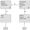
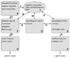
In addition to providing evidence for a second speech-language module and a second form of aphasia, Wernicke was the first to posit an information processing network when he suggested that posterior portion of the superior temporal lobe that is important in decoding speech sounds is connected to Broca's area in the frontal lobes that programs speech sounds. Based on his information processing system (Fig. 1), Wernicke suggested that this arc, from the posterior portion of the superior temporal gyrus to Broca's area would have to be used when speaking and he predicted that if a patient lost the connection between the posterior portion of the superior temporal gyrus (Wernicke's area) and Broca's area (Fig. 1, pathway C), because of a brain injury, this patient would also be aphasic. Since the area that stores word sounds (the phonological lexicon) could not supply Broca's area with the information about the phonological composition of words, the production of words would be impaired with patients making phonological errors and this patients would also have problems with repetition, but since the area that stores word sounds is intact these patients should be able to comprehend speech. Unlike the patients with sensory (Wernicke's) aphasia who have destroyed their knowledge of word sounds (phonological lexicon) and therefore, cannot monitor their errors, the patients with this disconnection disorder should be able to monitor their errors and may attempt to correct these speech errors. After Wernicke posited such an aphasia that was based on his information processing model, patients who had injury to the supramarginal gyrus and the underlying arcuate fasciculus that connects Wernicke's area to Broca's area were described. As Wernicke predicted, these patients make frequent phonological errors when speaking and often try to correct these errors. They also have impaired repetition, but intact comprehension. This disorder is called 'conduction aphasia'.
The ability of Wernicke to predict a form of aphasia that had not yet been described clearly demonstrated heuristic value of the information processing model. However, Henry Head, as had been mentioned above, in his classic book "Aphasia and Kindred Disorders" dismissed the investigators who used these types of information processing models and called them, "diagram makers".1 Head also thought that making such models might have influenced their clinical observations. For example, in his book when Head discusses one of Wernicke's clinical reports he wrote, "No better example could be chosen of the manner in which the writers of this period were compelled to lop and twist their cases to fit the Procrustean bed of their hypotheses".
Subsequent reports have, however, replicated the 'diagram makers' in clinical observations and the information processing model approach has been demonstrated to have value. Just as it helped Wernicke predict conduction aphasia, information processing models might allow investigators to develop hypotheses about how the brain works and what type of dysfunction may be seen with a variety of brain lesions. In spite of Henry Head's admonitions, in the past four decades there has been a renewed interest in using information processing models to help explain cognitive deficits and to develop new a priori hypotheses.
A few years after Wernicke's influential paper, Bastian and Kussmaul described patients who like Wernicke's patients could not comprehend speech or repeat speech, but unlike those patients with Wernicke's aphasia could speak normally.7,8 They could also understand written language. Meynert demonstrated that auditory input from the thalamus projects to the primary auditory cortex. The primary auditory cortex (Heschl's gyrus) is primarily on the dorsal surface of the superior temporal gyrus and anterior to Wernicke's area. Based on the information processing model of Wernicke, this disorder described by Bastian and Kussmaul was thought to be caused by an inability of auditory (speech) information to access an intact Wernicke's area (Fig. 1, pathway A). This disorder is called 'pure word deafness'.7,8
SEMANTICS: LICHTHEIM's MODEL
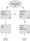
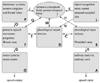
About eleven years after the publication of Wernicke's report there was another important paper written by Lichtheim who built on, and further advanced Wernicke's information processing model (Fig. 2).9 These modifications were based on Lichtheim's observations of two new aphasic syndromes. One type, now called transcortical sensory aphasia, is characterized by a reduced ability to comprehend speech similar to that observed with Wernicke's aphasia and pure word deafness. The patients with transcortical sensory aphasia are also fluent but have trouble expressing their thoughts. However, unlike patients with Wernicke's aphasia and pure word deafness, who cannot repeat or imitate speech, patients with transcortical sensory aphasia can repeat normally. Wernicke's information processing model could not explain how patients with impaired comprehension would be able to repeat normally. Lichtheim's new information processing model incorporated the Wernicke-Bastian speech arc that includes the primary auditory cortex (Heschl's gyrus), the posterior superior temporal gyrus (Wernicke's area), and the connections from Wernicke's area to the inferior frontal gyrus (Broca's) area and then to the motor cortex. In addition to Wernicke's arc, Lichtheim posited that the left hemisphere's cerebral cortex contains a region "where concepts are elaborated." the conceptual or semantic field. Thus, when a person hears another person speak, after auditory analysis (in Heschl's gyrus), auditory information is passed to Wernicke's area, where the representations of word sounds are activated and after these phonological word representations are activated this information is transmitted to the areas of the brain where concepts are elaborated (conceptual-semantic field).
Lichtheim suggested that when a person wants to speak they activate these conceptual-semantic representations and these conceptual representations directly access and activate Broca's area (Fig. 2, pathway G). Repetition, however, would take place as Wernicke and Bastian suggested in a sequence of auditory analysis, activation of phonological-word representations, and transmissions of this phonological information through the arcuate fasciculus to Broca's area and then to the motor cortex.
According to this model an injury to the left hemisphere's Heschl's gyrus, Wernicke's area and the connections between Wernicke's area and Broca's area, as well as damage to Broca's area would produce the speech deficits discussed above (e.g., pure word deafness, Wernicke's aphasia, conduction aphasia and Broca's aphasia). However, if the semantic-conceptual representations were degraded or if a patient could not access semantic representations, that patient would not be able to understand but still could repeat normally because his/her Wernicke arc would be intact enabling him/her to hear phonemes, activate phonological representations of words, and provide this phonological information to Broca's area that programs the motor neurons in the motor cortex for speech output. If there was a functional disconnection between the phonological word lexicon and the conceptual-semantic field, patients would not be able to comprehend speech, but unlike Wernicke's aphasics, these patients should be able to repeat because Wernicke's arc would remain intact.
LEXICAL ACCESS: KUSSMAUL's MODEL
Although Wernicke's schema could not account for aphasic patients who could not comprehend but could repeat (transcortical sensory aphasia), Lichtheim's (1895) model was able to account for this dissociation. However, Lichtheim's model could not explain the patients who had anomic aphasia with impaired confrontation naming (in all modalities), as well as impaired spontaneous speech with word finding difficulty, together with circumlocutions, but who had relative preservation of comprehension and speech repetition. Lichtheim was aware that his model could not account for anomic aphasia. He considered that the interruption of the pathway between the area of concepts and motor speech representations (Fig. 2, pathway G) could theoretically produce a defect in naming. However, this same defect that he posited would also cause decreased fluency with intact repetition and comprehension ('transcortical motor aphasia'). Since Lichtheim realized his model could not account for anomic aphasia, he tried to dispense with anomia as a specific aphasic subtype, "It seems to me questionable, however to place amnesia (anomic aphasia) on a par with the other phenomena of aphasic disturbance". He went on to state that "... it is not a sign of a focal lesion ..." and he concluded by suggesting, "The word amnesic aphasia had better be abandoned".
Lichtheim was partly correct, when he noted that anomia is often a residual of many different forms of aphasia, but it has also been clearly demonstrated that anomia, in isolation, can occur after a discreet lesion. Whereas patients with deficits in the semantic or conceptual field and in the phonological lexicon may be impaired at naming, such patients also have deficits in either comprehension or repetition or both. The patients with true 'isolated' anomic aphasia have normal comprehension and normal repetition.
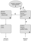
Whereas both Lichtheim's and Kussmaul's models suggest that the phonological lexicon has access to the conceptual-semantic representations, unlike Lichtheim's model, Kussmaul's model suggests that conceptual-semantic representation can directly access the phonological lexicon (Fig. 3, pathway E). Thus, an impairment of the ability of semantics to access the phonological lexicon might be an explanation of pure anomic aphasia. This explanation of anomic aphasia presumes that a patient can have a one way disconnection or dissociation between the conceptual-semantic field and the phonological lexicon.
Lichtheim, however, rejected Kussmaul's model. His rejection of this model was based upon an experiment he performed with a Broca's aphasic patient. After showing this patient some objects, he asked the subject to indicate by squeezing his hand how many syllables were in the words that denoted these objects. He found that Broca's aphasics could not adequately perform this task. According to Lichtheim's model (Fig. 2), conceptual representations cannot directly access the phonological lexicon. Therefore after seeing the object and activating the area of concepts, motor-phonetic representations are activated in concert with activation of the phonological lexical representations (Fig. 2, pathway C). Lichtheim's patient's motor-phonetic representations (in Broca's aphasia) were destroyed, and thus Lichtheim reasoned that this patient was unable to detect how many syllables a word may have, because Broca's area could not access the phonological lexicon where this information was stored. Lichtheim reasoned that according to Kussmaul's model even patients with destruction of the motor representations should have no difficulty in accessing the information stored in the phonological lexicon and since his patient with Broca's aphasia did have trouble accessing this information he rejected Kussmaul's model which suggested that conceptual representation could directly access the phonological lexicon without having to access Broca's area.
Although knowledge of how a word sounds (phonetic composition) might be stored in the phonological lexicon, deriving information about the number of sounds or syllables contained in a word might require additional processing. With each new speech sound or phoneme there is a new movement of the articulatory apparatus. Therefore, knowledge of the number of sounds (phonemes) or syllables in a word may depend upon the successful articulation of the word (Heilman et al., 1996) and patients with Broca's aphasia may be impaired at this articulatory process. Since patient with Broca's aphasia cannot normally articulate, they may not be able to parse words into its phonological or syllabic components.
If Lichtheim's argument for discounting Kussmaul's model was correct, when patients with Broca's or conduction aphasia are presented with pairs of pictures of objects and are required to determine whether the names of the two objects are the same (homophones), they should be unable to do so because lesions in Broca's area or its connections to Wernicke's area should prevent the conceptual field from accessing the phonological lexicon, where word sounds are stored. Feinberg et al, used this homophone versus non-homophone test to demonstrate that patients with conduction aphasia could successfully perform homophone judgments on words that they could not vocalize.10 The evidence that these patients could access their phonological lexicons provides support for Kussmaul's model (Fig. 3) and cannot be explained by Lichtheim's model.
Lichtheim's model has another possible flaw in that patients with a disconnection between the lexicon and the area of concepts would be impaired at comprehending speech but able to repeat, just as Lichtheim demonstrated in his case report of J. U. Schwarz. However, a review of this case revealed that when attempting to speak this patient used incorrect words and also had problems with naming. If as suggested by Lichtheim's model that the conceptual-semantic field has direct access to motor-phonetic representations, a disconnection that prevented phonological-lexical representations from gaining access to the conceptual semantic field (Fig. 2, pathway E) should not have produced this disorder. However, if as Kussmaul suggests the phonological lexicon both has input into the conceptual semantic field and also receives information from these conceptual-semantic representation, a disconnection of these two modules (Fig. 3, pathways G and E) might induce a disorder where patients cannot comprehend and have impaired spontaneous speech and naming but have a preserved intact repetition. Thus, Lichtheim's report of J.U. Schwarz provided stronger support for Kussmaul's model than for his own model.
As we mentioned above, according to Kussmaul's model, if a patient has a complete dissociation between the semantic-conceptual field and the phonological lexicon (Fig. 3, pathway G and E), this patient will have transcortical aphasia with impaired comprehension, anomia with impaired spontaneous speech, but preserved repetition.11 If the phonological lexicon can access the semantic-conceptual field (Fig. 3, pathway G), but not vice versa the patient would have anomia with intact comprehension and repetition abilities. What would happen, however, if there were the opposite dissociation, such that a patient could not access their semantic field from their phonological lexicon (Fig. 3, pathway G), but could access their phonological lexicon from their semantic-conceptual field (Fig. 3, pathway E) Heilman et al reported a patient who had impaired speech comprehension, but intact repetition, naming, and spontaneous speech.12 This patient's clinical profile ('transcortical sensory aphasia with intact naming and speech') provides support for such a one way dissociation between the phonological lexicon and the semantic-conceptual field and provides further support for Kussmaul's model.
INTENTION: KUSSMAUL's MODEL
Unlike Kussmaul's model, Lichtheim's schema (Fig. 2) has a connection between the regions where concepts are stored and where articulatory movements are programmed (Broca's area). Lichtheim thought that this part of the speech network was important for spontaneous speech and that an interruption between the area of concepts and motor phonetic representations stored in Broca's area (Fig. 2, pathway G) would also produce an aphasic disorder. According to Lichtheim's model, unlike patients with Broca's aphasia, who are non-fluent and are impaired when attempting to repeat speech, patients with this disorder should be non-fluent, but have spared repetition because Wernicke' arc is intact, therefore, have intact comprehension because the phonological lexicon can access the semantic-conceptual field. In support of this postulate, Lichtheim reported the patient Dr. C. K., who following a carriage accident, became non-fluent. This patient was non-fluent, and he could only say "Yes" or "No". He, however, was able to comprehend well and unlike patients with Broca's aphasia, Dr. C. K., was able to repeat flawlessly. This disorder is now known as transcortical motor aphasia.
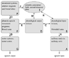
Unfortunately, Kussmaul's model cannot explain transcortical motor aphasia. The dorsolateral and medial frontal lobe together with the anterior cingulate gyrus and basal ganglia form what we have termed a conative-intentional network. Patients with damage to this network not only can demonstrate a transcortical motor aphasia, but can often show a contralateral limb akinesia or abulia. This network appears to be important in the spontaneous activation of both the motor systems and conceptual systems. There may be two forms of transcortical motor aphasia. In one there is an inability to activate the semantic-conceptual network ('adynamic aphasia') and in the other there is a deficit of motor activation ('speech akinesia' or 'extrasylvian motor aphasia Type II of Benson'). Nonfluency is the major sign in both of these disorders, but as mentioned before, unlike patients with Broca's aphasia these patients are able to normally repeat and also have good comprehension of speech. Whereas patients with adynamic aphasia may be mildly impaired at naming, those with speech akinesia can name normally. Benson and Ardila have suggested that whereas adynamic aphasia is associated with dorsolateral frontal lesions (superior to Broca's area), speech akinesia is more often associated with medial frontal lesions.13 Based on these postulates and observations we have modified the Wernicke-Kussmaul-model as illustrated in Fig. 4. According to this modification, a functional disconnection between the conative-intentional system and the conceptual-semantic field (Fig. 4, pathway P-J) would induce adynamic aphasia because patients with this disorder do not spontaneously activate their conceptual and semantic representations (they have nothing to say and no desire to speak), whereas, a functional disconnection between these intentional systems and motor speech areas (Fig. 4, pathway P-O) would produce speech akinesia, where they are impaired at initiating propositional speech.
THE INPUT AND OUTPUT PHONOLOGICAL LEXICONS
Michel and Andreewsky as well as Katz and Goodglass described an aphasic disorder similar to conduction aphasia that is called 'deep dysphasia'.14,15 Like the patients with conduction aphasia, when attempting spontaneous speech these patients' speech sounds like that produced by patients with conduction aphasia in that they are fluent but make frequent phonemic paraphasic errors. However, when asked to repeat, unlike the patients with classic conduction aphasic, who make phonological errors, these patients make semantic errors. When these patients are asked to repeat non-words they often produce real words that are phonologically related. The modified Wernicke-Kussmaul's model (Fig. 4) cannot account for this syndrome, but the Wernicke-Lichtheim model (Fig. 2) can. If, according to this model, patients are unable to repeat because there is a disconnection between the phonological lexicon (Wernicke's area) and the phonetic programs stored in Broca's area, a person could use an alternative route and after words are processed by the phonological lexicon they could activate semantic representations (Fig. 2, pathway E) that according to Wernicke-Lichtheim model could directly access the phonetic-articulatory programs stored in Broca's area (Fig. 2, pathway G). As mentioned, patients with deep dysphasia have more problems repeating non-words and this could be related to the inability of this pathway (Fig. 2, pathway B, E, F, G, D) to process words that have no meaning and therefore no semantic representation. As mentioned above, however, the Wernicke-Lichtheim model cannot account for pure anomia and if patients could use this alternative pathway to repeat, then how could we explain the classical conduction aphasia? To explain these aphasic syndromes, a further modification of the Wernicke-Lichtheim model or the Wernicke-Kussmaul model is required. This modification proposes that the phonological lexicon may be composed of two separate modules, a phonological input lexicon and a phonological output lexicon (Fig. 4). Whereas the input lexicon receives auditory information (Fig. 4, pathway A) and transmits lexical (word) information to the semantic conceptual field (Fig. 4, pathway H) the output lexicon receives input from both the auditory input lexicon (Fig. 4, pathway C) and the semantic conceptual field (Fig. 4, pathway M). This output lexicon provides lexical (word) phonological information to phonetic processor (Broca's area). According to this modified Wernicke-Kussmaul-Lichtheim model, after the auditory cortex performs a auditory analysis this information is fed into the input lexicon (that contains the memories of the phonological sequences of heard words) and this input lexicon feeds this information to the conceptual semantic field for comprehension (Fig. 4, pathway A, B, H, I) or to the output lexicon that in turn feeds words to the phonetic processor (Broca's area) (Fig. 4, pathways B, C, D, E, F). Based on this model there can be three loci of dysfunction that would allow a person to have preserved comprehension (intact auditory, phonological, input lexical and semantic processing) and fluent speech but make errors when repeating. Dissociation between the output lexicon and phonetic-articulatory module (Broca's area) (Fig. 4. pathway E) would produce the phonemic paraphasic errors when speaking spontaneously, naming and repeating, the signs that are characteristic of the classical conduction aphasia. Dissociation between the phonological input lexicon and the output lexicon (Fig. 4, pathway C) would still allow patients to repeat by means of the input lexicon, to semantic fields, to output lexicon and Broca's area (Fig. 4, pathway A, B, H, I, M, D, E, F, G). However, when using this alternative semantic route only real words that can access the semantic-conceptual field could be repeated and thus when relying solely this route a patient should be unable to repeat non-words, a sign that is associated with deep aphasia.
The semantic-conceptual field codes meanings and concepts not phonology. Thus, a person who solely uses this semantic-conceptual route might also make frequent semantic paraphasic errors because the semantic field is not constrained by phonology. For example, when a patient with deep dysphasia is asked to repeat the word "sea", the phonological representation, activated in the input lexicon, may activate the nodes that represent the concept of a large body of water. Subsequently, when the nodes in the semantic-conceptual field, that represent a large body of water, accesses the output lexicon they may activate the word "ocean" and the patient with deep dysphasia might say "ocean" rather than "sea".
Based on this alteration of the Wernicke-Kussmaul model anomia would be induced by an inability of the semantic-conceptual field to access the output lexicon (Fig. 4, pathway M), with a preservation of other pathways. Because patients with deep dysphasia have an impairment of naming and spontaneous speech, it is possible that that these also might be resulting from partial injury of this pathway.
PARALLEL PROCESSING
The Wernicke-Kussmaul's model suggested that naming is done by having the semantic-conceptual field access the phonological lexicon (Fig. 3) and in the revised Wernicke-Kussmaul model (Fig. 4) the lexicon has been divided into and input and output lexicon. In contrast, the Wernicke-Lichtheim model (Fig. 2) suggests that the semantic field can directly access the phonetic programs stored in Broca's area. It is possible that in normal people both these routes (Fig. 5, pathway I, M, D, E, F and pathway I, Q, F) are active and provide information to a phonetic buffer that computes these two inputs in preparation for phonetic-articulatory processing. Recently, Roth, Nadeau and Heilman reported a patient who was aphasic from an injury to his left carotid artery.16 When attempting to spontaneously speak he used all good words, but most of these words were incorrect (semantic paraphasic errors). However, they were often semantically related to the target words. During naming when he was provided with semantic cues, it did not appear to influence his naming. In contrast, when he was provided with phonological cues, his success in the retrieving correct name had increased, but now he made frequent phonemic errors. These observations suggest that he used two routes for naming, a whole word route (Fig. 5, pathway I, Q, F) and a phonological route (Fig. 5, pathway I, M, D, E, F). Although in this patient both routes were impaired, normal subjects might also use these two routes simultaneously, each sub-network, constraining the other, thereby reducing the probability of naming errors. The means by which each of these sub-networks constrain the other has not yet been determined, but the postulate that both these pathways are important in naming would suggest that a model which combines both the Wernicke-Kussmaul model and the Wernicke-Lichtheim model (the revised Wernicke-Kussmaul-Lichtheim model, Fig. 5) might best accounts for this patient's performance and normal naming.
There might also be another two parallel systems for visual confrontation naming. Four years after Lichtheim published his model, Freund described a patient with a right hemianopia who was unable to name objects presented in the visual modality, but was able to name objects in other modalities.17 A possible explanation of this patient's impaired visual naming is the presence of an object visual agnosia that can occur even with unilateral left ventral temporal-occipital lesions.18 However, visual agnosia is 'mind blindness' and patients with visual agnosia not only have trouble with naming seen objects, but also can not describe the use of these objects, whereas, patients with optic aphasia can describe and pantomime the use of the objects they see but cannot name them. Thus, rather than an agnosia these patients have an aphasic disorder. These patients, however, do not have anomic aphasia because they have no word finding problems when speaking spontaneously and can name in other modalities. Thus, optic aphasia is an another aphasic syndrome that even the modified Wernicke-Kussmaul-Lichtheim model (Fig. 5) cannot predict or explain. Freund thought that this disorder was caused by a disconnection between the visual areas in the occipital lobe and the speech areas in the left hemisphere, important in naming and his explanation might still appear to be valid.
People can be presented with several abstract figures that are paired with novel words and rapidly learn the association between these meaningless words and meaningless figures. This ability would suggest that even in the absence of meaning people can store new words in their output lexicon, store new visual forms in their object recognition units and link these new forms with new words. Thus, according to the modified Wernicke-Kussmaul-Lichtheim-Freund model presented in Fig. 6, patients with optic aphasia might have a functional dissociation between the portion of the brain that contains object recognition units and the phonological output lexicon (Fig. 6, pathway N). It's possible that normally, viewed objects are processed by parallel pathways, direct visual recognition unit to the output lexicon (Fig. 6, pathway L, N, D) and indirectly from object recognition units to the conceptual-semantic field and then to the output lexicon (Fig. 6, pathway L, K, I, M, D, E, F), or as mentioned above directly from the semantic-conceptual field to Broca's area (Fig. 6, pathway L, K, I, Q, F).
The postulate that there is direct input from the object recognition units to the output lexicon is supported by additional observations. Several years ago we saw several patients with a degenerative dementia in our clinic who demonstrated a naming disorder that was the reverse of optic aphasia.19 Unlike the patients with optic aphasia who can name to definition but not with visual confrontation these patients could name visually presented objects very well, but could not name the same objects to definition. We, therefore, called this disorder 'non-optic aphasia'. We think these patients could name because their object recognition units could access the phonological output lexicon (Fig. 6. pathway N) and this intact lexicon could access Broca's area. Since their repetition was flawless we think that the primary auditory cortex, phonological output lexicon, phonetic programmer (Broca's area) and motor cortex were all intact. When these patients spoke spontaneously they had semantic jargon and these patients also had poor comprehension. Naming to definition, comprehension and normal speech all requires an intact semantic-conceptual field and we think these patients' non-optic aphasia was caused by degradation of their semantic-conceptual field.
PARALLEL DISTRIBUTED PROCESSING (PDP)
The modified Wernicke-Kussmaul-Lichtheim information processing model, we developed, which is illustrated in Fig. 6, is able to explain almost all known aphasic syndromes, but this model does not describe how information in these processing modules might be stored, nor does it explain how these modules interact or transfer processed information. According to Nadeau's parallel distributed processing modification of the Wernicke-Lichtheim model, the acoustic module that is akin to Wernicke's area is located in the posterior auditory association areas and this module contain a large number of units that represent the acoustic features of phonemes.20 The phonetic motor, or what Nadeau calls the articulatory module, contains units that are located in the dominant frontal operculum (Broca's areas) and these units represent discrete articulatory features of speech. The semantic-conceptual field contains units that are widely distributed in unimodal, polymodal and supramodal cortices in the temporal, parietal and frontal lobes of both hemispheres.
Within these language-speech modules, all representations correspond to specific patterns of activity of all the units contained in this module. Many of the units in these modules are connected to the units of other modules by interposed hidden units and the entire set of connections between any two modules form what has been termed a pattern associator network.
During the process of learning a language, information is stored by alterations in the strength of the connections both between units, within and between modules. For example, the meaning of a spoken word is mediated by the connections between the acoustic module that determines the phonemic structure of the heard word and the module that contains the features of the concept. This acoustic (phonemic)-semantic pattern associator network would correspond to the phonological input lexicon that we mentioned above.
CONCLUSIONS
This paper discusses almost all of the major aphasic syndromes and attempts to explain these disorders using an information processing or a diagrammatic modeling approach. The classical models of Wernicke, Kussmaul and Lichtheim all had elements that could account for the behaviors associated with specific forms of aphasia, however, to account for all the major aphasic syndromes these three major models have to be combined. Even the combination of the three classic nineteenth century models, however, could not account for many of the aphasic syndromes described in the twentieth century and to help explain these syndromes several modifications to the Wernicke-Kussmaul-Lichtheim model were needed. In addition to the modifications of these classic models, this revised model also suggests that for several functions there are parallel processing systems or routes and that simultaneous processing might help constrain errors thus insuring accuracy and reliability.
The final diagrammatic model can be found in Fig. 6 along with markers that indicate the loci of dysfunction that might induce each specific aphasic syndrome. Although Fig. 6 is the final diagrammatic model in this paper, and this model does help explain many of the signs and symptoms associated with the aphasic syndromes, it is far from complete, but still a work in progress. For example, it does not even attempt to explain the disorders of syntax and other linguistic changes that are associated with aphasia and this model does not explain what happens as each of the hypothetic modules undergo degradation. There are many other approaches to describing and investigating the means by which the human brain processes language and the disorders associated with defective development and injury.21 Although we have discussed modular information processing models (the diagram makers approach), other approaches such as the psycholinguistic approach has been very successful and the more diverse the means by which phenomena can be viewed the more likely there will be creative insights.
In spite of Head's diatribes again the 'diagram makers' it is my hope that the diagrams and information processing models presented here will have heuristic value and aid both educational and research efforts.
 XML Download