

 PDF
PDF ePub
ePub Citation
 Print
Print


A discrete stroke may produce symptoms mimicking those from peripheral nerve diseases.1 The predominant involvement of a particular group of fingers due to brain lesion, or "pseudoperipheral palsy," was described as early as 1909.2 Since the development of the concept of the homunculus, it has been shown that the cortical representation of motor hand function is located in the superior part of the precentral gyrus.3 Small cortical strokes in the precentral gyrus can produce isolated weakness of the fingers, which may be prominent in ulnar fingers4 or radial fingers.5,6 The results from a previous study suggest that the ulnar fingers are located medially and radial ones laterally in the knob of the precentral gyrus.7 We present a case of isolated weakness of middle, ring, and little fingers due to a small cortical infarction in the medial precentral gyrus.
CASE REPORT
A 58-year-old right-handed man was admitted because of sudden weakness in his right middle, ring, and little fingers. He had had hypertension, diabetes mellitus, and hyperlipidemia since undergoing percutaneous transluminal coronary angioplasty with stenting for angina pectoris 8 years previously. However, he had never experienced paresthesia or weakness in the extremities. On admission, he was alert without dysarthria, aphasia, apraxia, or other higher cortical dysfunction. Cranial nerve examinations were normal. Motor examination revealed mild weakness (grade IV by manual muscle testing) of his right middle, ring, and little fingers. The weakness was severest at the distal interphalangeal joint. Flexion and extension movements were equally impaired. There was no significant difference in muscle power among the affected fingers. The strength of the other fingers, wrist, elbow, shoulder, and leg were normal. Sensations, including cortical sensation, were normally perceived in all modalities in his fingers as well as in other body parts.
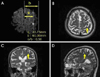
The electrocardiogram findings were normal. Transthoracic and transesophageal echocardiograms revealed severe atherosclerotic changes with mobile atheroma at the aortic arch area. Chest CT with enhancement showed multifocal atheromatous plaque in the thoracic and abdominal aorta. A nerve conduction study showed no evidence of peripheral neuropathy. Brain MRI performed on the onset day showed a small, discrete infarct in the medial precentral gyrus of the left frontal cortex (Fig. 1). MR angiogram showed hypoplastic changes in A1 of the right anterior cerebral artery, and focal luminal narrowing in the cavernous segment of the right internal carotid artery and the knee segment of the right middle cerebral artery. The patient was immediately treated with heparin and antiplatelet agents. He showed gradual improvement and regained his normal power 3 months later.
DISCUSSION
The motor topography of human fingers remains controversial. Traditional views have suggested point-to-point representations of each finger to neurons located in the motor cortex such that the radial fingers are located laterally and the ulnar fingers are located medially in the hand knob.7 Previous studies3,8 have calculated the ratio of the distance from the falx to the center of the stroke lesion in relation to that from the falx to the most lateral margin of the brain. The mean value in patients with predominant involvement of radial fingers (0.71) was greater than that in patients with predominant involvement of ulnar fingers (0.58). Therefore, lesions related to predominant involvement of ulnar fingers are located significantly more medially than those associated with predominant involvement of radial fingers in the presumed hand representation area of the motor cortex. These authors further suggested that the medial portion of the precentral knob representing topographically ulnar fingers correspond to the border-zone area associated with a hemodynamic mechanism, whereas the lateral portion representing radial fingers are supplied by distal middle cerebral artery branches associated with artery-to-artery embolism or cardiogenic embolism.8
In our patient, the location of the lesion was medially in the precentral hand knob with a ratio of 0.56 (Fig. 1-A). Therefore, MRI of our patient suggests that the medial portion of the precentral knob, representing topographically predominant involvement of ulnar fingers, corresponds to the situation reported previously.7 Some authors advocate multiple representation or spatially overlapping patterns of the cortical motor hand area with more sophisticated methods using intracortical microstimulation, or functional MRI.5,9,10
Regarding the pathogenesis of stroke, our patient had mobile aortic atheroma and normal MR angiogram findings except for the right anterior circulation. Therefore, artery-to-artery embolism from the aorta is a likely pathogenic mechanism. This observation appears to be inconsistent with the previous observation that medially located lesion was more often associated with a hemodynamic mechanism.8 Our case therefore suggests that the variable pathogenesis of stroke-induced weakness in some of the fingers.
 XML Download