

 PDF
PDF ePub
ePub Citation
 Print
Print


INTRODUCTION
Lateral medullary infarction is one of the most clearly recognized vascular syndromes of the vertebrobasilar territory, comprising about 2% of all admissions for acute stroke.1 Diagnosis of this syndrome generally depends on clinical symptoms and signs.2 However, the onset of a lateral medullary infarction is sudden in only about 40% of cases - more often the course is progressive over 24~48 h. Furthermore, unless paresis of the vocal cord or ipsilateral pharynx is present, the other findings do not unequivocally localize the lesion to the lateral medullary region.1 Magnetic resonance imaging (MRI) has become the preferred method to study lateral medullary infarction, since it has a high spatial resolution and sharp contrast in normal and pathologic tissues.3,4 Diffusion-weighted imaging (DWI) provides additional information;5 this technique is sensitive in detecting hyperacute infarction and can differentiate acute infarction from chronic cases, especially in supratentorial stroke.6 Nevertheless, several investigators have reported that DWI is less sensitive in detecting infratentorial infarcts than supratentorial ones.5-9 To our knowledge, there have been few reports on findings concerning DWI and lateral medullary infarction.5,9,10 To remedy this, we present a series of cases of acute lateral medullary infarction in order to investigate the efficacy of DWI.
MATERIALS AND METHODS
Between January 2000 and November 2004 we investigated 26 patients who had been diagnosed as having lateral medullary infarction and who underwent DWI within 72 h of the onset. We excluded patients with clinical or laboratory findings suggestive of demyelinating disease, central nervous system infections, transient ischemic attacks, or progressive neurologic changes. Prior to DWI examination, all patients underwent clinical evaluation by a neurologist and underwent CT scanning to rule out the possibility of intracranial hemorrhage. In each case, the final diagnosis was made by the neurologist, who reviewed all available clinical and radiologic data. The MRI findings were assessed by one neuroradiologist and two neurologists who were blinded to the clinical data. Initial DWI was considered positive for the diagnosis of acute lateral medullary infarction when a high signal intensity correlated with neurologic findings. All MRI investigations were performed on a 1.5-T clinical-imaging system (Philips Gyroscan ACS-NT 15 whole-body system). DWI was performed with echo-planar imaging in the axial plane. The parameters were TR/TE: 4000~4500/120 ms; number of acquisitions: 1; matrix: 256×256, slice thickness: 5 mm; number of slices: 22; and scan time: 10 min. Because 12 apparent diffusion constant (ADC) maps may not be reliable for small lesions,7 ADC values were not taken into account. In patients with negative initial DWI, follow-up MRI including fast fluid attenuated inversion recovery (FLAIR) (TR/TE: 6700~7000/130 ms) and DWI was performed before hospital discharge. Lesion volume was estimated from FLAIR images. The abnormality was manually outlined on each axial image using an image analysis package, and the area of abnormality on each section was multiplied by the image thickness. The clinical symptoms and signs when the DWI was performed were determined by the frequencies of neurologic symptoms and findings in patients with lateral medullary syndrome.1 Autonomic and respiratory dysfunctions were not included because the frequencies of those signs in patients with lateral medullary infarction have not been precisely determined.1,11 Our statistical analysis compared the initial DWI findings with time-to-MRI, number of clinical symptoms and signs when DWI was performed, and final lesion volume. The Mann-Whitney U-test was used to assess these variables. A probability value of P<0.05 was considered statistically significant. Analyses were performed using SPSS software (version 10.0), with the results of all calculations expressed as mean±SD values.
RESULTS
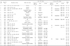
Our 26 patients (18 males, 8 females) in whom definite acute lateral medullary infarction was finally diagnosed. Table 1 lists demographic characteristics and neurologic symptoms and signs when DWI was performed, time-to-MRI (time between the onset of symptoms and initial DWI), and MRI findings. An initial DWI was performed within 5~69 h (30.5±21.6 h) of the onset of acute clinical symptoms. Of the 26 patients, 8 (31%) showed initial false-negative DWI results, of which 7 underwent follow-up DWI, which revealed clear hyperintense lesions that matched the location of the follow-up FLAIR abnormalities (Cases 1, 3, 4, 7, 9, 13, and 22 in Table 1; Fig. 1). The 8th case is listed as Case 22 (Fig. 2). Concomitant follow-up FLAIR images showing a focal hyperintensity in clinically relevant medullary regions in all 8 patients. Most of the false-negative cases occurred in patients imaged within the first 24 h of the onset of symptoms (except Case 2). Among the 14 patients imaged within 24 h of the onset, 7 (50%) had initial false-negative DWI results. Table 2 summarizes the mean time-to-MRI, lesion volume, and number of symptoms and signs in each group. During the first 72 h of stroke onset, the occurrence of false-negative DWI findings decreased significantly as time-to-MRI increased for the acute lateral medullary infarctions (P=0.014). There were 4.4±1.9 clinical symptoms and signs when initial DWI was performed, and the lesion volume was 0.41±0.27 cm3. There was no statistical significance between the false-negative rate and the number of clinical symptoms and signs at the time when the initial DWI was performed (P=0.67) or in the final lesion volume (P= 0.72).
DISCUSSION
DWI is an important diagnostic tool in stroke management.7,9,10 However, due to technical limitations, some lesions may be unidentified in the acute stage, particularly those in the medulla oblongata.7,9,10 Kitis et al. reported that DWI is a valuable technique for examining patients presenting with the signs and symptoms of Wallenberg's syndrome.5 Because most of their cases were in either the acute or subacute stage, the true sensitivity of the method in the acute stage of the syndrome remains unclear. Our study is the first to ascertain the proportion of false negatives in the acute stage (all within 72 h) of lateral medullary infarction. The rate of false-negative DWI (31%) in our study population was higher than that in the studies of Oppenheim et al.7 (19%) and Kitis et al. (8%).5
There are several potential mechanisms that could explain the lack of diffusion changes in these patients during the acute phase. First, cerebral blood-flow may be at an intermediate level - below the threshold for neuronal dysfunction (onset of symptoms) but above that of reduced diffusion.13 Generally, suppression of EEG activity (corresponding to deficit onset) occurs when the cerebral blood flow per 100 g of brain tissue is 15~20 mL/min (30~40% of the normal value of 50 mL/min), whereas membrane pump failure (the phenomenon associated with the bulk of the diffusion changes) does not occur until the blood flow falls below 10~15 mL per min.12,13,14,15 This hypothesis is supported by the onset of a lateral medullary infarction often progressing over a 24~48 h period.1,4,11 Transient ischemic attacks that precede lateral medullary infarction, usually related to lateral medullary syndrome, are recorded in about 25% of patients.1,4 The second possible mechanism is that the lesion is too small for the DWI echo-planar sequence to be clearly detected.4,7 This hypothesis is supported by high-resolution FLAIR images displaying a signal abnormality in lesions that are not visible on DWI (Case 22).7 Finally, magnetic susceptibility artifacts cause brain-stem distortions that could blur DWI images.7 DWI performed with turbo spin-echo sequences provides high-resolution images with few susceptibility artifacts and might overcome these technical limitations, particularly in the posterior fossa.16
Our data suggest that (1) false-negative DWI results are not uncommon during the acute stage of lateral medullary infarction, (2) a negative initial DWI finding obtained within 72 h of the onset of symptoms cannot reliably rule out lateral medullary infarction, and (3) false-negative DWI findings do not correlate with the number of clinical symptoms and signs or with the final lesion volume when DWI is performed. Thus, the diagnosis of lateral medullary infarction should not be ruled out on the basis of early negative DWI results: follow-up DWI or further MRI investigation should be performed in the event of a negative initial DWI result in patients with acute lateral medullary infarction.
Several potential limitations of our study should be noted. The selection of patients who underwent brain MRI could introduce bias into the study population because not all the patients with acute lateral medullary infarction underwent initial DWI within 72 h of the onset. The mode of onset in lateral medullary infarction is variable, and the onset time is not defined clearly in some patients. Clinical symptoms in acute lateral medullary infarction sometimes reveal atypical presentation, such as isolated ataxia or isolated dysphagia, and the clinical standards are therefore obscure. In addition, we did not study the relationship between the false-negative rate and the slice thickness in DWI. Future studies should investigate these problems.


 XML Download