

 PDF
PDF ePub
ePub Citation
 Print
Print


Dear Editor,
Primary central nervous system lymphoma (PCNSL), which is typically of B-cell lymphocytic origin, normally remains confined to the brain, spinal cord, and/or eyes, rarely spreading outside the nervous system. The appearance of a homogeneously enhancing periventricular rounded mass in MRI is suggestive of the diagnosis in an immunocompetent host. The case reported here illustrates an unusual presentation of PCNSL, initially as a neurolymphomatosis with multiple cranial neuropathy and evolving into a lymphomatosis cerebri with parenchymal infiltration over a 3-year span, without the typical periventricular enhancing mass.
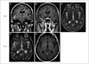
A 67-year-old previously healthy man suffered a 3-year history of alternating peripheral facial nerve and right abducens paresis followed by several months of lancinating right cheek pain. Three months previously he had been admitted with drowsiness and a weight loss of 9 kg. An examination performed at that time revealed residual mild left facial synkinetic movement with complete resolution of the right abducens and ipsilateral facial nerve palsy. Brain MRI revealed enhancement of the right middle temporal gyrus indicative of a 1.5-cm linear lesion without edema (Fig. 1A), of the right maxillary branch of trigeminal nerve (Fig. 1B), and of the bilateral facial nerves without parenchymal signal changes (Fig. 1C). Spinal fluid was acellular with an elevated protein level of 78 mg/dL, negative for viral PCR, and unremarkable cytology. A repeated spinal tap performed 1 month later was consistent with a traumatic tap. Worsening drowsiness and confusion despite an empiric steroid trial prompted follow-up MRI, which revealed bilateral deep white-matter and patchy cortical signal changes (Fig. 1D) accompanied by minimal enhancement (Fig. 1E). A brain biopsy revealed diffuse large B-cell lymphoma. Chemotherapy and steroid treatments were started. At a 3-month follow-up the patient exhibited a marked overall improvement, remaining alert and being able to speak appropriately.
Cerebral lymphoma can be categorized into systemic lymphoma with secondary spread or a PCNSL, representing 3% of all brain tumors.1 The recently discovered lymphatic system in human meninges2 helps to explain several unusual manifestations of PCNSL: 1) why most such tumors remain confined within the CNS, 2) frequent multifocal origin at the initial clinical presentation, 3) isolated meningeal lymphoma, and 4) lymphomatosis cerebri, which is a subtype indistinguishable from a primary glial infiltrative tumor referred to as gliomatosis cerebri. Thus broad range of MRI appearances cause difficulty in recognizing PCNSL in MRI. The clinical recognition of PCNSL can be even more problematic since its fluctuating clinical course that can occasionally span months or even several years without intervening steroid use makes it difficult to recognize the illness as a CNS malignancy.3
In an effort to facilitate the recognition of PCNSL, several categories have been proposed: 1) neurolymphomatosis characterized by cranial nerve involvement, 2) lymphomatous meningitis-ventriculitis, 3) intravascular lymphoma presenting mainly as strokes, and 4) lymphomatosis cerebri, which is a rare subtype, with less than 20 cases reported as of 2012.4 This last subtype is characterized pathologically by diffusely infiltrative lymphoma mostly involving deep structures. An MRI appearance of bilateral predominant deep white-matter signals with absent or mild contrast enhancement (as seen in the present case) is typical, which is identical to better-recognized gliomatosis cerebri and hence differentiating the two types requires a tissue biopsy.
This case highlights an unusual brain tumor presenting with an atypical clinicoradiologic evolution over a 3-year span. Although sequential facial nerve palsy is frequently related to viruses, subsequent involvements of the abducens and trigeminal maxillary branches have been rare. The delayed appearance of “parenchymal” clinical signs and evolving signal changes in the predominant deep white matter are suggestive of underlying infiltrative malignancy. This evolution from neurolymphomatosis to lymphomatosis cerebri that is not accompanied by a periventricular contrast-enhanced lesion in an immunocompetent patient has not been reported previously. Recognizing these unusual subtypes of PCNSL remains important since this condition is treatable. Steroid use and methotrexate-based chemotherapy extend the average survival from 1.5 to 44 months, and leading to complete remission in some cases.5
 XML Download