Dear Editor,
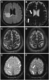
A 55-year-old man was referred to our tertiary stroke center for the investigation of gradually progressing weakness of the left upper and lower limbs. The patient had a history of smoking and hypertension, and had previously been diagnosed with ischemic stroke of undetermined etiology at another institution. During 4 months since that diagnosis, mild paresis in the left upper limb had evolved into severe left-side hemiparesis. Upon examination the patient was mildly dysarthric with labile affect and was unable to stand due to the marked left-limb weakness with ipsilateral pyramidal signs. A careful retaking of the patient's history revealed gradual progression of the symptoms as well as recent recurrent lower respiratory tract infections, which had not been noted previously. This newly revealed history combined with the illness course cast doubt on the diagnosis of stroke, and so a broad workup of the patient was performed. He was found to be HIV-positive with an extremely low CD4+ count (7/µL). Brain MRI (3T, MAGNETOM Skyra, Siemens Healthineers, Erlangden, Germany) revealed bilateral T2-weighted/fluid attenuated inversion recovery hyperintense lesions in the U-fibers, deep white matter of the right hemisphere, and the splenium of the corpus callosum (Fig. 1A), with cortical sparing in a well-defined scalloped pattern. Diffusion weighted imaging/apparent diffusion coefficient sequences showed true restriction of diffusion at the lesion borders (Fig. 1B) and there was no enhancement after the IV infusion of contrast agent. Of special note was the presence of conspicuous hypointensities with a laminar pattern in the otherwise normal-appearing cortex adjacent to the lesions in susceptibility-weighted imaging (SWI; white arrows in Fig. 1E and F) and fainter hypointense ribboning of the cortex over areas seemingly unaffected in other sequences (Fig. 1A-D). In filtered phase images, these areas appeared at similar intensities to the venous structures (appearing bright in our grayscale configuration), indicative of paramagnetic effects. There were minimal dipolar patterns. These neuroimaging findings–the location of nonenhancing white-matter hyperintensities, diffusion restriction at the lesion border, and a low-signal-intensity rim along deep layers of the cerebral cortex in SWI–were highly suggestive of progressive multifocal leukoencephalopathy (PML) based on recently described PML imaging characteristics.1
Prompt PCR of the CSF revealed high copy numbers (6.8×10⁵) for the JC virus, thus confirming the diagnosis of definite PML according to the AAN criteria.2 The patient was immediately started on highly active antiretroviral therapy, but died 29 days after the diagnosis following a continuous neurological deterioration. Consent for an autopsy was unfortunately not given, and so we were unable to investigate the neuropathology of the affected areas further.
PML is a rapidly debilitating demyelinating disease with a generally poor prognosis that is caused by the emergence of a latent JC virus in immunocompromised patients. The severity of the disease and the possibility of a better outcome in early-diagnosed cases3 mandate vigilance in identifying PML as early as possible using any available biomarker. Recent findings suggest the cortical and/or subcortical involvement of paramagnetic depositions in PML, as demonstrated by SWI and quantitative susceptibility mapping,45 despite ribboning SWI hypointensities having also been reported in other conditions.6 Pure involvement of an otherwise normal-appearing cortex overlying the white matter lesions, as in our case, remains an issue to be explained in future studies.
Figures and Tables
Fig. 1
Three Tesla Brain MRI findings. A: Brain FLAIR sequence showing hyperintense lesions in the U-fibers, deep white matter of the right hemisphere, and the splenium of the corpus callosum. B: Brain apparent diffusion coefficient sequence showing restricted diffusivity at the lesion borders (arrows), which is a characteristic neuroimaging finding of PML. C and D: T2-weighted MRI sequence showing sparing of the cortex at the involved right precentral and postcentral gyri. E and F: SWI images revealing laminar hypointensities of the cortex (arrows) at the involved sites and also over normal-appearing white matter. FLAIR: fluid attenuated inversion recovery, PML: progressive multifocal leukoencephalopathy, SWI: susceptibility-weighted imaging.

References
1. Wattjes MP, Richert ND, Killestein J, de Vos M, Sanchez E, Snaebjornsson P, et al. The chameleon of neuroinflammation: magnetic resonance imaging characteristics of natalizumab-associated progressive multifocal leukoencephalopathy. Mult Scler. 2013; 19:1826–1840.

2. Berger JR, Aksamit AJ, Clifford DB, Davis L, Koralnik IJ, Sejvar JJ, et al. PML diagnostic criteria: consensus statement from the AAN Neuroinfectious Disease Section. Neurology. 2013; 80:1430–1438.
3. Dong-Si T, Richman S, Wattjes MP, Wenten M, Gheuens S, Philip J, et al. Outcome and survival of asymptomatic PML in natalizumab-treated MS patients. Ann Clin Transl Neurol. 2014; 1:755–764.

4. Labauge P, Carra-Dalliere C, Ayrignac X, Menjot de Champfleur N. Low signals on T2* and SWI sequences in patients with MS with progressive multifocal leukoencephalopathy. AJNR Am J Neuroradiol. 2016; 37:E11.


 PDF
PDF ePub
ePub Citation
 Print
Print


 XML Download