

 PDF
PDF ePub
ePub Citation
 Print
Print


INTRODUCTION
Drug-induced parkinsonism (DIP) is the second most common cause of parkinsonism in the elderly after Parkinson's disease (PD).1 DIP is traditionally associated with old-generation (typical) antipsychotic agents, though it can also occur during the administration of several other classes of drugs including the newer (atypical) antipsychotic agents, gastrointestinal prokinetics, and calcium-channel-blocking agents.234
The clinical presentations of DIP and PD are very similar, and therefore patients with DIP are frequently misdiagnosed as having PD.1 Although most DIP patients experience a full and long-lasting recovery from DIP with no subsequent PD after discontinuing the offending drugs, some patients develop persistent and worsening parkinsonian symptoms after drug discontinuation (i.e., subclinical parkinsonism or DIP unmasks PD) or experience the reappearance of PD after full remission from DIP (i.e., DIP antedates PD).5
Cardiovascular autonomic dysfunction is relatively common in patients with PD, and can occur independently of levodopa treatment and early in the course of the disease.6 The manifestations of this dysfunction include orthostatic hypotension (OH), supine hypertension (SH), nocturnal hypertension (NH), and absence of a decrease in blood pressure (BP) during the night (nondipping).67
Recent analyses utilizing a nonmotor symptom questionnaire found that several nonmotor symptoms such as urinary and sleep symptoms and disturbances of taste or smell autonomic dysfunction occurred more frequent in patients with PD than in those with DIP.89 However, cardiovascular autonomic function has not previously been assessed in patients with DIP.
In this study we evaluated the cardiovascular autonomic function during head-up tilt-table testing and performed 24-h ambulatory BP monitoring in PD and DIP patients as well as control subjects.
METHODS
Patients
This study was approved by the Institutional Review Board of Seoul St. Mary's Hospital, and each subject provided written informed consent to participate. Twenty patients with DIP were enrolled consecutively, in addition to 99 with PD who met the clinical diagnostic criteria of the UK Brain Bank between March 2014 and February 2015.10 A clinical diagnosis of DIP was made based on the following diagnostic criteria: 4 1) presence of at least two of the four cardinal signs (tremor, rigidity, bradykinesia, and impaired postural reflexes), 2) absence of a personal history of extrapyramidal disorders before receiving treatment with an offending drug, 3) onset of symptoms during the course of treatment with an offending drug, 4) reversal of parkinsonian symptoms (though not necessarily completely) after discontinuing offending drugs during a follow-up lasting more than 6 months, and 5) normal dopamine transporter positron-emission tomography scan using 18F-N-(3-fluoropropyl)-2beta-carbon ethoxy-3beta-( 4-iodophenyl) nortropane (FP-CIT). All of the PD patients exhibited a decreased uptake of FP-CIT in the basal ganglia.
The DIP patients were subgrouped according to their underlying diseases (psychiatric diseases vs. functional dyspepsia or gastrointestinal prophylaxis) since patients with a psychiatric disease may experience some degree of autonomic dysfunction and most neuroleptics have the side effect of OH and act on dopaminergic, noradrenergic, cholinergic, and serotonergic pathways,1112131415161718 whereas most prokinetics mainly inhibit the dopamine D2 receptor in the central nervous system. 192021 Twenty-five age-matched healthy subjects free from neur,logical disease were enrolled as controls.
Clinical information was collected, including age, sex, symptom duration, history of arterial hypertension, diabetes mellitus, cigarette smoking, and current medications. All patients were evaluated using the Unified Parkinson's Disease Rating Scale (UPDRS) Parts 1–3 and classified according to the modified Hoehn and Yahr (H&Y) stage. Patients with the following characteristics were excluded from the study: 1) history of diabetic neuropathy or other peripheral/autonomic neuropathy, 2) history of previous relevant cardiac disease, or any abnormalities on routine chest radiography or electrocardiography, and 3) taking medications known to influence cardiovascular autonomic function.
Suspected offending drugs were continued in DIP patients during all of the tests. Antihypertensive medications were discontinued 7 days before the BP tests in all subjects, and no serious clinical problems were observed during this period.
Tilt-table testing
The patients were examined in a temperature-controlled clinical investigation room after having fasted overnight (except for water). Electrocardiographic and noninvasive continuous-BP-monitoring leads were connected (YM6000, Mediana Tech, Redmond, WA, USA). After 30 minutes of supine resting, head-up tilt-table testing (20 minutes at 60°) was performed using a Manumed Special Tilt1-section tilt-table (ENRAF NONIUS, Rotterdam, the Netherlands). The BP was measured manually every 5 minutes before tilting, at 1, 3, 5, 10, 15, and 20 minutes during head-up tilting, at 1 minute after tilting, and as deemed necessary to ensure subject safety. The mean supine baseline and lowest tilt values for BP were recorded. Statistical analysis was applied to the lower values from 3 and 5 minutes.
Ambulatory blood pressure monitoring
An automated 24-h BP recording instrument (Mobil-O-Graph NG, I.E.M., Stolberg, Germany) was used to measure the ambulatory BP every 15 minutes during the day and every 30 minutes during the night. The mean values of SBP, DBP, and heart rate during the daytime, nighttime, and over 24-h periods were evaluated. Nocturnal decreases in BP and heart rate were calculated as percentage changes between the daytime and nighttime mean values. Subjects with a <10% nocturnal decrease in mean BP were considered nondippers.27 NH was defined according to the 2013 European Hypertension Society/European Cardiology Society guidelines (i.e., mean nighttime SBP/DBP ≥120/70 mm Hg).28
Data analysis
Statistical analyses were performed using SPSS (version 15.0 for Windows, SPSS Incorporated, Chicago, IL, USA). One-way analysis of variance was used to compare means between groups, and Pearson's χ2 test was used to compare the frequencies of categorical variables. The Mann-Whitney non-parametric U test was also used to analyze DIP subgroups. Given the potential influence of age, sex, hypertension, and diabetes on the results of group comparisons, such comparisons were performed using analysis of covariance. Statistical significance was defined by a p value of less than 0.05.
RESULTS
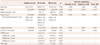
Table 1 summarizes the clinical characteristics in the DIP, PD, and control groups. Men comprised 4 of the 20 DIP patients. The age at examination was similar among groups, but the disease duration was shorter in the DIP group than in the PD group. The UPDRS score and H&Y stage tended to be higher in DIP than in PD patients. In the DIP group, gastrointestinal prokinetics were the most common offending drugs (n=13), while antipsychotics were the second most common cause of DIP (n=7). Underlying diseases were functional dyspepsia (n=7) or psychiatric diseases (n=7). Six patients were prescribed a prokinetic drug for gastrointestinal prophylaxis. Exposure times and symptom durations varied among patients, but complete remission was observed in all of them. The dosage of each offending drug was within the therapeutic range of pharmaceutical introduction in all DIP patients.
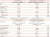
PD and DIP patients frequently had OH (controls vs. PD vs. DIP, 4.0% vs. 27.1% vs. 25.0%; χ2=6.236, p=0.016). The orthostatic change in SBP (ΔSBP) was also greater in the PD and DIP groups than in the control group. SH and NH were more frequent in PD patients than in controls. Supine BP was higher in patients with PD than in controls and patients with DIP (Table 2). The proportions of SH, NH, and non-dipping were similar in the DIP and control groups, and supine BP, nighttime BP, or nocturnal BP dipping did not differ significantly between these two groups (Table 2).
Among patients with DIP, OH was associated with underlying psychiatric disease and concurrent neuroleptics use (neuroleptic-induced parkinsonism, in 5 of 7 patients), whereas prokinetics were not related to OH (in 0 of 13 patients). Among patients with neuroleptic-induced parkinsonism, OH was related to mood disorders (four patients with depressive disorder and one with bipolar disorder). Patients with neuroleptic-induced parkinsonism displayed a higher ΔSBP during tilting than those with prokinetic-induced parkinsonism (Table 3). ΔSBP values during tilting did not differ significantly between patients with prokinetic-induced parkinsonism and normal healthy controls (6.7±7.3 mm Hg vs. 3.3±9.0 mm Hg, mean±standard deviation; p=0.210 in the Mann-Whitney U test).
DISCUSSION
Cardiovascular autonomic dysfunction occurs frequently in patients with PD, manifesting mainly as OH, SH, NH, nondipping, and heart rate variability.329 These abnormalities are associated with various nonmotor features such as cognitive impairment, dementia, depression, and sleep problems.30313233 These dysfunctions in PD are related to cardiac and extracardiac neuronal degeneration and the α-synuclein-related Braak pathological sequence.3435
In contrast, although the pathology underlying DIP is not clear, temporary blockade of the dopamine D2 receptor by offending drugs can induce parkinsonian symptoms and other extrapyramidal manifestations.36 We therefore speculated that patients with DIP would not have any other cardiovascular autonomic manifestations; however, to our surprise this was not the case—instead we frequently observed OH in patients with DIP. In particular, underlying psychiatric diseases and concurrent use of neuroleptics were more strongly associated with OH than gastrointestinal prophylaxis or treatment for functional dyspepsia and gastrointestinal prokinetics.
The DIP patients in this study who were taking neuroleptics exhibited a marked reduction in BP during orthostasis compared to healthy controls and DIP patients taking a prokinetic drug. The supine BP and nocturnal BP were higher in patients with PD—but not in those with DIP—compared to healthy controls, although the actual prevalence of SH/NH was higher in the DIP group than in the control group. The exposure time to the offending drug was significantly longer in patients with neuroleptic-induced parkinsonism than in those with prokinetic-induced parkinsonism. The psychiatric diseases are associated with dysfunction of the hypothalamic— pituitary axis, inducing increased secretion of cortisol, adrenaline, and noradrenaline, and eventually leading to increased sympathetic tone and decreased parasympathetic tone of the autonomic nervous system.37 Therefore, depressive disorders and other mood disorders can be reportedly related to autonomic manifestations such as OH.111213141516 In addition, we should also consider the properties of each offending drug. Because most neuroleptics have pluripotent activities, including antidopaminergic activity as well as anticholinergic, antiserotonergic, and antinoradrenergic properties, they affect autonomic and neurobehavioral systems and can have many side effects such as autonomic dysfunction and cognitive problems.171838 Antidopaminergic gastrointestinal prokinetics [e.g., bromopride, clebopride, domperidone, levosulpiride, metoclopramide, and DA-9701 (Motilitone®, Donga ST, Seoul, Korea)] are understandably able to interact with other receptor systems [e.g., 5-hydroxytryptamine 3 (5-HT) and 5-HT4 receptors for metoclopramide, 5-HT4 receptors for levosulpiride, 5-HT1A, and adrenergic α2 receptors for DA-9701]. Although the antiserotonergic/antiadrenergic properties are mild, they can induce central side effects.2021 Finally, we cannot fully exclude the effect of the cumulative dosage burden and the exposure time to the offending drug.
Our study had several methodological limitations. First, because the study design was cross-sectional, we did not assess the autonomic function after complete remission of DIP. Future longitudinal studies need to confirm the hemodynamic effects of offending drugs. Second, the sample for neuroleptic-induced parkinsonism was too small (n=7), which impairs the ability to generalize the results. To reduce selection bias, we enrolled consecutive PD and DIP patients who visited our movement disorder clinic during only 1 year. Third, our results should be interpreted with caution since the chronic use of antihypertensive medication can contribute to OH. In addition, because patients were tested after discontinuing antihypertensive medications for more than 7 days, the tested autonomic parameters could have been affected by drug withdrawal. In this study, none of the patients with neurolepticinduced parkinsonism had arterial hypertension, and so the OH observed in patients with DIP was clearly associated with the underlying diseases and properties of the offending drugs.
In conclusion, the DIP patients in this study frequently had OH, and OH in DIP was associated with the underlying diseases and action mechanisms of the offending drugs. These findings have important implications for the diagnosis of PD. The simple application of autonomic function tests to differentiate DIP from PD can be frustrating—to care properly for patients with parkinsonism, the manifestation of DIP should be individualized according to the action mechanism of the offending drug.

 XML Download