Journal List > J Clin Neurol > v.13(1) > 1048007

Vidale, Consoli, Arnaboldi, and Consoli: Postischemic Inflammation in Acute Stroke

Abstract

Cerebral ischemia is caused by arterial occlusion due to a thrombus or an embolus. Such occlusion induces multiple and concomitant pathophysiological processes that involve bioenergetic failure, acidosis, loss of cell homeostasis, excitotoxicity, and disruption of the blood-brain barrier. All of these mechanisms contribute to neuronal death, mainly via apoptosis or necrosis. The immune system is involved in this process in the early phases after brain injury, which contributes to potential enlargement of the infarct size and involves the penumbra area. Whereas inflammation and the immune system both exert deleterious effects, they also contribute to brain protection by stimulating a preconditioning status and to the concomitant repair of the injured parenchyma. This review describes the main phases of the inflammatory process occurring after arterial cerebral occlusion, with an emphasis on the role of single mediators.

INTRODUCTION

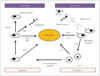
Stroke is a leading cause of disability in adults that has a heavy social burden worldwide. This disease is the third highest cause of mortality, resulting in approximately six million deaths annually.1 Acute cerebral ischemia accounts for more than 80% of all strokes and is due to brain arterial occlusion resulting from a thrombus or embolus. The pathophysiological processes following ischemic stroke are complex, involving bioenergetic failure, acidosis, loss of cell homeostasis, excitotoxicity, activation of neuronal and glial cells, and disruption of the blood-brain barrier (BBB) with infiltration of leukocytes.2 There is evidence that factors of the immune system are involved in all stages of acute cerebral ischemia (Fig. 1).3 The ischemic brain promotes a potent suppressive effect on lymphoid organs via the autonomic nervous system, which increases the risk of the poststroke infections that are major determinants of morbidity and mortality.4 On the other hand, the innate immune system contributes to subsequent repair of the damaged cerebral tissue.5
In this review we describe the main phases of the inflammatory processes during the early postischemic period, with an emphasis on the role of single mediators.

INFLAMMATION, ENDOTHELIUM, AND CLOT FORMATION

A growing amount of attention is being paid to the mechanisms of clot formation, particularly in the field of endovascular treatment of acute ischemic stroke. Although most of the focus has been on intervention devices (with developments from first- to second-generation devices, thrombus aspiration, and balloon-occlusion guiding catheters), some research groups have studied the physiopathological mechanisms of clot formation. The relation between inflammation and clot formation has been described previously, and it indicates that some cytokines (e.g., the RANTES)6 are responsible for the activation of a biochemical cascade or, indirectly, supports the concept that infections/inflammation promote atherogenesis and that some endothelial modifications that can lead to a prothrombotic status.7 Several research groups are currently focusing on the clot structure, with many findings supporting the concept of an inflammation-induced process. In particular, fibrinogen is susceptible to oxidation, and chronic exposure to oxidative stress supported by inflammation may lead to prothrombotic alterations in fibrin formation and architecture. Previous studies concerning air pollution have shown that particulate matter contributes to modulation of the fibrin structure.891011 Furthermore, some interesting studies that have used electron microscopy to investigate the clot surface (in myocardial infarction) highlight the less-investigated issue of the relation between the clot and the endothelium.12

EARLY POSTISCHEMIA TIME: THE ISCHEMIC CASCADE

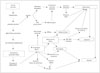
The ischemic cascade is represented by a complex series of interlinked molecular and cellular mechanisms that contribute to ischemic cell death via necrosis or apoptosis (Fig. 2). The primary insult after arterial occlusion is hypoperfusion, which dramatically reduces the availability of both glucose and oxygen in the brain, with particular vulnerability to ischemic injury being evident in specific regions: the caudate body, putamen, insular ribbon, paracentral lobule, precentral, and middle and inferior frontal gyri.13 This situation contributes to bioenergetic failure by stopping or slowing adenosine triphosphate (ATP) production.1415 A few minutes after an arterial occlusion, an ionic imbalance occurs with the abnormal influx of Na+ and efflux of K+, contributing to a widespread anoxic depolarization in the membranes of neurons and glial cells.16 The increased influx of Na+ into neurons causes the osmotic transport of water into cells that leads to cytotoxic edema, cell lysis, and necrosis. A recent neuroimaging study using 23Na MRI and quantitative histochemical K+ staining revealed heterogeneity in the rate of Na concentration increase and in the K+ distribution within the ischemic core.17 The reduced ATP production following Na/K imbalance (due to Na/K ATPase) also contributes to reduce the reuptake of glutamate, which is the main excitatory neurotransmitter.18 This condition overstimulates the glutamate receptors so as to influence the Ca2+ influx, producing a series of nuclear and cytoplasmic events (excitotoxicity) that lead to mitochondrial failure and apoptosis.1920 At the same time, the Ca2+ influx triggers the activation of catabolic enzymes with the production of arachidonic acid, and increases the formation of reactive oxygen species (ROS), mainly in neurons rather than astrocytes.21
Previous studies found that mitochondrial failure could be predicted from the K+ concentration. Indeed, the mitochondrial ATP-dependent K+ (mitoKATP) channel plays a critical role in the neuroprotective action, contributing to the so-called ischemic pre- and postconditioning states.22 The opening of mitoKATP channels attenuates the Ca2+ overload and inhibits the formation of free radicals and ROS that contribute to necrotic or apoptotic cell death.2324 The depolarization of other neurons produces a further Ca2+ influx and additional glutamate release, leading to local amplification of the ischemic damage.25 Contemporary with those processes, the persistence of arterial occlusion contributes to a critical reduction of pO2 and a concomitant increase in pCO2. In the case of hypercapnia, the tissue pH could fall to around 6.6 or lower if severe ischemia and tissue hypoxia occur; in the last situation, anaerobic glycolysis leads to lactic acid accumulation with signs of irreversible injury identifiable in the cell morphology.26 The acidosis state increases necrosis and cell death via a mechanism called acidotoxicity and mediated by Ca2+-permeable acid-sensing ion channels.272829 Other deleterious effects of acidosis influence the synthesis and degradation of cellular constituents, the mitochondrial function, the cell volume control, the postischemic flow, and the stimulation of ROS production, all conditions that occur also in the ischemic penumbra.30 Acidosis and ROS contribute to trigger a subsequent and concomitant phase represented by the activation of innate immunity and involving both resident cells (microglia) and circulating cells.

INFLAMMATORY AND INNATE IMMUNITY ACTIVATIONS

Inflammation in the ischemic brain: cell infiltration and damage

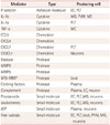
Postischemic inflammation begins in the vascular compartment immediately after arterial occlusion. The production of ROS leads to an increase in the procoagulant state involving the activation of complement, platelet, and endothelial cells.3132 The increased activity of cyclooxygenase-2 in inflammatory cells and neurons may lead to tissue damage due to excessive ROS production and toxic prostanoids.3334 ROS contribute also to reduce the availability of NO, leading to platelet aggregation and the adhesion of leukocytes, which aggravate the ischemic damage.35 There is evidence of iNOS (the inducible isoform of NO) being a critical effector and amplifier of tissue damage related to postischemic inflammation (Table 1).36 The oxidative stress and inflammatory mediators affect the permeability of the BBB, impairing the socalled neurovascular unit that includes endothelial cells, astrocytes, and neurons [involving matrix metalloproteinases (MMPs) or ROS], and allowing the extravasation of proteins and endothelial cells along with the activation of macrophages and mast cells via the ischemia and reperfusion mechanisms.37 This extravasation is also supported by a breakdown of the BBB-secondary to the pericyte death-leading to a long-lasting decrease in capillary blood flow.38
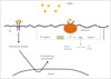
A few minutes after arterial occlusion and the associated intra- and extracellular modifications, the acute local damage is detected also by pattern-recognition receptors (PRR) (including Toll-like receptors) that respond to microbial structures (pathogen-associated molecular patterns) and host-derived danger signals (damage-associated molecular patterns).394041 These molecules can be released by stressed cells, such as during the ischemic cascade. There is new evidence that the PRR of neurons and glial cells can play a fundamental role in activating intracellular signaling pathways so as to enhance the proinflammatory expression of different genes (Fig. 3).4243 This mechanism activates the immune system elements, resulting in mast cells releasing vasoactive mediators (e.g., histamine), proteases, and tumor necrosis factor (TNF), while macrophages release proinflammatory cytokines.44
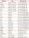
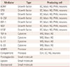
Adhesion-molecule P- and E-selectins (Table 2) and intercellular adhesion molecule-1 are expressed on the membrane after their intracellular translocation and with the rapid generation of proinflammatory signals.4546 The adhesion receptors mediate interactions between adhesion molecules and integrins, contributing to an initial rolling mechanism of leukocytes and leading to adhesion to the endothelium and to a subsequent transmigration into the brain parenchyma (leukocyte infiltration). Following ischemia, these cells rapidly release proinflammatory mediators into the area, which promotes ischemic injury via different pathways: 1) the cerebral no-reflow phenomenon by impeding the flow of red blood cells, 2) increased production of ROS and proteases at the endothelium surface, 3) platelet aggregation by leukotriene, prostaglandin, or eicosanoid production due to activation of leukocyte phospholipases, and 4) deleterious activity of proinflammatory cytokines in the penumbra area. In particular, during the acute phase of brain ischemia, microglia and macrophages release interleukin (IL)-1b, IL-6, and IL-18 from the transcriptional intracellular pathways activated by nucleotides from injured cells.47 These cytokines contribute to leukocyte infiltration in the damaged tissue, and they activate the presentation of antigens between dendritic cells and T-cells.48 T-cells lead to tissue damage by innate immunity, through interferon-gamma and ROS. T-cells activated by IL-23 released from microglia and macrophages produce IL-17, which worsens the acute ischemic cerebral injury.49 This unbalanced interplay between the immune and sympathetic nervous systems contributes to an early down-regulation of systemic cellular immune responses that leads to a functional deactivating of monocytes, T-helper cells, and invariant natural-killer T-cells.50
Previous studies have demonstrated the predictive role of certain acute immune and stress biomarkers on clinical outcomes: copeptin and mid-regional proatrial natriuretic peptide.5152 Even if the immune system is activated with recruiting elements in the focal ischemic area, specific injured cerebral sites contribute to different down-regulating responses being exhibited by the autonomous nervous system. In particular, involvement of the right frontoparietal cortex, insula, or brainstem could lead to increased cerebral inflammation and concomitant systemic immunosuppression.53 This process is characterized by the increased apoptosis of lymphocytes, suppression of peripheral cytokine release, and finally the inhibition of T-helper-1 cells and alteration of the T-helper-1/T-helper-2 ratio.54 The stroke-induced immunodepression contributes to increasing the risk of infection (influenced also by comorbidities such as age, diabetes mellitus, and atrial fibrillation) and consequently a poor functional outcome.

The activity of microglia

The microglia represent the resident immune cells of the central nervous system and can be activated by local and systemic infections, neurodegenerative conditions, and injury. Microglia are able to modify their morphology from a resting (ramified) to an active (amoeboid) state.55 Microglia respond rapidly to ischemic stroke and other injuring conditions. By entering the ischemic core within 60 minutes after the induction of focal ischemia without reperfusion, microglia significantly increase the number of their processes, while 24 hours later they are reduced in both number and distance from the insult. Previous studies have detected various forms of activated microglia.56 M1 is the proinflammatory type and is able to release TNF-a, IL-1b, IL-18, and IL-6. On the other hand, the M2 type is the healing cell involved in neuroprotection and repair, producing transforming growth factor (TGF)-b, nerve growth factor, and IL-4.57 The M1 type is observed for the first 24 hours in the ischemic core, and it increases in number over the first 2 weeks after the ischemic injury.58 The M2 type has been found at the end of the first 24 hours, entering the area during the first week before declining in number.55
Recent studies have provided evidence of microglia activation in the penumbra area. Indeed, the pattern of activity differs between the peri-infarct zone microglia and the ischemic core. During the first week, the M2 type was found to predominate in the ischemic core while the M1 type predominated in the peri-infarct zone.59 These observations suggest that the peri-infarct region is dominated by proinflammatory and activated microglia whose abundance increases during the first days after ischemic injury.60 The spatial distribution of the microglia phenotypes changes over time, which suggests the enlargement of injured and damaged cerebral tissue.

The role of inflammasomes

The above-mentioned inflammatory response leads to the production of proinflammatory cytokines and neuronal and glial cell death mediated by large intracellular multiprotein complexes called inflammasomes.45 Recent studies have demonstrated that the nod-like receptor pyrin (NLRP) and NLRP3 inflammasomes in neurons and glial cells allow detection of cellular damage and mediation of the inflammatory responses to aseptic tissue injury during cerebral ischemia.42 An increased activity of inflammasomes is also associated with neuron and glial cell death. Experimental studies and human data suggest that the NLRP1 and NLRP3 inflammasomes in brain cells could activate pro-caspase-1 (cleaving to caspase-1) to produce IL-1b and IL-18, which are proinflammatory cytokines, and lead to a particular type of cell death called pyroptosis (Fig. 3).61 In this way, the inflammasomes contribute to activating and supporting the innate immunity but also to worsening the tissue injury.

RESOLUTION OF INFLAMMATION AND REPAIR OF TISSUE

The inflammatory postischemia process is self-limiting, and its resolution is mediated by numerous factors that suppress the immune activity (Table 3). The termination of inflammation triggers structural and functional reorganization of the injured brain. The first mechanism involved in this phase is the removal of dead cells, which is performed by microglia and infiltrating macrophages, mainly comprising phagocytes.5962 The principal factors driving these cells to the ischemic site are purines released from injured cells and chemokines. Immunoglobulins directed against antigens of the central nervous system may also promote the release of IL-10 and TGF-b, which contribute to suppressing the immune process and to inhibiting the expression of adhesion molecules and the production of proinflammatory cytokines (Fig. 1).63 These pleiotropic immunoregulatory cytokines can facilitate tissue repair after promoting the resolution of inflammation, and they exert cytoprotective effects on the surviving cells in the ischemic area.35 Concomitant growth factors released by inflammatory cells, neurons, and astrocytes64 support cell sprouting, neurogenesis, and angiogenesis as well as matrix reorganization after ischemic injury. Insulin-like growth factor-1 is a critical factor in the sprouting of neurons after cerebral ischemia, while the reactivity of astrocytes is mandatory for the functional recovery of damaged tissue.65 Concomitant actions of vascular endothelial growth factor and neutrophil MMPs are required in angiogenesis, supporting the need for the combined activity of inflammatory cells and astrocytes.66

NEURONAL PRECONDITIONING AND INFLAMMATORY MEDIATORS

The brain is the most metabolically active organ in the body, consuming about 25% of total glucose and oxygen, and so a high oxidative stress is generated by its metabolism. Neurons would therefore be expected to respond actively to stress, have low capacities for replacement, and strong potential strategies for protection and repair. While most of the underlying neuroprotective and repairing pathways are still under investigation, this adaptive response to an insult by the activating intracellular signals is well known, and called preconditioning.67 Preconditioning occurs in two distinct phases. The first happens early and is associated with posttranslational modifications of proteins. This period lasts from minutes to 1–2 hours, and involves certain protective proteins (mainly protein chaperones) being rapidly released from stressed cells. The second phase requires the synthesis of new proteins. During this phase, which lasts several days, the proinflammatory cytokines IL-1b and TNF activate intracellular signaling pathways that lead to a tolerant state by mediating the stress responses. In particular, ROS and cytokines are able to activate Ras, Raf, and kinases of the cellular membrane, with the subsequent intracellular transcription and production of survival proteins. These last mechanisms contribute to preserving the endothelium via vascular protection and angiogenesis [mitogen-activated protein kinase p38 and extracellular signal-regulated kinase 1/2 (ERK 1/2)], the neurons via neuroprotection and neurogenesis [ERK 1/2 and nuclear factor kB (NF-kB)], and the glia via an anti-inflammatory action (serine/threonine kinase).68 Other proteins, called heat-shock proteins (Hsp), contribute to cellular maintenance.69 In particular, Hsp70 leads to neuronal survival, mitochondrial stabilization, and cell-death blocking, mainly by facilitating the activation of transcription factor and NF-kB.7071 NF-kB is affected also by the phosphorylation of Akt secondary to the activation of phosphoinositide-3-kinase72 This enzyme is also able to attenuate cellular apoptosis in the neurovascular unit, but it is inhibited after ischemia or reperfusion so as to induce cell death.73 Several studies have indicated that any stimulus that modifies brain function appears to increase the resistance of the brain to further injuries, including to different types of injury.74 The results of some studies involving patients with previous transient ischemic attacks suggest that brain preconditioning and tolerance occur because these patients have a more favorable clinical outcome and smaller infarcts.7576

CONCLUSIONS

Inflammatory responses during the acute stage of ischemic stroke have effects ranging from deleterious to beneficial. A first reaction is negative due to cytotoxicity, which leads to tissue damage by cellular death. At the same time, inflammation exerts protective effects by stimulating a preconditioning status that preserves brain tissue by adapting the brain itself after an insult. Finally, inflammatory mediators contribute to autolimit the pathological process and to repair the injured parenchyma by remodeling the tissue.
Future observations will contribute to a better understanding of other inflammatory mechanisms involved in these early stages of ischemic stroke, and this information will potentially lead to the development of effective neuroprotective agents.

SEARCH STRATEGY AND SELECTION CRITERIA

References for use in this review were identified by searching the PubMed and EMBASE databases for articles published between 1976 and June 2016, as well as references in relevant articles. The search terms used were “inflammation,” “immune system,” “immunity,” “brain ischemia,” “cerebral ischemia,” “stroke,” “cerebral occlusion,” and “cerebrovascular disease.” There were no language restrictions. The final reference list was generated based on the relevance to the topics covered in this review.

Figures and Tables

Fig. 1

Postischemic inflammation. Necrotic neurons release damage-associated molecular patterns (DAMPs), and these molecules activate macrophages via pattern-recognition receptors and inflammasomes. The activated macrophages contribute to enhance the inflammatory process via the release of proinflammatory cytokines and recruiting T-cells that contribute to maintain inflammation by interleukin (IL)-17. At several days after the acute injury, the cellular elements of the innate immune system change to an anti-inflammatory phenotype, contributing to inhibit the inflammation (dashed lines). In particular, anti-inflammatory cytokines (e.g., IL-10) are released. During this phase, the postischemic inflammation is resolved by the clearance of debris as well as angiogenesis supported by the release of growth factors. IGF: insulin-like growth factor, TGF, transforming growth factor, TNF: tumor necrosis factor, VEGF: vascular endothelial growth factor.

jcn-13-1-g001
Fig. 2

Cerebral ischemic cascade. AMPA: α-amino-3-hydroxy-5-methyl-4-isoxazolepropionic acid, BAD: Bcl-2-associated death promoter, BBB: blood-brain barrier, COX: cyclo-oxigenase, IL: interleukin, NMDA: N-methyl-D-aspartate, TNF: tumor necrosis factor.

jcn-13-1-g002
Fig. 3

DAMP receptors and signaling pathways. Cells detect DAMPs via DAMP receptors in two ways: (1) activation of a type of pattern-recognition receptor [Toll-like receptor (TLR)] and (2) activation of inflammasomes. The first mechanism involves proinflammatory factors being released by the nuclear gene expression mediated by transcriptional mediators activated by TLR. The second mechanism involves the activation of caspase-1 leading to the clivation of the proinflammatory cytokines IL-1 and IL-18, converting them into their activated forms. DAMP: damage-associated molecular pattern, IL: interleukin, NLRP: nod-like receptor pyrin.

jcn-13-1-g003
Table 1

Mediators of amplification of postischemic inflammation

jcn-13-1-i001
Mediator Type Producing cell
ICAM1 Adhesion molecule EC, Leuk, PVM, MG, AG
VCAM-1 Adhesion molecule EC, Leuk, PVM, MG, AG
P-selectin Adhesion molecule EC, Leuk, PVM, MG, AG
E-selectin Adhesion molecule EC, Leuk, PVM, MG, AG
Mac-1 Adhesion molecule EC, Leuk, PVM, MG, AG
VLA-1 Adhesion molecule EC, Leuk, PVM, MG, AG
IL-1 Cytokine EC, neurons, PVM, MG, AG
IL-6 Cytokine EC, neurons, PVM, MG, AG
IL-10 Cytokine EC, neurons, PVM, MG, AG
IL-17 Cytokine EC, neurons, PVM, MG, AG
IL-20 Cytokine EC, neurons, PVM, MG, AG
TNF Cytokine EC, neurons, PVM, MG, AG
CCL2 Chemokine EC, neurons, PVM, MG, AG
CCL3 Chemokine EC, neurons, PVM, MG, AG
CCL5 Chemokine EC, neurons, PVM, MG, AG
CXCL2/3 Chemokine EC, neurons, PVM, MG, AG
CXCL8 Chemokine EC, neurons, PVM, MG, AG
MMP2 Protease Circ, EC, AG, neurons
MMP9 Protease Circ, EC, AG, neurons
Complement Protease Circ, EC, AG, neurons
iNOS Other mediator MG, Leuk, EC
COX-2 Other mediator Neurons, MG, Leuk, EC

Modified from Iadecola et al. Nat Med 2011;17:796-808, with permission of Springer Nature.45

AG: astroglia, CCL: chemokine ligand, Circ: plasma, COX: cyclo-oxigenase, CX: d-chemokine ligand, EC: endothelial cell, ICAM: intercellular adhesion molecule, IL: interleukin, iNOS: inducible isoform of NO, Mac: macrophage antigen, MG: microglia, MMP: matrix metalloproteinase, PVM: perivascular macrophages, TNF: tumor necrosis factor, VCAM: vascular cell adhesion molecule, VLA: very late antigen.

Table 2

Mediators of initial postischemic inflammation

jcn-13-1-i002
Mediator Type Producing cell
P-selectin Adhesion molecule EC, PLT
IL-1b Cytokine MG, PVM, MC
IL-1a Cytokine PLT
TNF-a Cytokine MC
CCL5 Chemokine
CXCL4 Chemokine
CXCL7 Chemokine PLT
CX3CL1 Chemokine Neurons
Elastase Protease
MMP8 Protease
MMP9 Protease
MT6-MMP Protease Leuk
Clotting factors Protease Plasma
Complement Protease Plasma, EC, neuron
Prostanoids Small molecule EC, PLT, MG, neurons
Leukotrienes Small molecule EC, PLT, MG, neurons
ATP Small molecule Plasma, neurons
Free radicals Small molecule EC, PLT, Leuk, PVM, MG, neurons

Modified from Iadecola et al. Nat Med 2011;17:796-808, with permission of Springer Nature.45

ATP: adenosine triphosphate, CCL: chemokine ligand, CX: d-chemokine ligand, EC: endothelial cell, IL: interleukin, Leuk: leukocytes, MC: mast cells, MG: microglia, MMP: matrix metalloproteinase, PLT: platelets, PVM: perivascular macrophages, TNF: tumor necrosis factor.

Table 3

Mediators of resolution of postischemic inflammation

jcn-13-1-i003
Mediator Type Producing cell
BDNF Growth factor EC, Macr, AG, PVM, neurons
EPO Growth factor EC, Macr, AG, PVM, neurons
FGF Growth factor EC, Macr, AG, PVM, neurons
G-CSF Growth factor EC, Macr, AG, PVM, neurons
IGF-1 Growth factor EC, Macr, AG, PVM, neurons
NGF Growth factor EC, Macr, AG, PVM, neurons
VEGF Growth factor EC, Macr, AG, PVM, neurons
TGF-b Cytokine MG, Macr, AG
IL-10 Cytokine MG, Macr, AG
IL-17 Cytokine MG, Macr, AG
IL-23 Cytokine MG, Macr, AG
MMP9 Protease AG, neurons
Complement Protease Circ, EC, AG, neurons
Prostaglandin Small molecule
Lipoxin Small molecule
Docosanoid Small molecule

Modified from Iadecola et al. Nat Med 2011;17:796-808, with permission of Springer Nature.45

AG: astroglia, BDNF: brain-derived neurotrophic factor, Circ: plasma, EC: endothelial cell, EPO: erythropoietin, FGF: fibroblast growth factor, G-CSF: granulocyte colony-stimulating factor, IGF: insulin-like growth factor, IL: interleukin, Macr: macrophages, MG: microglia, MMP: matrix metalloproteinase, NGF: nerve growth factor, PVM: perivascular macrophages, TGF: transforming growth factor, VEGF: vascular endothelial growth factor.

Notes

Conflicts of Interest The authors have no financial conflicts of interest.

References

1. Writing Group Members. Mozaffarian D, Benjamin EJ, Go AS, Arnett DK, Blaha MJ, et al. Executive summary: heart disease and stroke statistics--2016 update: a report from the American Heart Association. Circulation. 2016; 133:447–454.
crossref
2. Woodruff TM, Thundyil J, Tang SC, Sobey CG, Taylor SM, Arumugam TV. Pathophysiology, treatment, and animal and cellular models of human ischemic stroke. Mol Neurodegener. 2011; 6:11.
crossref
3. Meisel C, Schwab JM, Prass K, Meisel A, Dirnagl U. Central nervous system injury-induced immune deficiency syndrome. Nat Rev Neurosci. 2005; 6:775–786.
crossref
4. Famakin BM. The immune response to acute focal cerebral ischemia and associated post-stroke immunodepression: a focused review. Aging Dis. 2014; 5:307–326.
crossref
5. Wattananit S, Tornero D, Graubardt N, Memanishvili T, Monni E, Tatarishvili J, et al. Monocyte-derived macrophages contribute to spontaneous long-term functional recovery after stroke in mice. J Neurosci. 2016; 36:4182–4195.
crossref
6. Libby P, Simon DI. Inflammation and thrombosis: the clot thickens. Circulation. 2001; 103:1718–1720.
7. Libby P, Aikawa M, Jain MK. Vascular endothelium and atherosclerosis. Handb Exp Pharmacol. 2006; (176 Pt 2):285–306.
crossref
8. Undas A, Ariëns RA. Fibrin clot structure and function: a role in the pathophysiology of arterial and venous thromboembolic diseases. Arterioscler Thromb Vasc Biol. 2011; 31:e88–e99.
9. Mehta BP, Nogueira RG. Should clot composition affect choice of endovascular therapy? Neurology. 2012; 79:13 Suppl 1. S63–S67.
crossref
10. Yuki I, Kan I, Vinters HV, Kim RH, Golshan A, Vinuela FA, et al. The impact of thromboemboli histology on the performance of a mechanical thrombectomy device. AJNR Am J Neuroradiol. 2012; 33:643–648.
crossref
11. Singh P, Kaur R, Kaur A. Clot composition and treatment approach to acute ischemic stroke: the road so far. Ann Indian Acad Neurol. 2013; 16:494–497.
crossref
12. Becatti M, Marcucci R, Bruschi G, Taddei N, Bani D, Gori AM, et al. Oxidative modification of fibrinogen is associated with altered function and structure in the subacute phase of myocardial infarction. Arterioscler Thromb Vasc Biol. 2014; 34:1355–1361.
crossref
13. Payabvash S, Souza LC, Wang Y, Schaefer PW, Furie KL, Halpern EF, et al. Regional ischemic vulnerability of the brain to hypoperfusion: the need for location specific computed tomography perfusion thresholds in acute stroke patients. Stroke. 2011; 42:1255–1260.
crossref
14. Hertz L. Bioenergetics of cerebral ischemia: a cellular perspective. Neuropharmacology. 2008; 55:289–309.
crossref
15. Rossi DJ, Brady JD, Mohr C. Astrocyte metabolism and signaling during brain ischemia. Nat Neurosci. 2007; 10:1377–1386.
crossref
16. Kaplan JH. Biochemistry of Na,K-ATPase. Annu Rev Biochem. 2002; 71:511–535.
crossref
17. Yushmanov VE, Kharlamov A, Yanovski B, LaVerde G, Boada FE, Jones SC. Correlated sodium and potassium imbalances within the ischemic core in experimental stroke: a 23Na MRI and histochemical imaging study. Brain Res. 2013; 1527:199–208.
crossref
18. Swanson RA, Farrell K, Simon RP. Acidosis causes failure of astrocyte glutamate uptake during hypoxia. J Cereb Blood Flow Metab. 1995; 15:417–424.
crossref
19. Li XM, Yang JM, Hu DH, Hou FQ, Zhao M, Zhu XH, et al. Contribution of downregulation of L-type calcium currents to delayed neuronal death in rat hippocampus after global cerebral ischemia and reperfusion. J Neurosci. 2007; 27:5249–5259.
crossref
20. Zhang QG, Xu YL, Li HC, Han D, Zhang GY. NMDA receptor/L-VGCC-dependent expression and AMPA/KA receptor-dependent activation of c-Jun induced by cerebral ischemia in rat hippocampus. Neurosci Lett. 2006; 398:268–273.
crossref
21. Kahlert S, Zündorf G, Reiser G. Glutamate-mediated influx of extracellular Ca2+ is coupled with reactive oxygen species generation in cultured hippocampal neurons but not in astrocytes. J Neurosci Res. 2005; 79:262–271.
crossref
22. Robin E, Simerabet M, Hassoun SM, Adamczyk S, Tavernier B, Vallet B, et al. Postconditioning in focal cerebral ischemia: role of the mitochondrial ATP-dependent potassium channel. Brain Res. 2011; 1375:137–146.
crossref
23. Teshima Y, Akao M, Li RA, Chong TH, Baumgartner WA, Johnston MV, et al. Mitochondrial ATP-sensitive potassium channel activation protects cerebellar granule neurons from apoptosis induced by oxidative stress. Stroke. 2003; 34:1796–1802.
crossref
24. McCabe RD, Bakarich MA, Srivastava K, Young DB. Potassium inhibits free radical formation. Hypertension. 1994; 24:77–82.
crossref
25. Sattler R, Tymianski M. Molecular mechanisms of calcium-dependent excitotoxicity. J Mol Med (Berl). 2000; 78:3–13.
crossref
26. Rehncrona S. Brain acidosis. Ann Emerg Med. 1985; 14:770–776.
crossref
27. Sherwood TW, Lee KG, Gormley MG, Askwith CC. Heteromeric acid-sensing ion channels (ASICs) composed of ASIC2b and ASIC1a display novel channel properties and contribute to acidosis-induced neuronal death. J Neurosci. 2011; 31:9723–9734.
crossref
28. Xiang Z, Yuan M, Hassen GW, Gampel M, Bergold PJ. Lactate induced excitotoxicity in hippocampal slice cultures. Exp Neurol. 2004; 186:70–77.
crossref
29. Xiong ZG, Zhu XM, Chu XP, Minami M, Hey J, Wei WL, et al. Neuroprotection in ischemia: blocking calcium-permeable acid-sensing ion channels. Cell. 2004; 118:687–698.
30. Anderson RE, Tan WK, Meyer FB. Brain acidosis, cerebral blood flow, capillary bed density, and mitochondrial function in the ischemic penumbra. J Stroke Cerebrovasc Dis. 1999; 8:368–379.
crossref
31. Carden DL, Granger DN. Pathophysiology of ischaemia-reperfusion injury. J Pathol. 2000; 190:255–266.
crossref
32. Peerschke EI, Yin W, Ghebrehiwet B. Complement activation on platelets: implications for vascular inflammation and thrombosis. Mol Immunol. 2010; 47:2170–2175.
crossref
33. Hurley SD, Olschowka JA, O'Banion MK. Cyclooxygenase inhibition as a strategy to ameliorate brain injury. J Neurotrauma. 2002; 19:1–15.
crossref
34. Minghetti L. Role of COX-2 in inflammatory and degenerative brain diseases. Subcell Biochem. 2007; 42:127–141.
crossref
35. Iadecola C, Abe T, Kunz A, Hallembeck J. Cerebral ischemia and inflammation. In : Mohr JP, Wolf PA, Grotta JC, Moskowitz MA, Mayberg M, von Kummer R, editors. Stroke: Pathophysiology, Diagnosis and Management. 5th ed. Philadelphia: Elsevier Health Sciences;2011. p. 138–153.
36. Garcia-Bonilla L, Moore JM, Racchumi G, Zhou P, Butler JM, Iadecola C, et al. Inducible nitric oxide synthase in neutrophils and endothelium contributes to ischemic brain injury in mice. J Immunol. 2014; 193:2531–2537.
crossref
37. Kim JY, Kawabori M, Yenari MA. Innate inflammatory responses in stroke: mechanisms and potential therapeutic targets. Curr Med Chem. 2014; 21:2076–2097.
crossref
38. Hall CN, Reynell C, Gesslein B, Hamilton NB, Mishra A, Sutherland BA, et al. Capillary pericytes regulate cerebral blood flow in health and disease. Nature. 2014; 508:55–60.
crossref
39. Zhang Q, Raoof M, Chen Y, Sumi Y, Sursal T, Junger W, et al. Circulating mitochondrial DAMPs cause inflammatory responses to injury. Nature. 2010; 464:104–107.
crossref
40. Matzinger P. The evolution of the danger theory. Interview by Lauren Constable, commissioning editor. Expert Rev Clin Immunol. 2012; 8:311–317.
41. Caso JR, Pradillo JM, Hurtado O, Lorenzo P, Moro MA, Lizasoain I. Toll-like receptor 4 is involved in brain damage and inflammation after experimental stroke. Circulation. 2007; 115:1599–1608.
crossref
42. Savage CD, Lopez-Castejon G, Denes A, Brough D. NLRP3-Inflammasome activating DAMPs stimulate an inflammatory response in glia in the absence of priming which contributes to brain inflammation after injury. Front Immunol. 2012; 3:288.
crossref
43. Fann DY, Lee SY, Manzanero S, Chunduri P, Sobey CG, Arumugam TV. Pathogenesis of acute stroke and the role of inflammasomes. Ageing Res Rev. 2013; 12:941–966.
crossref
44. Strbian D, Karjalainen-Lindsberg ML, Tatlisumak T, Lindsberg PJ. Cerebral mast cells regulate early ischemic brain swelling and neutrophil accumulation. J Cereb Blood Flow Metab. 2006; 26:605–612.
crossref
45. Iadecola C, Anrather J. The immunology of stroke: from mechanisms to translation. Nat Med. 2011; 17:796–808.
crossref
46. Huang J, Choudhri TF, Winfree CJ, McTaggart RA, Kiss S, Mocco J, et al. Postischemic cerebrovascular E-selectin expression mediates tissue injury in murine stroke. Stroke. 2000; 31:3047–3053.
crossref
47. Allan SM, Rothwell NJ. Cytokines and acute neurodegeneration. Nat Rev Neurosci. 2001; 2:734–744.
crossref
48. Burnstock G. Purinergic signalling and disorders of the central nervous system. Nat Rev Drug Discov. 2008; 7:575–590.
crossref
49. Shichita T, Sugiyama Y, Ooboshi H, Sugimori H, Nakagawa R, Takada I, et al. Pivotal role of cerebral interleukin-17-producing gammadeltaT cells in the delayed phase of ischemic brain injury. Nat Med. 2009; 15:946–950.
crossref
50. Meisel A, Meisel C, Harms H, Hartmann O, Ulm L. Predicting post-stroke infections and outcome with blood-based immune and stress markers. Cerebrovasc Dis. 2012; 33:580–588.
crossref
51. Katan M, Fluri F, Morgenthaler NG, Schuetz P, Zweifel C, Bingisser R, et al. Copeptin: a novel, independent prognostic marker in patients with ischemic stroke. Ann Neurol. 2009; 66:799–808.
crossref
52. Katan M, Fluri F, Schuetz P, Morgenthaler NG, Zweifel C, Bingisser R, et al. Midregional pro-atrial natriuretic peptide and outcome in patients with acute ischemic stroke. J Am Coll Cardiol. 2010; 56:1045–1053.
crossref
53. Günther A, Witte OW, Hoyer D. Autonomic dysfunction and risk stratification assessed from heart rate pattern. Open Neurol J. 2010; 4:39–49.
crossref
54. Hannawi Y, Hannawi B, Rao CP, Suarez JI, Bershad EM. Stroke-associated pneumonia: major advances and obstacles. Cerebrovasc Dis. 2013; 35:430–443.
crossref
55. Taylor RA, Sansing LH. Microglial responses after ischemic stroke and intracerebral hemorrhage. Clin Dev Immunol. 2013; 2013:746068.
crossref
56. Starossom SC, Mascanfroni ID, Imitola J, Cao L, Raddassi K, Hernandez SF, et al. Galectin-1 deactivates classically activated microglia and protects from inflammation-induced neurodegeneration. Immunity. 2012; 37:249–263.
crossref
57. Kawanokuchi J, Shimizu K, Nitta A, Yamada K, Mizuno T, Takeuchi H, et al. Production and functions of IL-17 in microglia. J Neuroimmunol. 2008; 194:54–61.
crossref
58. Morrison HW, Filosa JA. A quantitative spatiotemporal analysis of microglia morphology during ischemic stroke and reperfusion. J Neuroinflammation. 2013; 10:4.
crossref
59. Denes A, Vidyasagar R, Feng J, Narvainen J, McColl BW, Kauppinen RA, et al. Proliferating resident microglia after focal cerebral ischaemia in mice. J Cereb Blood Flow Metab. 2007; 27:1941–1953.
crossref
60. Villarreal A, Rosciszewski G, Murta V, Cadena V, Usach V, Dodes-Traian MM, et al. Isolation and characterization of ischemia-derived astrocytes (IDAs) with ability to transactivate quiescent astrocytes. Front Cell Neurosci. 2016; 10:139.
crossref
61. Fink SL, Cookson BT. Caspase-1-dependent pore formation during pyroptosis leads to osmotic lysis of infected host macrophages. Cell Microbiol. 2006; 8:1812–1825.
crossref
62. Schilling M, Besselmann M, Müller M, Strecker JK, Ringelstein EB, Kiefer R. Predominant phagocytic activity of resident microglia over hematogenous macrophages following transient focal cerebral ischemia: an investigation using green fluorescent protein transgenic bone marrow chimeric mice. Exp Neurol. 2005; 196:290–297.
crossref
63. Nathan C, Ding A. Nonresolving inflammation. Cell. 2010; 140:871–882.
crossref
64. Li S, Overman JJ, Katsman D, Kozlov SV, Donnelly CJ, Twiss JL, et al. An age-related sprouting transcriptome provides molecular control of axonal sprouting after stroke. Nat Neurosci. 2010; 13:1496–1504.
crossref
65. Hayakawa K, Nakano T, Irie K, Higuchi S, Fujioka M, Orito K, et al. Inhibition of reactive astrocytes with fluorocitrate retards neurovascular remodeling and recovery after focal cerebral ischemia in mice. J Cereb Blood Flow Metab. 2010; 30:871–882.
crossref
66. Hao Q, Chen Y, Zhu Y, Fan Y, Palmer D, Su H, et al. Neutrophil depletion decreases VEGF-induced focal angiogenesis in the mature mouse brain. J Cereb Blood Flow Metab. 2007; 27:1853–1860.
crossref
67. Obrenovitch TP. Molecular physiology of preconditioning-induced brain tolerance to ischemia. Physiol Rev. 2008; 88:211–247.
crossref
68. Dawson DA, Furuya K, Gotoh J, Nakao Y, Hallenbeck JM. Cerebrovascular hemodynamics and ischemic tolerance: lipopolysaccharide-induced resistance to focal cerebral ischemia is not due to changes in severity of the initial ischemic insult, but is associated with preservation of microvascular perfusion. J Cereb Blood Flow Metab. 1999; 19:616–623.
crossref
69. Daugaard M, Rohde M, Jäättelä M. The heat shock protein 70 family: highly homologous proteins with overlapping and distinct functions. FEBS Lett. 2007; 581:3702–3710.
crossref
70. Giffard RG, Han RQ, Emery JF, Duan M, Pittet JF. Regulation of apoptotic and inflammatory cell signaling in cerebral ischemia: the complex roles of heat shock protein 70. Anesthesiology. 2008; 109:339–348.
crossref
71. Yenari MA, Liu J, Zheng Z, Vexler ZS, Lee JE, Giffard RG. Antiapoptotic and anti-inflammatory mechanisms of heat-shock protein protection. Ann N Y Acad Sci. 2005; 1053:74–83.
crossref
72. Koh SH, Lo EH. The role of the PI3K pathway in the regeneration of the damaged brain by neural stem cells after cerebral infarction. J Clin Neurol. 2015; 11:297–304.
crossref
73. Lan R, Xiang J, Zhang Y, Wang GH, Bao J, Li WW, et al. PI3K/Akt pathway contributes to neurovascular unit protection of Xiao-Xu-Ming decoction against focal cerebral ischemia and reperfusion injury in rats. Evid Based Complement Alternat Med. 2013; 2013:459467.
74. Schaller B. Ischemic preconditioning as induction of ischemic tolerance after transient ischemic attacks in human brain: its clinical relevance. Neurosci Lett. 2005; 377:206–211.
crossref
75. Fu Y, Sun JL, Ma JF, Geng X, Sun J, Liu JR, et al. The neuroprotection of prodromal transient ischaemic attack on cerebral infarction. Eur J Neurol. 2008; 15:797–801.
crossref
76. Moncayo J, de Freitas GR, Bogousslavsky J, Altieri M, van Melle G. Do transient ischemic attacks have a neuroprotective effect? Neurology. 2000; 54:2089–2094.
crossref
TOOLS
Similar articles