

 PDF
PDF ePub
ePub Citation
 Print
Print


INTRODUCTION
Idiopathic normal-pressure hydrocephalus (INPH) is an uncommon neurological disorder. Of 563 cases showing the neuropathology of a dementing illness at autopsy, INPH was suspected only in 9 (1.6%).1 Nevertheless, the diagnosis and understanding of INPH are important because INPH is regarded as a potentially treatable neurological disorder.2 INPH is an adult-onset syndrome of uncertain origin, with symptoms of gait disturbance, cognitive impairment, and urinary dysfunction, that involves nonobstructive enlargement of the cerebral ventricles, along with normal cerebrospinal fluid (CSF) pressure at lumbar puncture.3 Although patients with INPH may present with varying combinations or degrees of each of these classic clinical symptoms, the most frequent and important clinical feature of INPH is that of gait disturbance.4
The CSF tap test (CSFTT) has been considered as a valuable examination for the diagnosis and prediction of shunt effectiveness in patients with INPH.5 Surgical treatment by placement of a ventricular shunt is indicated for patients with INPH who show a positive CSFTT response.5 Clinical improvement after the CSFTT is an important criterion that enhances diagnostic certainty from possible to probable, following the Japanese guideline.5
Parkinsonism is one of the most prevalent, chronic neurological syndromes facing the elderly.6 Differential diagnosis of patients with parkinsonism is important because prognosis and treatment options can differ substantially for Parkinson's disease (PD) and other parkinsonian disorders.7 A better understanding of parkinsonism in INPH is necessary, because parkinsonism is also observed in patients with INPH.8 In fact, in one report, INPH is described as shunt-responsive parkinsonism.8
The motor section of the Unified Parkinson's Disease Rating Scale (UPDRS-III) is well known as the gold standard for evaluating motor symptoms in PD and provides a semiquantitative analysis of the severity of parkinsonian signs and symptoms.9 Parkinsonian motor deficits also have been assessed with the UPDRS-III in other neurodegenerative diseases, such as Alzheimer's disease.9 Clinicians and researchers have often used the UPDRS-III scale to determine the standard of response in some interventions for parkinsonian patients. And, to the authors' knowledge, their changes on the UPDRS-III following the CSFTT have not yet been reported in INPH patients.
Our aims were to analyze the characteristics of parkinsonian features and to characterize changes in parkinsonian motor symptoms before and after the CSFTT in INPH patients who had a positive response to the CSFTT. We also explored whether a relationship exists between gait function and parkinsonism severity in these patients.
METHODS
Participants
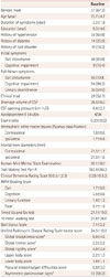
INPH participants were prospectively recruited from patients at the Center for Neurodegenerative Diseases of Kyungpook National University Medical Center, Korea from July 2011 to November 2014. This study was approved by our local Institutional Review Board. The criteria proposed by Relkin et al.10 was used to diagnose INPH. A lumbar tap removing 30–50 mL of CSF was performed on all 72 patients with INPH. All patients were re-evaluated after the tap using the INPH Grading Scale (INPHGS), the Korean-Mini Mental State Examination (K-MMSE) and the Timed Up and Go Test (TUG). Gait change was evaluated repeatedly over 7 days after the tap.1112 During the follow-up period, results with the greatest improvement were used for comparisons between baseline and follow-up measurements.1112 Changes in cognition and urination were evaluated at one week.13 Response to the CSFTT was defined using these three major scales.5 INPH patients having a positive response to the CSFTT were enrolled to increase diagnostic certainty, and the following criteria were used to identify responders: improvement of one point or more on the INPHGS, more than 10% improvement in time on the TUG test, or more than 3 points improvement on the K-MMSE.514 The final sample for analysis was 55 INPH patients. The demographic and clinical baseline characteristics are given in Table 1.
Assessing illness severity
The patients' general cognitive state and severity of dementia were evaluated with the K-MMSE and Clinical Dementia Rating Scale.1516 The Trail Making Test Part A (TMT-A) is a common neuropsychological test to evaluate psychomotor speed and is often used for patients with INPH.17 In this study, the amount of time taken to complete the TMT-A was recorded.
The INPHGS is a clinician-rated scale to assess the severity of the fundamental symptoms of INPH (cognitive impairment, gait disturbance, and urinary disturbance) after an unstructured interview with patients and caregivers.18 The score of each domain ranges from 0 to 4.1318 Grade 0 indicates normal, and grade 1 indicates subjective symptoms but no objective disturbance.1318 Grade 2, 3, and 4 indicate mild, moderate and severe disturbances, respectively.1318
Gait assessment included measurements of time on the TUG and 10-meter walking test.18 They were performed four times consecutively and the mean score was determined. Features of gait disturbance were also estimated using the Gait Status Scale (GSS).18
An experienced rater, who was blinded to the patient's diagnosis, performed UPDRS-III evaluations. We also used the following subscores based on the UPDRS-III: global bradykinesia score (items 23–26 and 31),19 global tremor score (items 20 and 21),20 global rigidity score (item 22),19 and postural instability/gait difficulties (PIGD) score (items 27–30).21 The upper body score was calculated as the sum of the mean score for right and left upper limbs on item 20, the mean score for right and left upper limbs on item 22, and the mean score for right and left upper limbs on items 23–25.19 The lower body score was calculated as the sum of the mean score for right and left lower limbs on item 20, the mean score for right and left lower limbs on item 22, and the mean score for right and left lower limbs on item 26.19 The right score was calculated as the sum of the scores for right upper and lower limbs on items 20 and 22–26.19 The left score was calculated as the sum of the scores for left upper and lower limbs on items 20 and 22–26.19 An asymmetry index was calculated as the absolute value of the right minus left scores from the UPDRS-III score.19 A UPDRS-III asymmetry index difference of at least two points was used as the threshold for defining clinical asymmetry.19 The UPDRS-III was applied again 24 hours after tap by the same rater.
Magnetic resonance imaging acquisition
MRI data were obtained using a 3.0 Tesla system (GE Discovery MR750, GE Healthcare). We performed MRI in INPH patients before the CSFTT. The evaluation of white matter lesions (WML) was provided by T2 weighted and fluid attenuated inversion recovery images. Hemispheric WML were rated using the Fazekas scale, scoring 0–3 (for deep and periventricular WML, where 0=none and 3=severe).22 For total hemispheric WML, we added scores in the deep and periventricular regions and obtained the average.2223 The diameter of the each frontal horn was measured electronically at the level of the head of the caudate nucleus and the measurements were used to assess the degree of the asymmetric lateral ventricle.24
Statistical analyses
The IBM SPSS Statistics for Windows version 21.0.0 was used for analyses of data. A paired t-test was used to compare the upper and lower body scores. The comparisons of the hemispheric WML and frontal horn diameters within subjects (i.e., between the contra- and ipsilateral hemispheres in accordance with the body side of the dominant motor symptoms) were also done using the paired t-test. Pearson's or Spearman's correlations were employed to investigate the relationship between gait function and the severity of parkinsonism at baseline in INPH. The changes in parkinsonian motor symptoms before and after the CSFTT were analyzed using the paired t-test or McNemar's test. The paired t-test was used for comparison of the continuous variables, including the TUG, 10-meter walking test, GSS and total motor score, global bradykinesia score, global tremor score, global rigidity score, upper body score, lower body score, and PIGD score of UPDRS-III. We used McNemar's test to compare the frequency of asymmetric parkinsonism in our INPH patients between baseline and follow-up. Statistical significance was set at p<0.05.
RESULTS
Baseline clinical characteristics and MRI findings (Table 1)
The initial mean UPDRS-III score was 24.5±10.2. There was no significant difference between the upper and lower body scores (paired t-test, p=0.174). Higher lower body scores correlated significantly with a higher upper body score (r=0.506, p<0.001). The parkinsonian signs were asymmetrical in 58.2% of the patients. No association was found between handedness and the side of symptom dominance. There were no significant differences in the hemispheric WML and frontal horn diameters between hemispheres ipsilateral and contralateral to the body side of the dominant motor symptoms (paired t-test, p=0.572 for the hemispheric WML and p=0.253 for the frontal horn diameters).
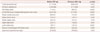
Correlations between gait function and parkinsonism severity in INPH (Table 2)
At baseline, the TUG and 10-meter walking test scores were positively correlated with the total motor score, global bradykinesia score, global rigidity score, upper body score, lower body score, and PIGD score of UPDRS-III. The GSS and INPHGS gait scores were positively correlated with the total motor score, global bradykinesia score, lower body score, and PIGD score of UPDRS-III. Not surprisingly, the TUG, 10-meter walking test, GSS, and INPHGS gait scores were more strongly correlated with the lower body score than with the upper body score.
Gait parameters and UPDRS-III measures in patients with INPH before and after the CSFTT
Differences in the gait parameters and UPDRS-III measures before and 24 hours after the CSFTT are shown in Table 3. The TUG score improved significantly (p<0.05). The 10-meter walking test and GSS results also improved significantly (p<0.01).
The total motor score, global bradykinesia score, upper body score, and lower body score of UPDRS-III significantly improved (p<0.01). The PIGD score of UPDRS-III improved, but less significantly (p<0.05). Asymmetric presentation of parkinsonian features was significantly less frequent at follow-up than at baseline evaluation (p<0.05). The global tremor score of UPDRS-III marginally improved (p=0.047). The global rigidity score did not significantly improve.
DISCUSSION
One of the most important findings of this study was that there was no significant difference in the severity of parkinsonism between the upper and lower extremities in our INPH patients, as measured by the UPDRS-III. Furthermore, asymmetric parkinsonism was observed in more than half of the INPH patients.
Generally, lower body parkinsonism is characteristic in INPH.25 Lower body parkinsonism classically presents as a slow, wide-based gait, short shuffling steps, and difficulty in turning and tandem walking.26 However, the clinical presentation of INPH seems to be commonly not limited to the classical triad.27 In a previous study, upper extremity bradykinesia was also present in 62 percent of INPH patients.28 It was suggested that the features of upper limb motor disability found in INPH patients seem to resemble those encountered in PD.2728 Our finding is consistent with the aforementioned studies. Using a strategy reported in a previous study,19 we found that patients with INPH suffered from a comparable degree of parkinsonism between the upper and lower extremities. As a possible explanation for this result, we can speculate as follows. The basal ganglia circuitry processes the signals that flow from the cortex, allowing the correct execution of voluntary movements.29 Dysfunction of basal ganglia circuitry is known to be mainly responsible for the development of the cardinal features of PD.3031 Considering the connection between cerebral perfusion (also referred to as cerebral blood flow) and brain function,432 and the fact that significant reductions in mean cerebral blood flow of the basal ganglia and the thalamus were found in INPH patients compared with controls,4 this may explain the evident upper body parkinsonism also observed in our patients (as seen in PD).
In general, asymmetry with regard to parkinsonian features is considered as strong evidence toward a PD diagnosis. In addition, it was reported that of the 4,057 right-handed patients who experienced asymmetrical onset of PD motor symptoms, 2,413 (59.5%) had right-dominant and 1,644 (40.5%) had left-dominant PD symptoms.33 A careful clinical evaluation revealing asymmetry of symptoms and signs has been known as the one of the best methods for differentiating PD from other parkinsonian diseases, such as INPH.34 However, in INPH, information about motor asymmetry has been unclear. Interestingly, the frequency of asymmetric parkinsonism in our INPH patients was comparable with previous reports, showing between 50% and 60% of PD patients with asymmetric disease,3536 although inconsistent classification criteria limit comparisons across studies. In our study, similar right or left distribution of sidedness among patients was observed. One potential explanation of the asymmetry is that some neurodegenerative diseases are believed to progress asymmetrically.37 For example, brain atrophy in Alzheimer's disease is asymmetric but not lateralized (i.e., asymmetry directed toward one hemisphere).37 And this asymmetry seemed to account for the overt asymmetric symptoms.37 Additionally, in a report on INPH cases, most of them were also known to have Alzheimer's disease pathology, and the comorbidity of such pathology has been shown to greatly influence the symptomatology of INPH.38 Further studies comparing the frequency and degree of motor asymmetry in INPH and those in PD would be needed to confirm our findings. WML are commonly observed on brain imaging studies in older adults, often presenting as signal hyperintensities in MRI images.39 These WML might be associated with balance and gait impairment in aging populations.39 Enlargement of the cerebral ventricles is known to play an important role in the diagnosis of INPH.40 For patients with asymmetric parkinsonian signs, we found no significant differences in the hemispheric WML and frontal horn diameters between hemispheres ipsilateral and contralateral to the body side of the dominant motor symptoms. However, these results should be interpreted cautiously because of the limited number of participants. And the study of advanced quantitative magnetic resonance imaging techniques, such as diffusion tensor imaging, may give some insight into the pathophysiologic mechanisms underlying the clinical manifestations of INPH in the future.
Unexpectedly, tremor was a common symptom in our INPH patients. Pathophysiologically, tremor is linked to altered activity in not one, but two distinct circuits: the basal ganglia and cerebellum.4142 Furthermore, tremor seems to be more directly produced by the cerebellar pathways.42 Considering the fact that a significant reduction in mean cerebral blood flow of the cerebellum was found for INPH patients compared with controls,4 our finding is not surprising. In fact, one previous study reported that tremor was observed in 28 of 65 patients with INPH (43%).28 The possible association of tremor with hydrocephalus needs further clarification.28
Our data showed that all gait measures were correlated with the total motor score, global bradykinesia score, lower body score, and PIGD score of UPDRS-III. The TUG and 10-meter walking test scores were also correlated with the upper body score of UPDRS-III. The TUG, 10-meter walking test, GSS, and INPHGS gait scores have been commonly used as a clinical measure of gait in INPH patients. The origin of the gait disturbance in INPH is not fully understood. The typical gait disturbances observed in INPH patients had characteristics of basal ganglia gait disorder.43 And a previous PET study reported that [11C]raclopride binding in the dorsal putamen significantly correlated with gait performance in INPH patients.44 Considering the aforementioned information about the basal ganglia circuitry and parkinsonian signs, we can hypothesize that some degree of association may exist between gait function and parkinsonism severity in INPH. And these results may suggest potentially co-affected basal ganglia circuits simultaneously producing gait and parkinsonian symptoms in INPH patients.
The CSFTT is considered to represent an acute treatment of INPH.45 And, the clinical parameters improving during the CSFTT might be very specific to the condition of INPH.45 Furthermore, not only gait patterns can improve after CSF removal, but other areas as well such as finger motor performance.46 Interestingly, our INPH patients showed significant improvements in the various subscores of UPDRS-III (especially in the global bradykinesia score, upper body score, lower body score, and PIGD score). At the same time, according to the criteria defined in a previous study,19 there was a significant decrease in the number of patients with asymmetric parkinsonism in our study after CSF removal. Although these improvements in our patients further imply that distinct asymmetric and upper body parkinsonism might be caused by INPH, no previous study has analyzed changes in UPDRS motor score after CSF removal.
The basal ganglia is known to interact closely with the cortex.47 It is possible that a complex network of the basal ganglia may exist.48 A functional magnetic resonance imaging study reported that reduced brain activity occurred in the basal ganglia and cortex of patients with nontremor-dominant PD compared with patients with tremor-dominant PD.49 Considering the fact that in a previous study, brain function and local connectivity were linked,50 it seems that several parkinsonian symptoms are not generated by identical neuronal circuits.48 Furthermore, tremor, rigidity, bradykinesia, and gait dysfunction in PD may respond differently to levodopa treatment or surgical procedures, presumably because motor control of these functions is mediated by somewhat different anatomical-functional pathways.5152 It was suggested that motor function recovery in INPH patients after CSF removal was related to a reversible suppression of frontal periventricular cortico-basal ganglia-thalamocortical circuits.46 In our study, the global tremor score of UPDRS-III only marginally improved and the global rigidity score did not significantly improve. The question remains why several parkinsonian symptoms in INPH may respond differently to the CSFTT. Further studies of the complex network of the basal ganglia and their related neuronal structures in INPH will shed more light on their role in INPH.
INPH subjects were selected in consecutive order from our prospectively enrolled INPH registry. In a relatively large sample of INPH patients, we tried to reduce potential bias related to clinical evaluation before and after the CSFTT through using various objective grading scales. One limitation of this study is that we did not include INPH patients who had a negative response to the CSFTT. However, we were motivated to enhance diagnostic certainty of INPH by restricting our study to CSFTT responders. Additionally, INPH patients with a negative response to the CSFTT were more likely to have other cerebral comorbidities.53 Moreover, from a clinical perspective, it seemed that information about parkinsonian signs in INPH patients with a positive response to the CSFTT was especially important in the differential diagnosis between INPH and other neurological disorders. Additional studies with CSFTT non-responders are also necessary to achieve a further understanding of parkinsonism in INPH. A second limitation was that we did not utilize the quantitative human motion analysis using motion sensor systems in our INPH patients. Although the clinical rating scales we used may not be as precise as kinematic analysis, these clinical measures are considered to be sufficiently validated and easy to administer.818 And raters were blinded to the outcome of CSFTT. Third, dopaminergic system imaging was not performed in our INPH patients. The dopamine transporter imaging is an effective tool in the identification of diseases involving presynaptic dopaminergic system neurodegeneration. It is possible that INPH and PD may coexist in this age group. However, we observed an apparent improvement in upper body parkinsonism and a significant decrease in the number of patients with asymmetric parkinsonism following CSF removal.
In the differential diagnosis of elderly patients presenting with asymmetric and upper body parkinsonism, we also need to consider a diagnosis of INPH. Association between gait function and parkinsonism severity suggests the involvement of similar circuits producing gait and parkinsonian symptoms in INPH.

 XML Download