Journal List > J Clin Neurol > v.12(4) > 1047979

Koo and Nam: Clinical Considerations of Obstructive Sleep Apnea with Little REM Sleep

Abstract

Background and Purpose

Obstructive sleep apnea (OSA) is more severe during rapid eye movement (REM) sleep than during non-REM sleep. We aimed to determine the features of patients with OSA who experience little REM sleep.

Methods

Patients with a chief complaint of sleep-disordered breathing were enrolled. All subjects underwent overnight polysomnography (PSG) and completed questionnaires on sleep quality. Patients were divided into the following three groups according to the proportion of REM sleep detected in overnight PSG: little REM sleep [REM sleep <20% of total sleep time (TST)], normal REM sleep (20–25% of TST), and excessive REM sleep (>25% of TST). Multiple logistic regression analyses were applied to the data. The success rate of continuous positive airway pressure (CPAP) titration was estimated in these groups.

Results

The age and body mass index of the patients were 47.9±15.9 years (mean±SD) and 25.2±4.1 kg/m2, respectively. The 902 patients comprised 684 (76%) men and 218 (24%) women. The apnea-hypopnea index (AHI) in the little-REM-sleep group was 22.1±24.4 events/hour, which was significantly higher than those in the other two groups (p<0.05). Multiple logistic regression showed that a higher AHI (p<0.001; odds ratio, 1.512; 95% confidence interval, 1.020–1.812) was independently predictive of little REM sleep. The titration success rate was lower in the little-REM-sleep group than in the normal-REM-sleep group (p=0.038).

Conclusions

The AHI is higher and the success rate of CPAP titration is lower in OSA patients with little REM sleep than those with normal REM sleep.

INTRODUCTION

Obstructive sleep apnea (OSA) is a very common condition characterized by recurrent episodes of complete or partial obstruction of the upper airway.1 OSA causes intermittent hypoxemia, hypercapnia, microarousals, and fragmented sleep.23 These consequences of OSA have adverse effects on the cardiovascular system,45 even when the OSA is only mild.678 OSA is thought to be independently associated with hypertension, stroke, and cardiovascular mortality.91011 The risk factors for OSA include high body mass index (BMI), male sex, old age, supine positioning during sleep, and anatomical pathologies in the upper airway.1213 Sleep-disordered breathing can be present in both rapid eye movement (REM) sleep and non-REM (NREM) sleep, and OSA has been reported to be more severe in REM sleep than in NREM sleep, although this is controversial.14 Apnea-hypopnea events last much longer in REM sleep than in NREM sleep.1516 Several studies have shown that the apnea-hypopnea index (AHI) does not differ between REM sleep and NREM sleep.161718 In some patients with OSA, the proportion of time spent in REM or NREM sleep can be modified to reduce the severity of the OSA. However, no previous study has focused on the impact of clinical or polysomnographic factors on the alteration of the proportion of REM sleep. The exact relationship between the proportion of REM sleep and sleep quality and the severity of OSA remains largely unexplored.
We used polysomnography (PSG) to examine the proportion of REM sleep in patients who were diagnosed with OSA. The patients were divided into the following three groups according to their percentage of REM sleep: little REM sleep, normal REM sleep, and excessive REM sleep. This study aimed to differentiate the clinical and polysomnographic characteristics of these three groups and determine the features of the little-REM-sleep group.

METHODS

Subjects

We screened individuals who underwent PSG at the Boramae Hospital of Seoul National University between June 2007 and March 2014. The chief complaint of all of these patients was sleep-disordered breathing, including snoring, shortness of breath, or observed apnea during sleep. We obtained a detailed sleep history, past medical history (including medications), and family history, and performed a physical examination, including determining the BMI. Of the 1,141 subjects who completed overnight PSG, 239 (21%) patients were excluded due to following reasons: 174 had an insufficient total sleep time (TST; <4 hours) during the study night, and 65 (6%) patients used REM suppressants such as tricyclic antidepressants or selective serotonin-reuptake inhibitors. Approval for this study was obtained from the institutional review board at the Boramae Hospital of Seoul National University (IRB No. 26-2016-70). We obtained a written informed consent for participation in this study from each patient or his/her legal representative.

Overnight PSG and continuous positive airway pressure titration

Subjective daytime sleepiness was measured with the Epworth Sleepiness Scale (ESS) and Stanford Sleepiness Scale (SSS). The Pittsburgh Sleep Quality Index (PSQI) was used to measure the quality and disturbances of sleep during the last month wherein a total score of greater than 5 indicated poor sleep quality.19 The subjects were asked not to drink alcohol or caffeinated beverages and to go to sleep and wake up at their habitual hours for a week before the study. The sleep studies were recorded with Twin-PSG software (Natus Neurology Incorporated, West Warwick, RI, USA). Overnight PSG was performed with a six-channel electroencephalogram (F3/A2, F4/A1, C3/A2, C4/A1, O1/A2, and O2/A1), a four-channel electrooculogram, an electromyogram (submental and anterior tibialis muscles), and an electrocardiogram with surface electrodes. A thermistor, nasal air pressure monitor, oximeter, piezoelectric bands, and body position sensor were also attached to the patients. The sleep architecture was analyzed in 30-s epochs, and sleep staging was scored according to the standard criteria of Rechtschaffen and Kales. The apnea and hypopnea events were scored, and the AHI was calculated for each patient.20 Obstructive apnea was defined as a reduction in airflow of 90% or more lasting for at least 10 s, during which there was evidence of persistent respiratory effort. Hypopnea was defined as a reduction in airflow of 30% or more lasting for at least 10 s and accompanied by a decrease in oxygen saturation (SpO2) of 4% or greater compared with the pre-event baseline. The time for which the blood SpO2 was below 90% was estimated. Overnight continuous positive airway pressure (CPAP) titration was performed on another night, and the optimal pressure was estimated according to the clinical guideline for manual titration of a positive airway pressure.21 We regarded the CPAP titration as successful when the AHI was ≤50% of baseline and ≤10 events/hour.22

Statistical analysis

The subjects in this study were divided into the following three groups according to their proportion of REM sleep during the TST: little REM sleep (REM sleep <20% of TST), normal REM sleep (20–25% of TST), and excessive REM sleep (>25% of TST).23 All of the continuous quantitative variables are presented as mean±SD values. The differences in continuous variables between three groups were assessed by one-way analysis of variance with adjustments for multiple comparisons made using post-hoc analyses with Bonferroni's correction. A multiple logistic regression was applied to further evaluate certain factors in the little-REM-sleep group. Variables that were significant (p<0.05) in the preliminary univariate analyses and those previously reported as significant were included in the multivariate analysis. Additionally, partial correlation analysis was applied to the variables after controlling confounding factors. All of the analyses were performed with the SPSS statistical software (version 19.0, IBM Corporation, Armonk, NY, USA). Probability values less than 0.05 were considered indicative of statistical significance.

RESULTS

Clinical and polysomnographic data

In total, 902 patients with sleep-disordered breathing were enrolled in the study, comprising 684 (76%) men and 218 (24%) women. The age and BMI of the patients were 47.9±15.9 years and 25.2±4.1 kg/m2, respectively. The ESS, SSS, and PSQI scores were 7.5±5.2, 2.7±0.9, and 8.6±3.7, respectively. Age, sex, BMI, and the scores in the questionnaires did not differ significantly among the three groups. Table 1 summarizes the clinical demographics in each group.
In PSG, the TST was 354.1±53.0 min, AHI was 18.7±22.0 events/hour, and the respiratory disturbance index was 22.9±22.3 events/hour. The AHI was significantly higher in the little-REM-sleep group (22.1±24.4 events/hour) than in the normal- and excessive-REM-sleep groups (p=0.004, p<0.001). The AHIs in the supine and lateral positions were significant higher in the little-REM-sleep group than in the other groups (Table 2). The little-REM-sleep group showed a significant reduction of the lowest SpO2 and an increased time for which the SpO2 was below 90% compared to other groups. The little-REM-sleep group had a significantly lower sleep efficiency and higher arousal index (p<0.001). The proportion of time spent in a supine position during the study night did not differ significantly among the three groups.

Descriptive features according to sex

In all three groups the women were older than the men (p<0.001). The BMI was significantly higher in men than in women in the little- and normal-REM-sleep groups. Men had higher AHI, longest apnea, and prolonged time below 90% SpO2 in all three groups. In addition, the AHI was significantly higher in the little-REM-sleep group than in the other two groups in both sexes. Arousals were more frequent in men than in women. The detailed information is presented in Table 3.

Factors associated with little REM sleep

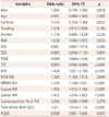
The clinical variables and PSG data were evaluated in order to determine whether they were related to patients with OSA and little REM sleep. Higher BMI and AHI values were significantly related to little REM sleep in univariate logistic regression (p=0.025 and p<0.001, respectively). In consideration of the sleep position (supine or lateral) and sleep state (REM or NREM sleep), higher AHI was significantly predictive of little REM sleep. A short TST was also significantly related to little REM sleep (p<0.001).
Table 4 summarizes the results from univariate logistic regression models that estimated the risk of a decrease in the proportion of REM sleep in patients with OSA. We performed a multivariate analysis of the predictive factors that were found to be significant in the univariate analysis. Reduced TST and reduced REM sleep, increased AHI as a total, and increased AHI in NREM sleep seemed to be codependent variables. In the multiple logistic regression, codependent variable overlap was avoided. The multiple logistic regression showed that a higher AHI (p<0.001; odds ratio, 1.512; 95% confidence interval, 1.020–1.812) was independently predictive of little REM sleep. Sleep position in patients with OSA had no predictive value for the reduction of REM sleep. The detailed results of the multivariate logistic regression are presented in Table 5. The partial correlation analysis produced similar results. AHI was negatively correlated with the proportion of REM sleep after adjusting confounding factors including BMI, age, and sleep position.

Treatment response to CPAP titration

Moderate-to-severe OSA was present in 193 of the 461 patients in the little-REM-sleep group. Thirty patients underwent overnight CPAP titration, of which 21 (70%) completed CPAP titration successfully with an optimal pressure (Table 6). Moderate-to-severe OSA was also present in 95 of the 264 patients in the normal-REM-sleep group. Twenty of the 21 (95%) subjects successfully completed CPAP titration with an optimal pressure. The titration success rate was lower in the little-REM-sleep group than in the normal-REM-sleep group (p=0.038). There was no significant difference in the optimal pressure between the little- and normal-REM-sleep groups.

DISCUSSION

This study found distinct features in patients with OSA who had little REM sleep. These patients showed a significantly increased AHI. We applied multiple logistic regression models and a partial correlation analysis to determine the independent factors related to the diminished REM sleep. The following potential confounding factors were adjusted: age, sex, caffeine intake, smoking status, alcohol intake, BMI, and sleep parameters. To the best of our knowledge, this is the first study to focus on the proportion of REM sleep and to estimate predictive factors of little REM sleep in patients with OSA.
The muscle tone of the upper airway is usually thought to be suppressed more during REM sleep than during NREM sleep. Previous studies have found that longer durations of apnea and larger decreases in SpO2 occurred more often after apnea events in REM sleep than in NREM sleep.1524 During REM sleep, cholinergic-system-mediated inhibition of the hypoglossal nerve may suppress the genioglossus muscle tone, potentially increasing the collapsibility of the upper airway.2526 Decreased upper airway muscle activation, impaired genioglossus reflex responsiveness to negative pressure, and reduced chemosensitivity are potential explanations for the worsened apnea during REM sleep. Some studies have suggested that OSA patients are more likely to have higher AHI during NREM sleep than during REM sleep.161718 However, the present consensus is that OSA is worse in REM sleep than in NREM sleep.152427 The subjects in our little-REM-sleep group showed a higher AHI and larger decreases in SpO2 compared with patients in the other two groups. Our findings suggest that the decreased exposure time to REM sleep might be a compensatory mechanism to reduce the severity of the apnea events or a pathologic process itself, especially in patients with higher AHIs. This plausible hypothesis of reduced REM sleep being an adaptation to severe OSA is supported by the phenomenon of REM sleep rebound after applying CPAP.28
Patients in our little-REM-sleep group demonstrated more severely fragmented sleep as well as more sleep-disordered breathing compared with patients in the other two groups. This phenomenon was also found when each sex was analyzed separately. Men showed a worse status of sleep fragmentation and sleep-disordered breathing compared to women in all REM sleep groups. The severity of OSA was worse in both men and women who experienced little REM sleep. Given that the women in the three REM sleep groups were much older than the men, it seems likely that the differences in sleep parameters could be due to sex-related differences rather than to age-related differences.
A previous study involving 99 obese patients showed that a reduction in the proportion of REM sleep was best predicted by an increased BMI.29 However, the reduction of REM sleep was not significantly correlated with OSA severity in that study. The present study involved a larger sample of 492 obese and 410 nonobese subjects, and regression models and partial correlation analysis were applied to determine the factors related to the decreased proportion of REM sleep in the patients with OSA. Higher BMI, higher AHI, and longer time below 90% SpO2 were significantly associated with little REM sleep in a univariate regression analysis. Furthermore, we analyzed the predictive value of AHI for the reduction of REM sleep according to various combinations of sleep stage (REM and NREM) and position (supine and lateral). In all situations, increased AHI had a robust impact on little REM sleep. However, sleep position did not affect the REM sleep reduction. To estimate the independent effect of AHI on the proportion of REM sleep, codependent variables associated with AHI and REM sleep were avoided in multivariate analysis. TST, sleep efficiency, and arousal index were considered as codependent variables. Despite the reduced TST in the little-REM-sleep group, the habitual sleep time reported in the sleep questionnaires did not differ significantly between the three groups, being 6.3±2.1, 6.1±1.8, and 6.1±1.7 hours in the little-, normal-, and excessive-REM-sleep groups, respectively. The first-night effect during the PSG study is a possible reason for this discrepancy. Among the possible candidates, a higher AHI was the predictive factor for the reduction of REM sleep. Unlike a previous study,29 BMI after adjusting for AHI was not associated with the proportion of REM sleep in the present study, which could be due to the BMI being lower in Asian than in Western populations. Our results suggest that worse sleep apnea could decrease the proportion of REM sleep, but BMI and sleep position might not affect the REM sleep reduction.
Nasal CPAP therapy has been the most effective and generally used treatment for OSA.30 However, 25% to 50% of patients with OSA reportedly either refuse to try or fail to maintain CPAP therapy.31 Some patients show a poor therapeutic response to CPAP treatment, either without symptom improvements or without reductions in overall respiratory events. The success rate of CPAP titration was significantly lower in our OSA patients who experienced little REM sleep than in those who experienced normal REM sleep. This present finding suggests that the small proportion of REM sleep in the baseline PSG is a reason for the poor effectiveness in the CPAP treatment.
The amount of REM sleep is significantly positively correlated with the formation and performance of procedural memory.32 In an animal study, rats showed greater memory retention and behavioral performance on memory-requiring tasks after they experienced an increased amount of REM sleep.33 Another study involving rats found that decreased REM sleep seemed to impair the performance of complex memory tasks.34 In the theories based on human studies, sleep stages play different roles in memory.35363738 REM sleep has been associated with the consolidation of nondeclarative memories such as procedural skills and emotional memories, whereas slow-wave sleep has been closely related to hippocampus-dependent declarative memories (episodic and semantic memories).36 A recent study demonstrated that REM sleep had a role in the consolidation of spatial navigational memory in human subjects, and that apnea-induced disruption of REM sleep had a negative effect on cognition.39 This is an additional reason why patients with OSA—especially those who experience little REM sleep—should be treated.
In conclusion, the AHI is higher in OSA patients with little REM sleep than in those with normal REM sleep. OSA patients with little REM sleep also tend to have a lower arousal threshold, which predisposes them to disrupted and fragmented sleep. Furthermore, the success rate of CPAP titration was significantly lower in the little-REM-sleep group than in the normal-REM-sleep group in the present study. Clinicians need to be aware of and also be able to identify these unique features of patients with little REM sleep in order to ensure good clinical practices.

Figures and Tables

Table 1

Baseline characteristics of the groups with different proportions of REM sleep

jcn-12-426-i001
Variables Little REM sleep group Normal REM sleep group Excessive REM sleep group p
Subjects, No. 461 264 177
Age, years, mean±SD 47.5±17.0 47.7±15.2 49.0±14.1 NS
Female, No. (%) 111 (24.1) 67 (25.4) 40 (22.6) NS
BMI, kg/m2, mean±SD 25.5±4.6 24.9±3.8 24.7±3.2 NS
ESS score, mean±SD 7.4±5.3 7.4±5.0 8.0±5.6 NS
SSS score, mean±SD 2.7±0.9 2.7±0.9 2.8±1.0 NS
Global PSQI score, mean±SD 8.6±3.6 8.2±3.1 9.1±4.6 NS
Caffeine, No. (%) 333 (72.2) 197 (74.6) 127 (71.8) NS
Smoking, No. (%) 111 (24.1) 83 (31.4) 55 (31.1) NS
Alcohol, No. (%) 224 (48.6) 133 (50.4) 99 (55.9) NS

Post-hoc analysis with Bonferroni's comparison was used for statistics.

BMI: body mass index, ESS: Epworth sleepiness scale, No.: number, NS: not significant, PSQI: Pittsburgh Sleep Quality Index, REM: rapid eye movement, SD: standard deviation, SSS: Stanford sleepiness scale.

Table 2

Polysomnographic data of the groups with different proportions of REM sleep

jcn-12-426-i002
PSG variables Little REM sleep group
(mean±SD)
Normal REM sleep group
(mean±SD)
Excessive REM sleep group
(mean±SD)
TST, min 344.7±50.6*† 358.5±55.5 372.0±50.2
N1 sleep, % 17.7±13.8 13.7±10.6 10.4±7.5
N2 sleep, % 47.9±12.3 44.3±10.1 42.3±9.2
N3 sleep, % 20.0±12.3 19.9±10.2 18.2±9.4
REM sleep, % 14.4±4.3*† 22.3±1.4 28.9±2.9
Sleep latency, min 12.8±19.0 11.9±18.4 8.9±10.8
REM sleep latency, min 143.3±74.2*† 100.5±50.0 87.3±48.3
Sleep efficiency, % 80.1±10.8*† 83.1±10.1 85.1±8.7
Arousal index, events/h 28.8±25.3*† 23.3±18.9 20.0±17.9
PLMS index, events/h 6.2±19.2 5.4±15.4 9.0±21.7
RDI, events/h 26.6±24.5*† 20.6±19.0 17.0±18.9
AHI, events/h 22.1±24.4*† 16.7±19.0 12.8±17.2
 AHI during NREM sleep 22.1±25.4*† 16.4±21.4 11.4±17.9
 AHI during REM sleep 22.3±22.5*† 19.8±20.5 15.8±18.0
Supine AHI, events/h 29.7±29.5 25.6±25.4 20.6±23.7
 Supine AHI during NREM sleep 29.3±30.6 24.1±26.3 18.8±25.0
 Supine AHI during REM sleep 27.3±27.1 28.7±27.3 26.7±25.7
Lateral AHI, events/h 11.2±21.7*† 6.6±14.3 4.8±12.4
 Lateral AHI during NREM sleep 11.0±22.7*† 5.6±13.6 4.3±12.6
 Lateral AHI during REM sleep 10.7±19.6 8.4±16.9 5.6±12.8
Supine position, % of TST 62.3±27.0 58.8±27.7 59.5±28.2
Apnea index, events/h 10.4±17.2*† 7.3±12.3 5.3±11.1
Mixed apnea index, events/h 0.9±2.9 1.0±3.8 0.9±4.2
Hypopnea index, events/h 10.6±12.6*† 8.1±8.2 6.2±8.6
RERA, events/h 3.7±5.0 4.0±4.4 2.6±2.7
Longest apnea, sec 30.6±29.6 32.4±27.9 28.6±22.4
Lowest oxygen saturation, % 78.8±17.9*† 82.1±11.1 83.1±12.2
Time below 90% SpO2, % 18.7±28.5*† 13.4±23.2 11.0±22.3

*Significant difference compared with Normal REM sleep group (p<0.05, post-hoc analyses with Bonferroni's), Significant difference compared with Excessive REM sleep group (p<0.05, post-hoc analyses with Bonferroni's).

AHI: apnea-hypopnea index, NREM: non-REM, PLMS: periodic limb movement during sleep, PSG: polysomnography, RDI: respiratory disturbance index, REM: rapid eye movement, RERA: respiratory effort-related arousal, SD: standard deviation, SpO2: oxygen saturation, TST: total sleep time.

Table 3

Gender-wise characteristics of patients and polysomnography data for the groups with different proportions of REM sleep

jcn-12-426-i003
Variables Little REM sleep group (mean±SD) Normal REM sleep group (mean±SD) Excessive REM sleep group (mean±SD)
Men Women p Men Women p Men Women p
Subjects, No. 350 111 197 067 137 40
Age, years, mean±SD 45.3±16.2 54.5±17.6 <0.001 45.8±14.7 53.3±15.2 0.001 46.8±14.2 56.5±11.0 <0.001
BMI, kg/m2, mean±SD 25.9±4.7 24.2±4.0 <0.001 25.4±3.5 23.6±4.3 0.002 24.8±3.3 24.1±3.0 NS
ESS scores, mean±SD 7.7±5.4 6.6±4.7 NS 7.3±4.9 7.7±5.3 NS 8.0±5.6 8.1±5.7 NS
Global PSQI scores, mean±SD 8.6±3.7 8.5±3.4 NS 8.0±3.0 8.7±3.4 NS 8.8±3.1 10.2±7.9 NS
Total sleep time, min 345.9±49.7 341.1±53.3 NS 359.3±57.5 356.0±49.5 NS 372.2±48.3 371.2±56.8 NS
N1 sleep, % 18.7±14.4 14.5±11.4 0.003 14.9±11.4 10.0±6.6 0.004 11.0±8.0 8.3±5.2 0.013
N2 sleep, % 44.7±10.3 44.7±11.2 0.001 44.7±10.3 43.2±9.7 NS 42.9±8.6 40.4±11.1 NS
N3 sleep, % 18.0±11.3 26.4±13.2 <0.001 18.4±9.8 24.3±10.1 <0.001 17.1±8.9 22.0±10.2 0.008
REM sleep, % 14.4±4.3 14.5±4.5 NS 22.3±1.4 22.3±1.4 NS 28.9±2.9 28.9±3.0 NS
Sleep latency, min 10.5±15.0 19.9±27.1 0.001 11.6±19.5 12.8±14.9 NS 8.2±10.9 11.1±10.3 NS
REM sleep latency, min 138.7±71.4 158.0±80.9 0.025 97.0±49.5 111.1±50.2 0.049 86.4±45.7 90.3±56.6 NS
Sleep efficiency, % 80.7±11.0 78.3±10.0 0.030 83.3±10.4 82.8±9.1 NS 85.5±8.2 83.5±10.1 NS
Arousal index, events/h 31.1±26.1 21.3±21.3 <0.001 26.0±19.7 15.0±13.6 0.010 21.8±18.5 13.9±14.3 0.006
PLMS index, events/h 4.6±14.4 11.2±29.0 0.023 4.9±13.8 7.0±19.2 NS 8.6±21.8 10.2±21.4 NS
RDI, events/h 29.3±25.0 18.1±20.9 <0.001 23.1±19.9 11.9±12.0 <0.001 19.6±20.3 7.7±7.2 <0.001
AHI, events/h 25.2±25.2 12.2±18.7 <0.001 19.3±20.2 9.1±11.9 <0.001 14.6±18.5 6.6±9.8 <0.001
Supine AHI, events/h 33.9±30.0 16.3±23.6 <0.001 28.9±25.9 15.9±21.3 <0.001 23.5±25.2 10.4±13.6 <0.001
Lateral AHI, events/h 13.1±23.2 5.3±14.6 <0.001 7.9±15.9 3.0±7.1 0.001 5.8±13.8 1.5±2.8 0.002
NREM AHI, events/h 25.2±26.2 11.8±19.4 <0.001 19.4±23.2 7.6±11.0 <0.001 13.1±19.4 5.4±9.5 0.001
REM AHI, events/h 24.6±22.9 14.8±19.2 <0.001 21.1±21.2 16.0±17.9 NS 17.7±18.7 9.1±13.4 0.002
Supine position, % 62.0±27.2 63.4±26.4 NS 58.5±27.7 59.8±27.7 NS 60.2±27.6 56.9±30.4 NS
Apnea index, events/h 12.5±18.3 3.8±10.8 <0.001 8.8±13.5 2.9±6.5 <0.001 6.6±12.3 0.9±2.0 <0.001
Hypopnea index, events/h 11.4±12.7 8.0±12.2 0.012 8.6±8.0 6.8±8.5 NS 6.4±8.4 5.6±9.4 NS
RERA, events/h 3.9±5.1 3.2±4.6 NS 4.2±4.6 3.4±3.8 NS 2.8±2.7 1.8±2.1 0.042
Longest apnea, sec 35.4±31.1 15.5±17.3 <0.001 36.0±29.8 21.4±17.3 <0.001 33.1±22.4 12.4±13.4 <0.001
Lowest oxygen saturation, % 78.9±15.2 78.6±24.5 NS 81.1±12.0 85.1±7.1 0.001 81.8±13.4 87.8±4.6 <0.001
Time below 90% SpO2, % 21.2±29.4 10.8±23.9 <0.001 16.0±25.6 5.8±10.7 <0.001 12.8±23.9 4.9±14.2 0.011

AHI: apnea-hypopnea index, BMI: body mass index, ESS: Epworth sleepiness scale, No.: number, NS: not significant, PLMS: periodic limb movement during sleep, PSQI: Pittsburgh Sleep Quality Index, RDI: respiratory disturbance index, REM: rapid eye movement, RERA: respiratory effort-related arousal, SD: standard deviation.

Table 4

Univariate logistic regression results of the relationship of little REM sleep and variables in patients with OSA

jcn-12-426-i004
Variables Odds ratio 95% CI p
Men 1.004 0.740±1.363 0.978
Age 0.997 0.989±1.005 0.507
Caffeine 1.015 0.754±1.366 0.922
Smoking 1.216 0.812±1.656 0.160
Alcohol 1.176 0.905±1.528 0.226
BMI 1.038 1.005±1.073 0.025
ESS 0.991 0.967±1.016 0.486
SSS 0.985 0.854±1.135 0.831
PSQI 0.999 0.964±1.036 0.976
TST 0.993 0.990±0.996 <0.001
AHI 1.436 1.021±1.750 <0.001
REM AHI 1.309 1.103±1.516 0.004
NREM AHI 1.315 1.109±1.521 <0.001
Supine AHI 1.452 1.015±1.765 <0.001
Lateral AHI 1.412 1.018±1.652 <0.001
Supine position, % of TST 1.004 0.999±1.009 0.079
Time below 90% SpO2 1.021 1.014±1.135 <0.001
PLMS 0.998 0.991±1.005 0.625

AHI: apnea-hypopnea index, BMI: body mass index, CI: confidence interval, ESS: Epworth Sleepiness Scale, NREM: non-REM, OSA: obstructive sleep apnea, PLMS: periodic limb movements during sleep, PSQI: Pittsburgh Sleep Quality Index, REM: rapid eye movement, SpO2: oxygen saturation, SSS: Stanford Sleepiness Scale, TST: total sleep time.

Table 5

Multivariate logistic regression of risk factors of little REM sleep in patients with OSA

jcn-12-426-i005
Variables Odds ratio 95% CI p
Age 0.997 0.989–1.005 0.483
BMI 1.009 0.973–1.046 0.633
AHI 1.512 1.020–1.812 <0.001
Supine position 1.003 0.998–1.272 0.180

AHI: apnea-hypopnea index, BMI: body mass index, CI: confidence interval, OSA: obstructive sleep apnea, REM: rapid eye movement.

Table 6

Success rate of CPAP titration

jcn-12-426-i006
CPAP titration Little REM sleep group (n=30) Normal REM sleep group (n=21) p
Success, No. (%) 21 (70) 20 (95) 0.038
Failure, No. (%) 9 (30) 1 (5) 0.038
Optimal pressure, mean±SD 10.3±3.1 9.8±2.4 NS

CPAP: continuous positive airway pressure, No.: number, NS: not significant, REM: rapid eye movement, SD: standard deviation.

Acknowledgements

This research was supported by the Clinical Research Grant of Boramae Medical Center (03-2014-1) and partly supported by the Grant from the Seoul National University Hospital Research Fund (04-2014-0480).

Notes

Conflicts of Interest The authors have no financial conflicts of interest.

References

1. Young T, Palta M, Dempsey J, Skatrud J, Weber S, Badr S. The occurrence of sleep-disordered breathing among middle-aged adults. N Engl J Med. 1993; 328:1230–1235.
crossref
2. Mokhlesi B, Finn LA, Hagen EW, Young T, Hla KM, Van Cauter E, et al. Obstructive sleep apnea during REM sleep and hypertension. results of the Wisconsin Sleep Cohort. Am J Respir Crit Care Med. 2014; 190:1158–1167.
crossref
3. Peppard PE, Young T, Barnet JH, Palta M, Hagen EW, Hla KM. Increased prevalence of sleep-disordered breathing in adults. Am J Epidemiol. 2013; 177:1006–1014.
crossref
4. Young T, Palta M, Dempsey J, Peppard PE, Nieto FJ, Hla KM. Burden of sleep apnea: rationale, design, and major findings of the Wisconsin Sleep Cohort study. WMJ. 2009; 108:246–249.
5. Somers VK, White DP, Amin R, Abraham WT, Costa F, Culebras A, et al. Sleep apnea and cardiovascular disease: an American Heart Association/American College Of Cardiology Foundation Scientific Statement from the American Heart Association Council for High Blood Pressure Research Professional Education Committee, Council on Clinical Cardiology, Stroke Council, and Council On Cardiovascular Nursing. In collaboration with the National Heart, Lung, and Blood Institute National Center on Sleep Disorders Research (National Institutes of Health). Circulation. 2008; 118:1080–1111.
6. Kohler M, Craig S, Pepperell JC, Nicoll D, Bratton DJ, Nunn AJ, et al. CPAP improves endothelial function in patients with minimally symptomatic OSA: results from a subset study of the MOSAIC trial. Chest. 2013; 144:896–902.
crossref
7. Kohler M, Craig S, Nicoll D, Leeson P, Davies RJ, Stradling JR. Endothelial function and arterial stiffness in minimally symptomatic obstructive sleep apnea. Am J Respir Crit Care Med. 2008; 178:984–988.
crossref
8. Buchner NJ, Sanner BM, Borgel J, Rump LC. Continuous positive airway pressure treatment of mild to moderate obstructive sleep apnea reduces cardiovascular risk. Am J Respir Crit Care Med. 2007; 176:1274–1280.
crossref
9. Campos-Rodriguez F, Martinez-Garcia MA, de la Cruz-Moron I, Almeida-Gonzalez C, Catalan-Serra P, Montserrat JM. Cardiovascular mortality in women with obstructive sleep apnea with or without continuous positive airway pressure treatment: a cohort study. Ann Intern Med. 2012; 156:115–122.
crossref
10. Yaggi HK, Concato J, Kernan WN, Lichtman JH, Brass LM, Mohsenin V. Obstructive sleep apnea as a risk factor for stroke and death. N Engl J Med. 2005; 353:2034–2041.
crossref
11. Young T, Finn L, Peppard PE, Szklo-Coxe M, Austin D, Nieto FJ, et al. Sleep disordered breathing and mortality: eighteen-year follow-up of the Wisconsin sleep cohort. Sleep. 2008; 31:1071–1078.
12. Oksenberg A, Khamaysi I, Silverberg DS, Tarasiuk A. Association of body position with severity of apneic events in patients with severe nonpositional obstructive sleep apnea. Chest. 2000; 118:1018–1024.
crossref
13. Tufik S, Santos-Silva R, Taddei JA, Bittencourt LR. Obstructive sleep apnea syndrome in the Sao Paulo Epidemiologic Sleep Study. Sleep Med. 2010; 11:441–446.
crossref
14. Peregrim I, Grešová S, Pallayová M, Fulton BL, Štimmelová J, Bačová I, et al. Does obstructive sleep apnea worsen during REM sleep? Physiol Res. 2013; 62:569–575.
crossref
15. Findley LJ, Wilhoit SC, Suratt PM. Apnea duration and hypoxemia during REM sleep in patients with obstructive sleep apnea. Chest. 1985; 87:432–436.
crossref
16. Siddiqui F, Walters AS, Goldstein D, Lahey M, Desai H. Half of patients with obstructive sleep apnea have a higher NREM AHI than REM AHI. Sleep Med. 2006; 7:281–285.
crossref
17. Muraki M, Kitaguchi S, Ichihashi H, Haraguchi R, Iwanaga T, Kubo H, et al. Apnoea-hypopnoea index during rapid eye movement and non-rapid eye movement sleep in obstructive sleep apnoea. J Int Med Res. 2008; 36:906–913.
crossref
18. Loadsman JA, Wilcox I. Is obstructive sleep apnoea a rapid eye movement-predominant phenomenon? Br J Anaesth. 2000; 85:354–358.
crossref
19. Buysse DJ, Reynolds CF 3rd, Monk TH, Berman SR, Kupfer DJ. The Pittsburgh Sleep Quality Index: a new instrument for psychiatric practice and research. Psychiatry Res. 1989; 28:193–213.
crossref
20. American Academy of Sleep Medicine. The AASM Manual for the Scoring of Sleep and Associated Events: Rules, Terminology, and Technical Specifications. Westchester, IL: American Academy of Sleep Medicine;2007.
21. Kushida CA, Chediak A, Berry RB, Brown LK, Gozal D, Iber C, et al. Clinical guidelines for the manual titration of positive airway pressure in patients with obstructive sleep apnea. J Clin Sleep Med. 2008; 4:157–171.
22. Rowley JA, Tarbichi AG, Badr MS. The use of a predicted CPAP equation improves CPAP titration success. Sleep Breath. 2005; 9:26–32.
crossref
23. Carskadon MA, Dement WC. Normal human sleep: an overview. In : Kryger MH, Roth T, Dement WC, editors. Principles and Practice of Sleep Medicine. 5th ed. St. Louis, MO: Elsevier Saunders;2011. p. 16–26.
24. Sériès F, Cormier Y, La Forge J. Influence of apnea type and sleep stage on nocturnal postapneic desaturation. Am Rev Respir Dis. 1990; 141:1522–1526.
crossref
25. McSharry DG, Saboisky JP, Deyoung P, Jordan AS, Trinder J, Smales E, et al. Physiological mechanisms of upper airway hypotonia during REM sleep. Sleep. 2014; 37:561–569.
crossref
26. Grace KP, Hughes SW, Horner RL. Identification of the mechanism mediating genioglossus muscle suppression in REM sleep. Am J Respir Crit Care Med. 2013; 187:311–319.
crossref
27. Eckert DJ, Malhotra A. Pathophysiology of adult obstructive sleep apnea. Proc Am Thorac Soc. 2008; 5:144–153.
crossref
28. Brillante R, Cossa G, Liu PY, Laks L. Rapid eye movement and slow-wave sleep rebound after one night of continuous positive airway pressure for obstructive sleep apnoea. Respirology. 2012; 17:547–553.
crossref
29. Dixon JB, Schachter LM, O'Brien PE. Predicting sleep apnea and excessive day sleepiness in the severely obese: indicators for polysomnography. Chest. 2003; 123:1134–1141.
crossref
30. Marin JM, Carrizo SJ, Vicente E, Agusti AG. Long-term cardiovascular outcomes in men with obstructive sleep apnoea-hypopnoea with or without treatment with continuous positive airway pressure: an observational study. Lancet. 2005; 365:1046–1053.
crossref
31. Zozula R, Rosen R. Compliance with continuous positive airway pressure therapy: assessing and improving treatment outcomes. Curr Opin Pulm Med. 2001; 7:391–398.
crossref
32. Fischer S, Hallschmid M, Elsner AL, Born J. Sleep forms memory for finger skills. Proc Natl Acad Sci U S A. 2002; 99:11987–11991.
crossref
33. Wetzel W, Balschun D, Janke S, Vogel D, Wagner T. Effects of CLIP (corticotropin-like intermediate lobe peptide) and CLIP fragments on paradoxical sleep in rats. Peptides. 1994; 15:237–241.
crossref
34. Pearlman CA. Effect of rapid eye movement (dreaming) sleep deprivation on retention of avoidance learning in rats. Rep No 563. Rep US Nav Submar Med Cent. 1969; 1–4.
35. Ackermann S, Rasch B. Differential effects of non-REM and REM sleep on memory consolidation? Curr Neurol Neurosci Rep. 2014; 14:430.
crossref
36. Plihal W, Born J. Effects of early and late nocturnal sleep on declarative and procedural memory. J Cogn Neurosci. 1997; 9:534–547.
crossref
37. Smith C. Sleep states and memory processes in humans: procedural versus declarative memory systems. Sleep Med Rev. 2001; 5:491–506.
crossref
38. Rasch B, Born J. About sleep's role in memory. Physiol Rev. 2013; 93:681–766.
crossref
39. Varga AW, Kishi A, Mantua J, Lim J, Koushyk V, Leibert DP, et al. Apnea-induced rapid eye movement sleep disruption impairs human spatial navigational memory. J Neurosci. 2014; 34:14571–14577.
crossref
TOOLS
Similar articles