

 PDF
PDF ePub
ePub Citation
 Print
Print


INTRODUCTION
Herpes simplex virus (HSV) is a human herpes virus that causes herpes simplex encephalitis (HSE), a fatal type of sporadic encephalitis that is the most common form of encephalitis in humans.1 Prior to the emergence of effective antiviral treatments, the mortality rate was as high as 70%. Moreover, fewer than 3% of patients with HSE reportedly fully recover normal function.2 Although optimal antiviral therapy with acyclovir can significantly improve the prognosis, some reports have suggested that profound neurological deficits persist.34 Several studies have examined the long-term outcomes and relevant prognostic factors in patients with HSE,2567 but data on the prognosis based on radiological and electrophysiological characteristics are rare.
The present study examined whether initial brain MRI and EEG findings have a prognostic value at 6 months after the onset in patients who were diagnosed with HSE by positive results of HSV DNA when applying the polymerase chain reaction (PCR) test to cerebrospinal fluid (CSF), and who received appropriate antiviral therapy.
METHODS
Study subjects
We retrospectively reviewed the electronic medical records of all patients who were clinically diagnosed with encephalitis and exhibited CSF positivity for HSV DNA in the PCR analysis at Seoul National University Hospital from January 1, 2000 to July 31, 2014. All demographic data, initial neurological symptoms and signs, and laboratory findings were retrieved from medical records. The current study was approved by the Institutional Review Board of Seoul National University Hospital.
Clinical and biological characteristics
The following clinical and biological variables were collected: age, sex, HSV type (including type 1 and type 2), fever, serum leukocytosis, serum neutrophilia, C-reactive protein level, CSF leukocytosis, CSF protein, CSF glucose, initial Glasgow Coma Scale (GCS) score, mean duration to the initiation of acyclovir, epileptic seizures at the initial presentation, and response to epilepsy treatment.
MRI and lesion volume analysis
MRI examinations were performed with two 1.5-tesla MRI units using diffusion-weighted imaging (DWI) sequences [time of repetition/time of echo (TR/TE) of 6,500/110 ms for the Siemens device and 3,737/66 ms for the Intera device] and fluid-attenuated inversion recovery (FLAIR) sequences [TR/TE of 9,000/119 ms for the Siemens device (Siemens, Erlangen, Germany) and 11,000/ 140 ms for the Intera device (Philips, Amsterdam, The Netherlands)]. The volume of lesions with high FLAIR MRI signal intensities was extracted using a semiautomatic procedure based on in-house software developed as a plug-in for the Medical Image Processing, Analysis, and Visualization (MIPAV) software package.8
EEG
EEG data were recorded for at least 30 min using a 40-channel digital EEG machine (Comet®, Grass Technology, West Warwick, RI, USA), and 21 channel electrodes (Fp1/2, F7/8, T1/T2, T7/8, P7/8, F3/4, C3/4, P3/4, O1/2, Fz, Cz, and Pz) were placed on the scalp according to the International 10-20 system. The sampling rate was 800 Hz based on the standard EEG protocol of Seoul National University Hospital. Baseline EEG data were recorded, and the results were divided into the following categories:9 Grade I, dominant alpha activity with some scattered theta activity; Grade II, dominant theta activity and generally reactive; Grade III, dominant widespread delta activity or small-amplitude, diffuse, irregular delta activity, and nonreactive; Grade IV, burst suppression pattern, epileptiform discharges, low-output activity, nonreactive, alpha coma pattern, and theta coma pattern; and Grade V, isoelectric activity. EEG findings of Grades I & II and of Grades III-V were classified into favorable and poor results, respectively.
Outcome assessments
The 6-month (long-term) outcome was evaluated based on the degree of handicap and the quality of life according to the Glasgow Outcome Scale (GOS) as follows:10 Grade I, good recovery, allowing independent life without any neurological impairment; Grade II, mild disability, defined by the presence of minimal cognitive alterations (speech, memory, or attention impairments) and/or seizures (partially controlled with antiepileptic drugs) without consequences for socioprofessional life; Grade III, moderate disability, defined using criteria identical to those of Grade II but with consequences for socioprofessional life; Grade IV, severe disability defined as loss of autonomy requiring institutionalization or constant life support; and Grade V, death. Outcomes of Grades I & II and of Grades III-V were classified into favorable and poor results, respectively.
Statistical analysis
SPSS software (version 19.0 for Windows, SPSS Inc., Chicago, IL, USA) was used for statistical analysis. For the analysis of prognostic factors, the outcome at 6 months was the main end point and was dichotomized into favorable and poor outcomes. To compare clinical and biological data between the favorable and poor outcomes at 6 months, categorical variables were assessed using the χ2 test and continuous variables were assessed using Student's t-test. The significance cutoff was defined as p<0.05. A forward analysis was applied to the significant (p<0.05) and statistically important variables using a binary logistic regression model to control simultaneous interaction factors to generate odds ratios (ORs) and 95% confidence intervals.
RESULTS
Clinical and biological characteristics
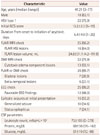
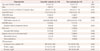
We initially enrolled 34 patients, and after excluding 5 patients with combined pathogens, 29 patients were analyzed in this study. The median patient age was 41.21 years (range, 3-77 years), 18 patients (62%) were male, and 22 patients (76%) were HSV type 1. The basic demographic and clinical characteristics of the patients with HSE are presented in Table 1. The initial neurological signs and symptoms on admission included seizure (55%), mental alteration or disorientation (25%), behavioral changes or psychosis (17%), and motor deficit or dysarthria (3%). All of the patients received an intravenous injection of acyclovir (10 mg/kg three times daily for 10-14 days), and the duration from symptom onset to the initiation of acyclovir was 5.4±4.0 (mean±SD; range, 1-25) days. The clinical outcome at 6 months after the onset of HSE was evaluated in all of the patients. At 6 months after onset, the patients exhibited complete recovery (34%), mild disability (14%), moderate disability (31%), or severe disability (21%), as assessed based on GOS criteria, and they were classified into the favorable and poor outcome groups. There were no cases of mortality. We found no significant correlation between any of the various clinical characteristics— including the mean duration to initiation of acyclovir—and the outcome (Table 2).
Initial manifestation of seizure and epilepsy
Seizure was an initial presentation in 16 patients (55%), with 10 (35%) presenting with generalized tonic-clonic seizures and 7 (24%) with status epilepticus. There were 22 patients (76%) who continued taking antiepileptic drugs (AEDs) after being diagnosed with epilepsy 6 months later. Of those who were taking AEDs, 36% and 64% received monotherapy and polytherapy, respectively, and 14% met the diagnostic criteria for drug-resistant epilepsy (DRE).11 The presence of epileptic seizures at the initial presentation was significantly correlated with the 6-month clinical outcome (p=0.009) (Table 2).
EEG
EEG recording was performed in 25 patients (86%), including 3 young patients (8, 13, and 15 years old). The period between the admission and the EEG recording was 1.6±2.1 (range, 0-8) days. The EEG findings were evaluated based on the 5-grade system proposed by Synek.9 Twelve patients (48%) had favorable EEG findings (Grades I & II) and 13 (52%) had poor EEG findings (Grades III-V). The 13 patients with poor EEG findings comprised 4 patients with periodic discharges such as periodic lateralized epileptiform discharges (PLEDs) or bilateral independent periodic lateralized epileptiform discharges (biPLEDs), 3 patients with continuous diffuse delta slow waves, 2 patients with frontal intermittent rhythmic delta slow waves, 2 patients with frequent rhythmic spike and wave complexes, and 2 patients who exhibited several episodes of subclinical ictal discharges. Favorable initial EEG findings were significantly correlated with favorable 6-month clinical outcomes (p=0.005) (Table 2). The logistic regression analysis showed that initial EEG findings independently predicted the 6-month outcome (OR=16.7, p=0.006) (Table 3).
MRI and lesion volume analysis
MRI was performed on 25 patients (86%), including 1 young patient (13 years old). The duration from the onset of symptoms to the MRI examination was 2.8±2.0 (range, 0-7) days. The presence of abnormal MRI findings was not significantly associated with the 6-month clinical outcomes. The small number of patients in each group meant that the relationships between MRI lesion localization, bilateral involvement, and 6-month clinical outcomes could not be analyzed. An analysis of the volume of lesions using the MIPAV software package8 found that the volume of lesions in FLAIR MRI was 24.8±31.1 (range, 4.2-111.9) mL, and this was not related to the clinical outcome. There was also no correlation between the extent of cytotoxic edema on the apparent diffusion coefficient (ADC) value and outcomes (Table 2).
DISCUSSION
HSE causes selective damage mainly to the mesial temporal lobe structures, including the hippocampus, which has highly epileptogenic properties.12 One previous study found that 40-60% of patients with early-stage HSE had epileptic seizures.12 EEG and MRI might therefore be essential for the early diagnosis and initial severity evaluation of HSE. There are continuing efforts to identify the prognostic factors in the early stage of HSE, but they have been hampered by several factors, including the lack of a definitive diagnosis and the variability of appropriate antiviral therapy. The present study found that the initial EEG findings and seizure presentations were the factors predictive of the 6-month outcome in patients with HSE.
Most MRI studies involving patients with HSE have demonstrated that MRI findings have early diagnostic value.131415 One case-series MRI study analyzed the signal changes in DWI MRI in five patients with HSE for prognostic prediction.16 The patients with cytotoxic types of lesions had fulminating disease with a severe clinical condition, and those with vasogenic types of lesions were in early stages of the disease and generally had a good outcome.16 The initial FLAIR MRI in the present study found that the distribution of lesions (only temporal versus extra-temporal lesions and unilateral versus bilateral lesions) and the mean volume of lesions were not significantly associated with the 6-month clinical outcome. We were unable to analyze the direct correlation of the volume of lesions with high DWI MRI signal intensities matched with the ADC map, because of the different durations from the disease onset to the MRI scan time for each patient. The presence of lesions with cytotoxic edema on DWI MRI was not related to the 6-month clinical outcome. Further prospective studies with predefined MRI time points are needed to clarify the association between the volumes of cytotoxic edema in DWI MRI matched with the ADC map and clinical outcome.
EEG studies have searched for the diagnostic yield in patients with HSE. It is highly probable for a diagnosis of HSE to be made if the periodic discharges appear or disappear at various disease stages.1718 Another study found no pathognomonic EEG findings, but it did find that the spike/sharp or slow waves in the temporal lobe had a diagnostic value and that the unilateral PLEDs from the temporal lobe were the key diagnostic clue for HSE.19 Studies of the prognostic value of EEG findings have found unilateral periodic discharges to be associated with a good prognosis and bilateral ones to be associated with a poor prognosis.51820 However, those studies had some limitations such as the variability in the timing of EEG recordings, the appropriateness of antiviral therapies, and the diagnostic criteria used for HSE. In our study, 4 of the 13 patients with poor EEG findings had periodic discharges such as PLEDs or biPLEDs, and the favorable EEG findings were significantly related to favorable outcomes. It can therefore be inferred that the initial EEG findings are a significant prognostic indicator for predicting the clinical outcomes in patients with HSE.
We found that 16 patients (55%) had epileptic seizures at the initial presentation, which is in agreement with the results of a previous study.12 It has been reported that the incidence of late unprovoked seizure reaches 40-65% in patients with HSE.2122 This higher incidence relative to other types of encephalitis has been attributed to involvement of the highly epileptogenic temporal lobe and its necrotizing nature. The current study included 22 patients (76%) who were taking AEDs, which is a higher proportion than in previous studies. Two-thirds of these 22 patients received AED polytherapy, while only 3 patients (14%) had DRE. The presence of epileptic seizures at the initial presentation was significantly correlated with poor clinical outcomes at 6 months. However, initial presentations with the generalized tonic-clonic seizure type and status epilepticus were not related to clinical outcomes. Unlike previous reports, our results showed that the age at onset, initial GCS scores, and the duration from the onset of symptoms to the initiation of antiviral therapy were not significant prognostic factors.
Our study had some limitations. First, its retrospective design meant that we could not evaluate the MRI and EEG recordings in all patients, and these recordings were made with various timings, although mostly in the acute stage. Also, the EEG analysis did not consider the effects of drugs, including AEDs or sedative agents, taken before making EEG recordings. Second, the timing of the initiation acyclovir therapy differed markedly, but mostly this occurred within 1 week. Third, although long-term review data were collected, the number of enrolled patients was relatively small. Finally, the data did not include any patient deaths, which represents an unusual result. Despite these limitations, the present findings are of clinical significance in that they confirm that the EEG severity and the presence of epileptic seizures at the initial presentation are the significant indicators for predicting the 6-month clinical outcome in patients with HSE.

 XML Download