

 PDF
PDF ePub
ePub Citation
 Print
Print


INTRODUCTION
White-matter hyperintensities (WMHs) are typically observed in patients affected by vascular dementia (VaD) but can also be present in patients who fulfil the clinical diagnostic criteria for typical Alzheimer's disease (AD). The impact of WMHs on cognition and the relationship between AD and cerebrovascular diseases are uncertain. However, there is an increasing body of evidence supporting that vascular factors can influence the risk of developing AD and its progression,1 affecting brain functions both directly and via neurodegenerative processes.2 Carotid diseases also reportedly play a relevant role. A greater intima-media thickness (IMT) has been consistently associated with AD, possibly as a marker of cerebral microcirculatory involvement,345 while severe carotid stenosis may increase the risk of dementia as a source of cerebral microembolisms and hemodynamic failure.678
In recent years there have been numerous diffusion-weighted imaging (DWI) studies describing an increase in water diffusivity in T2-weighted hyperintense areas and in normal-appearing white matter (NAWM) in patients with impaired cognitive performance.91011121314151617 Increased random water motion may derive from a degeneration of microstructural barriers, such as loss of membrane integrity and myelin or decreased cellular density. Water diffusivity was reported to be higher in demented patients than in controls, mainly in temporal and posterior regions.181920 In addition, an increase in white matter (WM) water diffusivity and high WMH load can both predict the clinical progression of patients with mild cognitive impairment to dementia.2122 The LADIS study showed that apparent diffusion coefficient (ADC) values within the NAWM are related to WMH severity and have a WMH-independent effect on cognitive function in patients with leukoaraiosis.23 More recently, ADC changes in NAWM were also demonstrated to precede the development of WMH lesions.2425 Finally, higher water ADCs were described in patients with carotid steno-occlusive diseases as the result of chronic low-grade ischemic damage due to an impaired cerebral hemodynamics.2627 Water ADC changes in the ipsilateral hemisphere were also found to be partly reversible after carotid revascularization.2829
These findings together raise the question of whether the increased water diffusion in NAWM observed in demented patients reflects an overt neuronal tissue disruption that leads to degenerative or microvascular lesions. To address this question, this study compared regional ADCs of NAWM in patients affected by AD or VaD. We also investigated the relationships of ADCs with WMH burden, carotid atherosclerosis, and cognitive performance.
METHODS
This single-cohort, cross-sectional study included consecutive patients admitted to our Neurology Unit from January 2010 to December 2011 for a first-ever evaluation of cognitive impairment according to the following inclusion criteria:
1) Diagnosis of probable AD according to NINCDS-ADRDA
criteria.30
2) Diagnosis of probable VaD according to NINDS-AIREN criteria with brain magnetic resonance imaging (MRI) evidence of small-vessel disease.3132
Patients who could not undergo MRI were excluded from enrollment. Screened subjects underwent neurological and neuropsychological evaluations, and brain imaging. We enrolled 80 patients (49 AD and 31 VaD) based on the inclusion criteria.
The clinical history were obtained from each patient using a structured clinical interview (with the involvement of caregivers where necessary) and hematochemical data with a focus on the main vascular risk factors of hypertension, diabetes, smoking habits, and hyperlipidemia. Hypertension was defined as a history of high blood pressure, a systolic blood pressure of ≥140 mm Hg, a diastolic blood pressure of ≥90 mm Hg, or the use of an antihypertensive; dyslipidemia was defined as a history of dyslipidemia, a fasting serum total cholesterol level of ≥6.22 mmol/L (2.4 g/L) or a triglycerides level of ≥2.26 mmol/L (2 g/L), or the use of a statin or fibrate; diabetes mellitus was defined as a history of diabetes mellitus, a fasting serum glucose level of ≥7.0 mmol/L (1.26 g/L), or the use of an oral antihyperglycemic or insulin; and smoking was defined as a history of active tobacco smoking.
All enrolled patients underwent brain MRI to assess regional ADCs and the WMH volume, and an ultrasound examination of neck vessels to investigate carotid atherosclerosis. All patients were naïve to dementia therapy. Patients displaying strategic infarct lesions in MRI were excluded from the study.
The experimental protocol was approved by our Hospital Ethical Committee and all subjects or their caregivers signed a written informed consent.
Neuropsychological evaluation
Subjects underwent cognitive testing in a single assessment during the week when the ultrasound and MRI investigations were performed. The neuropsychological evaluation battery aimed to provide a measure of general cognitive efficiency as well as to assess a wide range of definite cognitive areas. The Italian version of the Mini Mental State Examination (MMSE)33 was used as a global measure of cognitive status. Episodic long-term memory was assessed using the Rey Auditory Verbal Learning test, assessing the immediate and 15-min delayed recall of a 15-word list. Language was assessed based on oral denomination of real-life objects.34 Problem-solving and abstract reasoning were assessed using Raven's Coloured Progressive Matrices, focused attention was tested using a digit visual search task,35 and constructional praxia was tested by asking the subjects to copy some simple pictures.36 The raw scores of neuropsychological tasks in each test of the battery were corrected for age and education using normative validation for the Italian population.35 Each test used a normative cutoff score corresponding to the lower limit of the tolerance interval on a 95% one-tailed test (for a confidence level of 95%) based on the score obtained by healthy subjects in the standardization sample.
Neuroradiological evaluation
MRI and diffusion-weighted imaging studies were performed with a 1.5-T magnet (Magnetom Avanto B13, Siemens, Erlangen, Germany). The following imaging protocols and parameters were used: fluid-attenuated inversion recovery (FLAIR) sequences (TR=11,451 msec, TE=102 msec, TI=2,360 msec, slice thickness=3 mm) acquired on the axial plane, turbo spin echo T2-weighted fat-saturated sequences (TR=10,400 msec, TE=105 msec, slice thickness=3 mm) acquired on the coronal plane, spin echo T1-weighted sequences (TR=647 msec , TE=11 msec, slice thickness=4 mm) acquired on the sagittal plane, and echo planar imaging diffusion-weighted sequences (TR=3,927 msec, TE=106 msec, slice thickness=5 mm; one with b=0 and three orthogonal gradient directions at b=1,000) acquired on the axial plane. The total WMH volume was computed on FLAIR axial images at E0 after producing segmented lesional region of interests (ROIs) through a semiautomated local thresholding approach (Osirix® software, version 3.9.4 Pixmeo SARI, Bernex, Switzerland).37
Average ADC maps were computed on a pixel-by-pixel basis using the equation of Stejskal and Tanner.38 Elliptic ROIs were positioned bilaterally on the ADC maps as described by Stahl et al.18 in NAWM in the parietal, frontal, temporal, and occipital lobes of the corpus callosum (genu and splenium), and in the posterior limb of the internal capsule (between the pallidum and the thalamus). In contrast to Stahl et al.,18 the ROI size was kept consistent (30–35 mm2) across patients in order to obtain a stable number of voxels and decrease the variance of ADCs. The correct ROI position was confirmed by simultaneous visual comparison with the corresponding slices of the FLAIR axial data sets. ROIs were positioned on the NAWM, defined as areas without WMHs on the corresponding FLAIR image. If WMHs were present, the ROI was shifted to reduce type I errors.
Carotid ultrasound
Carotid arteries were assessed in all subjects and defined by continuous-wave Doppler ultrasound and color-flow B-mode Doppler ultrasound, as described elsewhere1 using high-resolution 7.5 MHz transducers (iU22 Philips Ultrasound, Bothell, WA, USA). IMT was measured according to standardized guidelines.39 In each arterial segment, the plaque severity was quantified as follows: 0, no plaque; 1, one plaque with stenosis of <30% (mild stenosis) of the vessel diameter; 2, one plaque with stenosis of 30–50% of the vessel diameter (moderate stenosis) or multiple plaques with stenosis of <30%; and 3, one plaque with stenosis of >50% of the vessel diameter (severe stenosis) or multiple plaques with at least one moderate stenosis. A plaque index was calculated by adding the scores of the right and left carotid arteries. The neurosonographers were blind to the MRI findings and cognitive performance of the patient.
Statistical analysis
The dimensionality of ADC measures (as indicated in the Methods) was reduced using principal-components analysis (PCA). The Kaiser-Meyer-Olkin measure of sampling adequacy was fair for PCA (0.72), with 74% of the total variance explained by the following six factors: frontal, parietal, temporal, and occipital lobes and the genu and splenium of the corpus callosum. We considered the mean value for the right and left sides of each area, since the results did not differ significantly between the two sides. Variables that did not conform to a normal distribution in the Kolmogorov-Smirnov test (i.e., years of education, ADCs in the frontal lobe and the genu and splenium of the corpus callosum, and WMH volume) underwent a logarithmic transformation in order to produce a better fit with a Gaussian distribution. Baseline characteristics of the sample were compared with respect to demographic, radiological, and clinical characteristics using the chi-square test, t-test, or Mann-Whitney test depending on the data type and distribution. Spearman's rho test was adopted to assess correlations between ADCs and cognitive performance.
Linear regression models were used to assess if regional ADCs in NAWM were related to the WMH load, ultrasound carotid findings, or vascular risk factors. Relationships between the WMH load, classical risk factors, and ultrasound carotid findings were assessed using different multiple linear regression (MLR) analyses. The first MRL model adopted the WMH load as the dependent variable and diagnosis, dyslipidemia, hypertension, diabetes, and smoking as independent factors. The second MLR model used the WMH load as the dependent factor and the mean IMT, diagnosis, and plaque index as independent variables. The third model assessed the correlation between WMH load and the PCA-selected ADCs. We included the features from the MLR models that were the most statistically significant in a general linear model (GLM) in order to assess the relationship between factors and the WMH load. A factor was deemed to be significantly associated with the dependent variable and selected for the GLM if its probability value was ≤0.1 in the regression analyses. Finally, we evaluated the associations of the WMH load, mean IMT, and ADC in the anterior corpus callosum in a GLM correcting for the following covariates that could influence the association: age, sex, diagnosis, education, and MMSE score. Statistical analyses were performed using the SPSS software (version 17.0, SPSS Inc., Chicago, IL, USA).
RESULTS
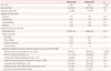
Table 1 summarizes the patient sociodemographic, vascular, and cognitive profiles. The VaD and AD patients did not differ in terms of age, education, prevalence of vascular risk factors, or ultrasound findings. AD patients were more frequently female (p=0.04) and presented more-severe memory deficits (p=0.001) than did VaD patients.
Table 2 summarizes the neuroradiological findings. The ADCs did not differ between VaD and AD patients, while the WMH volume was greater in VaD than in AD patients (p=0.04). We found no significant relationship between vascular risk factors, ADCs, and WMH volume (p>0.1).
Linear regression models showed that the mean IMT was associated with the temporal ADC (β=85.72, SE=37.175; t=1.947, p=0.24; R2=0.105, p=0.04) and WMH volume (β=4.504, SE=1.909; t=2.360, p=0.21; R2=0.122, p=0.036). No other associations were observed between radiological and ultrasound markers.
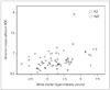
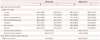
Table 3 presents the results obtained when applying the MLR model to test if ADCs can predict the WMH volume. This analysis demonstrated that ADC in the genu of the corpus callosum was related to the WMH volume (R2=0.272, p=0.008). Fig. 1 shows a scatterplot illustrating the relationship on a logarithmic scale between ADC in the anterior corpus callosum and the WMH volume in AD and VaD patients. The GLM was set up using the WMH load as the main dependent variable. The mean IMT and ADC in the anterior corpus callosum were both inserted in the model as predictors, since they were significantly associated with the WMH load in different regression analyses. The GLM results were significant (p<0.0001), confirming that both the mean IMT (p=0.029, partial η2=0.071, observed power=0.597) and ADC in the anterior corpus callosum (p=0.003, partial η2=0.122, observed power=0.848) were significantly associated with the WMH load. The corrected GLM results were also significant (p=0.010), again confirming that both the mean IMT (p=0.031, partial η2=0.076, observed power=0.586) and ADC in the anterior corpus callosum (p=0.008, partial η2=0.110, observed power=0.765) were significantly associated with the WMH load even after correcting for covariates.
Spearman's rho coefficients for the correlations between neuropsychological performances in different cognitive domains and neuroradiological markers are reported in Table 4. In brief, the memory performance was worse in patients with higher temporal ADC scores. Attention and language status were not associated with any neuroradiological marker. Constructional praxis test scores were related to frontal and occipital ADCs, ADCs of the genu and splenium of the corpus callosum, and the WMH volume. Abstract reasoning was related to frontal, parietal, and temporal ADCs.
DISCUSSION
Whether WM is primarily involved in the physiopathology of AD or secondarily in axonal loss is still a matter of debate. DWI is a promising MRI technique for evaluating ultrastructural abnormalities in the gray matter and the NAWM in demented patients and adults without dementia.40 We investigated if the extent and distribution of NAWM alterations, as analyzed by DWI MRI, differ between neurodegenerative processes and VaD.
In our cohort, the regional ADC in NAWM did not discriminate AD from VaD patients. Nevertheless, ADC in the anterior corpus callosum was associated with WMH severity (Fig. 1) in both groups. This finding is consistent with the results of a postmortem study of the prefrontal WM in a cohort of patients with concomitant vascular and degenerative pathological changes, which found that the free-radical damage related to increased diffusivity was more severe in cases with vascular brain injury and poor degenerative pathology.41 Similarly, another MRI study of AD patients observed changes in the diffusor tensor in the limbic network and an increase in ADCs in the cortico-cortical association tracts.42 Those authors speculated that while degeneration and myelin damage contributed to the changes in the MRI diffusor tensor of the limbic network, microvascular pathology subtended the WM damage in the association tracts. The main involvement of the genu of the corpus callosum is also consistent with the previous observations that degenerative and vascular processes contribute equally to the disruption of the anterior corpus callosum in AD patients, while the impact of neurodegeneration is prominent in the posterior corpus callosum.43 In fact, vascular pathology commonly affects the frontal and parietal WM regions to a greater extent than the temporal and occipital WM in both AD and VaD patients.44
This was recently confirmed by a very recent study that analyzed the impact of WMHs on the WM network.45 That study found that nodal efficiency—which is one of the parameters quantifying the importance of each node for communication within a network—was mostly affected by WMHs in the frontal regions.
On the other hand, the ADC in the temporal lobe was related to the carotid IMT, which in turn was associated with WMH severity. The carotid IMT is a surrogate of atherosclerosis that can be used to characterize the global vascular risk and, among the other markers, including carotid plaques, is the only one related to the predisposition to the development of cognitive decline in both normal subjects and AD patients.13 Vessel degenerative changes may be better reflected by the carotid IMT than by carotid plaques, also at the cerebral microcirculation level. An altered microcirculation may produce diffuse hypoperfusion with chronic hypoxia, which might ultimately also trigger neurodegenerative changes.2 The temporal lobe—more specifically the hippocampus—is known to be particularly vulnerable to hypoxia. In this view, the relationship between ADC in the temporal lobe and the carotid IMT can be explained by a neural derangement, probably with a mixed microcirculatory and degenerative origin. That is, cerebral hypoperfusion induced by small-vessel disease leads to ischemia in WM regions, causing demyelination and axonal loss in the WM network, which in turn results in secondary damage to neuronal cell bodies and atrophy of the gray matter.
Our results also indicated that the memory, abstract reasoning, and picture-copying test scores were associated with the regional NAWM ADCs in the brain areas involved in the corresponding cognitive domains. In addition, patients who experienced greater difficulties in copying pictures, which is a task involving multiple cognitive domains, presented moresevere WMHs.
However, the regional ADCs were not associated with the scores in the attention tests. One possible explanation is that the test we had selected to assess attention is specific but may not be adequately sensitive. The regional ADCs also were not correlated with language performances, suggesting that WM subtle alterations, such as those depicted by NAWM ADC analysis, do not have a relevant clinical effect on this cognitive domain.
One limitation of this study is that we did not perform an assessment of cortical atrophy. It would have been interesting to investigate the possible relationship between NAWM ADCs and cortical volumetric measures. In addition, we did not perform diffusion-tensor and fractional anisotropy imaging so as to study water diffusivity in the WM. Similarly, to better study cerebral microcirculation, other radiological techniques (e.g., perfusion MRI using contrast agents) would have yielded more informative data. On the other hand, we chose rather simple radiological markers that can be applied rapidly in a clinical setting, which allowed us to extend the evaluation to a larger number of demented patients, for whom applying MRI can often be difficulty.
Another limitation of our study is that the cohort comprised a rather cognitively homogeneous population of patients; this might have concealed some possible relationships between radiological markers and cognitive status. However, to test if ADCs could help in discriminating AD from VaD in an early stage of dementia, subjects were enrolled from among patients admitted to our department for a first evaluation of cognitive impairment.
The results of our study suggest that although the regional water diffusivity in NAWM does not discriminate AD from VaD patients, increased regional ADCs may reflect a derangement of neural tissue linked to a microcirculatory impairment, which is below the detection threshold of conventional MRI techniques but is associated with altered cognitive domains. It should be noted that these findings are consistent with previous studies demonstrating that water diffusivity measured in the cerebral cortex may be an early marker of neuronal degenerative damage.46


 XML Download