

 PDF
PDF ePub
ePub Citation
 Print
Print


INTRODUCTION
Central auditory processing disorder (CAPD) is a modality-specific perceptual dysfunction in which the peripheral hearing is not impaired.123 Patients with temporal lobe epilepsy (TLE) exhibit functional deficits in central auditory processing (CAP).4 We previously reported that patients with TLE might have a higher risk of CAP abnormalities, especially in those with hippocampal sclerosis or a longer duration of epilepsy.5
Previous studies have investigated the impact of temporal lobectomy or hemispherectomy on CAP.467 Boatman et al.4 reported that patients with right TLE exhibited an increased risk of speech recognition impairments, but that this association was not affected by surgery. In contrast, two children with Rasmussen's syndrome showed impaired word recognition in the presence of noise after a hemidecorticectomy was performed.6 Speech recognition scores under adverse listening conditions can decline in patients with epilepsy after right anterior temporal resection.7 Senbongi et al.8 suggested that unilateral anterior temporal lobectomy was not associated with detrimental effects, but did yield improvements in verbal auditory recognition in the ear ipsilateral to the epileptogenic focus. These reports indicate that the effects of temporal lobectomy on CAP remain controversial. Additionally, the factors that were previously associated with postoperative worsening of CAP have not been extensively characterized.
The current study aimed to determine the effects of anterior temporal lobectomy with amygdalohippocampectomy (ATL-AH) on CAP function in patients with mesial temporal lobe epilepsy with hippocampal sclerosis (mTLE-HS) by comparing patient CAP task scores between before and after surgery. Additionally, we identified factors that can contribute to postoperative worsening of CAP.
METHODS
Subjects
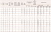
This prospective study enrolled 22 patients [10 women; age, 40.41±10.31 years (mean±SD), range 16–62 years] with medically intractable mTLE-HS who underwent ATL-AH at Asan Medical Center, Seoul, Korea (Table 1). The patients had been epileptic for 21.82±12.04 years (range 3–44 years). All of the patients were diagnosed with unilateral mTLE-HS based on the clinical history of seizures, MRI, PET, and video-EEG monitoring. MRI was performed using a 3.0-T MRI system (Gyroscan Intera Achieva, Philips Medical System, The Netherlands) equipped with a multichannel head coil. The MRI protocol included obtaining oblique coronal T2-weighted images perpendicular to the hippocampus, oblique axial FLAIR images, and both T1-weighted sagittal and coronal images. Ten of the study patients had right temporal lobe epilepsy (RTLE) and 12 had left temporal lobe epilepsy (LTLE). All of the patients had been taking between one and three appropriate antiepileptic drugs prior to surgery.
Preoperative intracarotid sodium amobarbital testing (IAT) revealed that 18 patients exhibited left-hemisphere dominance for language. Based on the IAT results, we determined that 10 patients had TLE in the dominant hemisphere (DTLE) and 12 had TLE in the nondominant hemisphere (NDTLE). Subjects were excluded if they had an intellectual deficit as indicated by a full-scale intelligence quotient (IQ) of <70 on the Korean version of the Wechsler-Bellevue Intelligence Scale (K-WAIS) prior to surgery. Table 1 provides other neuropsychological data that were used to estimate the preoperative temporal lobe function besides CAP in patients with TLE. None of the patients had any attention disorder or speech/language impairments before or after ATL-AH, or exhibited postoperative neurological deterioration or major complications after ATL-AH. The antiepileptic medication was not changed during the first 6 months after surgery. This study was reviewed and approved by the institutional review board of the Asan Medical Center (approval no. 2007-0348).
CAP tasks
In the 22 patients with intractable mTLE, frequency-pattern, duration-pattern, and dichotic tests as well as pure-tone audiometry were performed before and after epilepsy surgery. A postoperative follow-up study was performed 1–5 months after ATL-AH. Each test involved randomly presenting various test items on a computer to prevent educational effects both pre- and postoperatively. The pure-tone thresholds were within 20 dB of hearing level at all frequencies (0.5, 1, 2, and 4 kHz), and the speech discrimination score was >92% in all patients when 50 words were used in the test. The CAP tasks were performed in a sound booth according to previously described protocols.5 Our group previously developed diagnostic tests for CAPD in Korean speech and obtained norm data from a Korean right-handed population with normal hearing.9 Based on the normal right-handed population data obtained in that previous study, cutoff values were calculated as normal mean—[2×(standard deviation)]. Cutoff values of 77% in the frequency test, 81% in the duration test, 83% in the right-sided dichotic test, and 82% in the left-sided dichotic test were established.9
Frequency-pattern test
Frequency testing was performed in a sound booth using the method developed by Musiek.10 The prototype frequency pattern comprised three 150-ms pure tones with rise and fall times of 10-ms and two 200-ms intertone intervals. Pure tones at two frequencies (880 and 1122 Hz) were used, which were designated as the low-frequency tone (low) and high-frequency tone (high), respectively. There were six possible three-tone sequences (low-low-high, low-high-low, low-high-high, high-low-high, high-low-low, and high-high-low), and each tone sequence was randomly selected using a computer. Thirty of these sequences were presented to each ear, and the patients were instructed to identify the pattern that they heard by pressing an appropriate computer key. The total testing time was about 6 min per patient.
Duration-pattern test
The protocol for the duration-pattern test was similar to that for the frequency-pattern test except that the tone frequency was kept constant at 1000 Hz with two 300-ms intertone intervals, and patients were asked to identify the tone duration. Short (250-ms) and long (500-ms) pure tones were presented in three-tone sequences. Thirty of these sequences were presented to each ear, and the patients were asked to identify the pattern that they heard (i.e., short-short-long, long-short-long, etc.). The total time required for this test was about 7 min per patient.
Dichotic test
Dichotic speech tests were performed using common spondee words (e.g., scissor and peanut) selected from an appropriate Korean word list that had been used for speech tests. Spondee words recorded by a female speaker were delivered from a computer via headphones at a comfortable volume in each ear. Two spondee words were simultaneously presented to each ear, and the patient was instructed to repeat them. Sixty spondee words were used, and the two-word combinations were randomly selected using a computer.
Surgical procedures
ATL-HS was performed on 10 RTLE and 12 LTLE patients (10 DTLE and 12 NDTLE as determined by IAT). The hippocampus and amygdala were resected in all of the patients, with the resection length of the hippocampus not differing between the dominant and nondominant sides. The resection of the anterior temporal lobe extended 3.0 cm into the dominant temporal lobe and 4.0–5.0 cm into the nondominant temporal lobe, as measured from the temporal pole along the middle and inferior temporal gyri; care was taken to spare the superior temporal lobe. However, one LTLE (an NDTLE) patient underwent more extensive temporal lobectomies, including frontal lobe resection.
Data analysis
Scores from each test were compared between before and after surgery, and trend analyses were also performed. The Wilcoxon signed-rank test was used to detect statistically significant differences between pre- and postoperative scores. Comparisons of mean scores were also performed for each of the DTLE, NDTLE, RTLE, and LTLE subgroups. Second, patients with CAPD were identified based on normal cutoff values used in our laboratory before and after surgery, and whether surgery affected CAPD was analyzed using Fisher's exact test. In the trend analyses, patients were classified into two groups: with and without postoperative worsening. Post-operative worsening was defined as a reduction of more than a 5 points in the postoperative CAP percentage scores that were based on 1 SD of normative data for Korean subjects.9 Postoperative worsening of each CAP task was analyzed based on the surgery side (dominant or nondominant hemisphere, and left or right side), along with the verbal and nonverbal IQ scores. The normative cutoff value for the left-side dichotic test was applied to the dominant side, and the normative cutoff value of the right-side dichotic test was applied to the nondominant side. Thus, for patients with right-hemisphere language dominancy in the Wada test, the normative cutoff value for the left-side dichotic test was applied to the dominant side (right side), while the normative cutoff value for the right-side dichotic test was applied to nondominant side (left side). Logistic regression analysis was used to determine the strength of the associations between reduced performance after surgery and patient parameters. Statistical analyses were performed using SPSS (version 21.0, SPSS, Chicago, IL, USA). In all of the analyses the threshold for statistical significance was set at p<0.05.
RESULTS
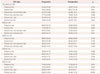
The test results for the patients are summarized in Table 2. All of the patients had normal hearing, with mean pure-tone thresholds within 20 dB of the hearing level at frequencies of 0.5, 1, 2, and 4 kHz; an "A"-type tympanogram in impedance audiometry; and normal findings in an otoscopy examination.
Changes in mean scores after surgery
The pre- and postoperative scores in each test are also presented in Table 2. The scores in the frequency test, duration test, and all types of dichotic tests did not differ significantly between before and after surgery (Table 2). Subgroup analysis according to the surgery side (dominant or nondominant hemisphere, and left or right side) showed no significant differences between pre- and postoperative scores in the CAP tests (Table 2).
CAPD before and after surgery
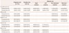
The frequencies of patients with pre- or postoperative CAPD in each test are presented in Table 3, CAPD were identified with the normal cutoff values used in our laboratory.9 For each of the CAP tasks, the frequencies of CAPD did not differ significantly between before and after surgery in all patients or in subgroups that were assigned according to the surgery site of dominancy (DTLD or NDTLD) and the surgery side (RTLE or LTLE). We detected no overall significant association between surgery and CAPD (Table 3).
Analysis of postoperative worsening
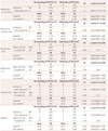
We performed a trend analysis of postoperative worsening and the following probable risk factors that are considered to be associated with CAP: dominancy of the surgery side, left or right surgery side, verbal IQ, and nonverbal IQ. Logistic regression analysis revealed a strong association between surgery in the language-dominant temporal lobe and postoperative worsening of CAP according to the non-dominant-side dichotic test (Table 4). That test indicated that the probability of a reduced performance after surgery among patients with surgery in the dominant temporal lobe was 7.5-fold greater than for patients with surgery in the nondominant temporal lobe. However, the frequency, duration, and dominant-side dichotic tests did not reveal a significant relationship between postoperative worsening of CAP and surgery in the language-dominant temporal lobe (Table 4). There were no significant associations between postoperative worsening of CAP on any test and other variables, such as the verbal IQ, nonverbal IQ, or right- or left-side lobectomy (Table 4).
DISCUSSION
We found that postoperative deterioration of CAP capability is associated with dominant ATL-AH in mTLE. TLE patients who underwent dominant ATL-AH showed a stronger trend for worsening CAP ability in non-dominant-side dichotic tests compared with those patients who underwent nondominant ATL-AH. The likelihood of worsening CAP in non-dominant-side dichotic tests for patients who underwent dominant ATL-AH was 7.5-fold greater than for patients who underwent nondominant ATL-AH.
Our analyses revealed that surgery on the dominant temporal lobe could lead to postoperative worsening of CAP compared with surgery on the nondominant temporal lobe, which may be a consequence of the testing modality used for the verbal stimuli-based dichotic test. We conducted the dichotic test by inputting a different word in each ear to evaluate binaural separation. While the frequency- and duration-pattern tests were performed using nonverbal stimuli, the dichotic test has been applying using verbal stimuli as a tool for linguistic assessment.111213 All but one patient in the present study underwent standard ATL-AH. To resect the lateral temporal structures, a posterior cortical incision at the lateral temporal gyri was extended 4.0–5.0 cm from the temporal tip on the nondominant temporal lobe and 3.0 cm from the temporal tip on the dominant temporal lobe. Thus, patients in whom surgery was performed in the dominant temporal lobe underwent less resection of lateral temporal areas compared with patients who underwent nondominant temporal lobectomy. However, surgery of the dominant temporal rather than the nondominant temporal lobe was more strongly associated with postoperative worsening in the non-dominant-side dichotic test, which means that the resection length alone cannot adequately explain any worsening. The postoperative worsening of CAP after surgery in the dominant temporal lobe could be due to complex interdependency and a richer linguistic role for deeper structures of the medial1415 and anterior161718 temporal lobes, together with the lateral neocortex on the dominant hemisphere. 19 Previous authors have suggested that the superior temporal gyrus is also critical for auditory speech processing. 720 However, we could not confirm such an association of CAPD with resection of the superior temporal lobe because this lobe was spared in all of the patients included in the present study.
A particularly interesting finding was the significant results obtained in the non-dominant-side dichotic test but not the dominant-side dichotic test, which is perhaps due to CAPD in the dichotic test being observed in the ear that was contralateral to the affected hemisphere.21 The initial report on CAPD21 indicated that temporal lobe lesions can alter a patient's perception of distorted speech presented to the ear contralateral to the lesion, and subsequently study have obtained similar findings.222324 Musiek25 demonstrated a markedly reduced performance in the ear that was contralateral to the hemispheric lesion. Sparks et al.26 reported a lesion effect wherein a loss of relative listening effectiveness occurred in the contralateral ear. The mechanism underlying this phenomenon remained unclear; however, because a dichotic test is a sensitive measure of the functioning of the transcallosal auditory pathways, a lesion on one side of a hemisphere may be related to CAPD in the ear contralateral to the affected side.25 These findings may support our observation of performance deterioration in the non-dominant-side dichotic test after dominant ATL-AH. Therefore, the laterality of temporal lobectomy was related to worsening of the CAP ability in contralateral-side dichotic tests in patients with dominant ATL-AH.
Our analyses further revealed that except for an association between surgery in the dominant temporal lobe and worsening in the non-dominant-side dichotic test, unilateral ATL-AH was not associated with deterioration of CAP function in any other test. This absence of an association might be due to patients with chronic mTLE-HS already exhibiting CAPD prior to temporal lobectomy, which could negate the "lesion effect" associated with surgery. Previous studies have found that individuals with mTLE-HS exhibit worse performance in temporal ordering and dichotic listening for both verbal and nonverbal sounds27 and show a reduced ability to process rapid auditory information.28 Our present finding that TLE patients have a high incidence of preoperative CAPD is consistent with these previous reports.
The current study was subject to some noteworthy limitations. First, the postoperative follow-up was performed between 1 and 5 months after ATL-AH. We had concerns that the use of different retest periods could affect the postoperative CAP results and introduce educational effects in the early postoperative follow-up group. However, a computer was used to randomly present test items on each test, so the possibility of educational effects was very low. Furthermore, we would argue that the differences in the duration of the follow-up period did not strongly affect our results. Indeed, Duffau et al.29 reported that nearly complete functional recovery could be observed within 1–3 months after surgical resection, and none of our patients exhibited postoperative neurological deterioration throughout the follow-up period. Additionally, we compared CAP scores with the post-operative variation in CAP scores between the early (within 2 months) and late (after 3 months) retest groups using the Mann-Whitney test, which revealed no significant differences in CAP scores between these groups (data not shown). Second, our data were obtained from a small number of patients with mTLE with ATL-AH, which could lead to ambiguous statistical findings in direct comparisons. Although no significant differences in mean CAP scores were observed according to surgery, the contralateral dichotic test results revealed a significant trend indicating that dominant ATL-AH contributes to postoperative worsening of CAP. Therefore, a larger patient cohort could more robustly identify differences between subgroups. To this end, the enrollment of additional participants is currently ongoing.
In conclusion, the current findings indicate that only surgery in the dominant temporal lobe can cause postoperative CAP deterioration. ATL-AH on the dominant rather than the nondominant side in patients with mTLE-HS was associated with worse postoperative CAP function in non-dominant-side dichotic tests. Other variables, including the surgery side (left or right), verbal IQ, and nonverbal IQ, did not have a postoperative effect on CAP. The present data suggest that the pre- and postoperative auditory function should be routinely investigated in patients with TLE, especially using verbal stimuli. Future large-scale studies should attempt to elucidate additional risk factors associated with ATL-AH on CAP abnormalities in patients with epilepsy.
 XML Download