Dear Editor,
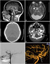
A 28-year-old woman presented with a 2-year history of headache, and magnetic resonance imaging (MRI) led to a diagnosis of suspected intracranial malformation. She had been in good health and had developed normally prior to developing the symptom. A neurological examination on admission revealed no anomaly. Brain MRI revealed agenesis of the inferior part of cerebellar vermis and a large posterior fossa cyst communicating with the fourth ventricle without hydrocephalus, suggestive of Dandy-Walker variant (DWv) (Fig. 1A and B). Paraclinoidal vascular dilation (Fig. 1B) and thinning of the occipital bone (Fig. 1C) were also observed. Catheter angiography revealed that the right internal carotid artery (ICA) was absent at the sixth segment, and the remnant of the intracranial clinoidal ICA and the left anterior cerebral artery territory were supplied by the right posterior communicating artery. The remnant segment of the ICA had an aneurysmal appearance, and was approximately 6.5 mm in diameter (Fig. 1E and F). The absence of the right ICA in the carotid canal confirmed the diagnosis (Fig. 1D). The patient's symptom was relieved by the administration of vasodilator agents.
Congenital aplasia of the ICA is an extremely rare vascular anomaly that occurs in less than 0.01% of the population, and is usually symptomless. Common symptoms and signs include recurrent headache, convulsions, blurred vision, hearing loss, hemiparesis with or without cranial nerve palsy, or transient ischemic attack and subarachnoid hemorrhage, depending on the collateral circulation and hemodynamics-related aneurysms, with an incidence of 25-34%.1 DWv consists of malformations with less severe cerebellar vermis hypoplasia, less notable or absent upward rotation of the vermis, and communication between the fourth ventricle and subarachnoid space, with no hydrocephalus.2 The etiology of the patient's headache remains to be established. The sporadic nature of the unilateral headaches along with the associated nausea is consistent with the diagnosis of either neurovascular insufficiency or intermittent high intracranial pressure.
This report describes a new association with DWv in a patient with no ICA, which has not been previously reported in the literature. Such patients must be followed clinically and radiographically in order to monitor for the possibility of cerebrovascular disorders, especially the development of aneurysms and hydrocephalus.
Figures and Tables
Fig. 1
Aplasia of the internal carotid artery (ICA) with Dandy-Walker variant. A: T1-weighted sagittal magnetic resonance imaging (MRI) showing a hypoplastic cerebellar vermis (long white arrow). B: T2-weighted axial MRI revealing a paraclinoidal aneurysmal appearance (short white arrow). C: Computed tomography (CT) scan showing a large posterior fossa cyst and thinning of the occipital bone. D: CT scan disclosing a pneumatized canal (asterisk). E: Right carotid arteriography revealing absence of the ICA. F: Three-dimensional image revealing that the anterior circulation is supplied by the posterior circulation (the white arrow indicates the aneurysmal appearance).


 PDF
PDF ePub
ePub Citation
 Print
Print


 XML Download