

 PDF
PDF ePub
ePub Citation
 Print
Print


INTRODUCTION
Human prion diseases are diagnosed by postmortem pathological examinations of tonsils and brain tissues.1 Current clinical diagnostic criteria are based on the clinical features and analytical results from electroencephalography (EEG), brain magnetic resonance imaging (MRI), and the presence of the 14-3-3 protein in the cerebrospinal fluid (CSF).23 The expression of the 14-3-3 protein in CSF is up-regulated not only in patients with CJD but also in patients with other neurodegenerative disorders.45 The only reliable molecular marker of human prion diseases is the pathogenic prion protein (PrPSc). From a diagnostic perspective, body fluids such as CSF, blood, and urine are valuable for the early and specific diagnosis of human prion diseases.6 Since the levels of PrPSc in these fluids are known to be extremely low, the efficient in vitro amplification of minute PrPSc is an encouraging approach for diagnosing human prion diseases at an early phase and with a higher level of precision.67 Recent studies have utilized ultrasensitive tests such as protein-misfolding cyclic amplification, quaking-induced conversion (QuIC), the amyloid seeding assay, and the surround optical fiber immunoassay for diagnosing prion diseases.89 In the QuIC assay, thioflavin T (ThT) dye displays an enhanced fluorescence and a characteristic red shift of its emission spectrum when it incorporates into the β-sheet-rich structures that are observed in amyloid aggregates.10 A version of the QuIC assay that is able to measure the ThT fluorescence in real time, called real-time QuIC (RT-QuIC), was recently developed for the diagnosis of sporadic CJD (sCJD) and genetic CJD in CSF samples.1112 In addition, RT-QuIC analysis may ameliorate the specificity weakness of 14-3-3 in diagnosing sCJD.8 In this study, we tried to check whether RT-QuIC assay is useful for the diagnosis of probable sCJD patients by using the CSF of the patients diagnosed by the presence of 14-3-3 and high levels of total tau protein in CSF.
METHODS
Patients
CSF specimens were obtained from 81 sCJD patients [37 females and 44 males; aged 27-86 years, 65.3±11.2 years (mean±SD)] diagnosed as definite or probable cases, as well as from 100 non-CJD subjects in the Republic of Korea. None of the patients carried the mutation in PRNP. The patients were diagnosed as sCJD based on the specific EEG findings, specific hyperintensity in the cerebral cortex in diffusion-weighted MRI, and the presence of the 14-3-3 protein in CSF. The CSF, blood samples, and personal information of all of the subjects were collected from data obtained during 2010-2014 in the nationwide CJD surveillance program that was established in the Republic of Korea in 2001. All of the experiments were performed blinded for personal information and clinical data. PrP genotyping was performed using genomic DNA extracted from peripheral blood leukocytes as described previously.13
Immunoblotting
Proteins of the CSF samples were separated using 12% sodium dodecyl sulfate-polyacrylamide gel electrophoresis and transferred to polyvinylidene difluoride membranes (Amersham Biosciences, Freiberg, Germany). After blocking with 5% skim milk in TBS with 0.1% Tween-20, the membranes were incubated with rabbit polyclonal anti-14-3-3 antibody (1:500; Santa Cruz Biotechnology, Santa Cruz, CA, USA) and sequentially with horseradish-peroxidase-conjugated anti-rabbit IgG (1:5,000; Thermo Fisher Scientific Inc., Waltham, MA, USA). The 14-3-3 protein bands were detected using an enhanced chemiluminescence detection system (Thermo Fisher Scientific Inc., Waltham, MA, USA).
Determination of the total tau protein level
The levels of total tau protein in CSF were measured using an ELISA kit following the manufacturer's instructions (Thermo Fisher Scientific Inc., Waltham, MA, USA). Briefly, each CSF sample (50 µL) was diluted in sample diluent (1:1 v/v) in the kit prior to incubation with the anti-tau antibody. The colorimetric reaction was measured at 450 nm with a microplate reader (SpectraMax 190; Molecular Devices, CA, USA). The total tau protein concentration is reported herein as the mean value of duplicate measurements.
Expression and purification of recombinant human PrP
Recombinant PrP using a plasmid containing a DNA sequence coding for residues 24-234 of the human PrP sequence was expressed, refolded into a soluble form, and purified as described previously.11 Briefly, the DNA sequence coding for human PrP was amplified by the polymerase chain reaction and ligated into the pET41 vector with Nde I-Hind III inserts (EMD Biosciences Inc., Madison, WI, USA), and the obtained sequence was verified by sequencing. After transforming the plasmids into E. coli BL21 (DE3) (EMD Biosciences Inc., Madison, WI, USA), the enriched recombinant human PrP (rec HuPrP) was purified using a previously described method with minor modifications.11 Briefly, the denatured rec HuPrP was incubated with a Ni-NTA Superflow resin (Qiagen, Valencia, CA, USA) and refolded with a linear gradient over 6 h at a flow rate of 1 mL/min using the AKTA Purifier system (GE Healthcare Europe GmbH, Germany) and then eluted with 100 mM sodium phosphate (pH 5.8) containing 500 mM imidazole and 10 mM Tris. Finally, the rec HuPrP was filtered and dialyzed against 10 mM phosphate (pH 5.8) before its concentration was determined. Purified aliquots of the proteins were stored at -80℃ prior to use. The plasmid containing a DNA sequence coding for human prion protein (residues 24-234) was provided by Dr. Noriyuki Nishida at Nagasaki University.
RT-QuIC assay
The RT-QuIC assay was performed as described previously.11 The composition of the RT-QuIC reaction buffer was as follows: 500 mM NaCl, 25 mM PIPES (pH 7.0), 1 mM EDTA, and 10 µM ThT. To perform the RT-QuIC assay, freshly thawed rec HuPrP (0.1 mg/mL) in 95 µL of the reaction buffer and 5 µL of CSF was transferred to each well in a 96-well optically black bottom plate (Nunc, Rochester, NY, USA) for CSF-seeded reactions. A sample of sCJD brain tissue from the "Korea CJD Autopsy Center" was used as a positive control, as shown in Fig. 1. Artificial CSF (A-CSF) was used as a negative control in the RT-QuIC assay. To prevent contamination, we used non-infectious materials and filter tips inside a biological safety cabinet in a prion-free laboratory. The 96-well plate was covered with sealing tape (Nunc) and incubated in a fluorescence plate reader (Infinite F200, TECAN, Männedorf, Switzerland) at 37℃ with intermittent shaking. The ThT fluorescence was measured (for excitation at 450 nm and emissions at 480 nm) every 10 min. Each experimental sample and control sample (not containing seeded A-CSF) was run in quadruplicate. The ThT fluorescence was measured in relative fluorescence units (i.e., rfu; i.e., relative to the negative control or A-CSF), and the average fluorescence for each quadruplicate sample was monitored over time. An rfu value of 250-400 for the brain homogenate (BH) of a normal human and A-CSF was considered as the baseline. All RT-QuIC assays were performed blinded for personal information and clinical data.
Statistical analysis
Statistical analysis was performed as described previously.14 Total tau protein levels in the CSF were compared between sCJD and non-CJD patients using the two-tailed t-test. The Mann-Whitney U test was used for the statistical comparisons of the age at onset. Differences were considered statistically significant when p<0.05. All analyses were performed using GraphPad Prism 4 software (GraphPad Software Inc., San Diego, CA, USA) and IBM SPSS Statistics (IBM, USA).
RESULTS
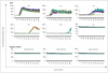
We first used the RT-QuIC assay to determine whether PrPSc in the BH of a sCJD patient can function as a prion seed. The sCJD BH was serially diluted (from 10-3 to 10-12 in 10-fold steps) with A-CSF and then used each of these dilutions as a prion seed in the RT-QuIC reaction. We observed the high peaks of ThT fluorescence in all four of the quadruplicate samples when the sCJD BH was used at dilutions of 10-3 to 10-7, in three of the quadruplicate samples at a dilution of 10-8, in one at dilutions of 10-9 and 10-10, and in none at dilutions of 10-11 and 10-12 (data for dilutions of 10-3 to 10-5 not shown) (Fig. 1). In addition, there was no significant ThT fluorescence peak in the negative control samples (at dilutions of 10-6 to 10-8), and so we used a 10-6 dilution of the 10% BH from a sCJD patient as a positive control for the RT-QuIC assay in this study.
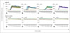
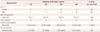
We next evaluated the availability of the RT-QuIC assay using CSF specimens of 81 sCJD patients with higher levels of total tau protein and the presence of the 14-3-3 protein in their CSF in order to improve the diagnostic accuracy of sCJD (Table 1). The RT-QuIC reaction was considered to be positive even though only one reading of the quadruplicate samples was 900 rfu or greater at 320 cycles. By 320 cycles (1 cycle takes 10 min) the number of sCJD CSF-seeded reactions showed increased fluorescence when compared to the approximate baseline of 250 rfu for A-CSF (the negative control) (Fig. 2). The fluorescence began to increase at a median of 60 cycles in the 40 CSF samples of sCJD patients and after 150 cycles in the 22 CSF samples (Fig. 2). Of the 81 specimens with 14-3-3-positive CSF, 62 were positive for the ThT fluorescence signal of RT-QuIC (sensitivity of 76.5%) (Supplementary Table 1 in the online-only Data Supplement), but there was no signal for the CSF specimens of the 100 non-CJD patients (Supplementary Table 2 in the online-only Data Supplement). These observations indicate that PrPSc in the CSF of sCJD patients functions as a prion seed, and that the rec HuPrP utilized as a substrate for prion conversion undergoes a conformational change into a β-sheet in the RT-QuIC reaction. In addition, we checked the expression levels of 14-3-3 and total tau proteins in the CSF samples of sCJD patients. All of the 81 CSF specimens were positive for 14-3-3, whereas the levels of total tau protein varied (Table 1). The distributions of sex and age did not differ significantly among the four quadruplicate samples, but the CSF produced four positive reactions in the quadruplicate samples in the RT-QuIC analysis had the highest average levels of total tau protein (Table 1).
DISCUSSION
In this study we evaluated the diagnostic application of the RT-QuIC assay in Korean patients with definite or probable sCJD. The RT-QuIC assay showed a sensitivity of 76.5% in 81 CSF samples of Korean patients with sCJD (Table 1). The sensitivity and specificity of CSF-based RT-QuIC analysis of this study were similar to the results obtained in previous studies involving sCJD patients, which demonstrated a sensitivity of >80% and a specificity of 100% in CJD specimens from Japan and Australia.1112 In another previous study, RT-QuIC analysis showed a sensitivity of 84% and a specificity of 100% in the CSF of sCJD patients.1115 One other CSF-based RT-QuIC analysis revealed a sensitivity of 85-90% and a specificity of nearly 100%, which suggests the superiority of RT-QuIC analysis over other diagnostic methods based on the expression levels of total tau and 14-3-3 proteins that have thus far been used as the diagnostic markers of human prion diseases.8
The level of the 14-3-3 protein in CSF is already commonly used as a helpful biomarker in the diagnostics of human prion diseases in many countries,7 including as a determinant biomarker for diagnosing CJD. Several groups have recently begun to apply the RT-QuIC method in the diagnosis of human prion diseases.8151617 We therefore measured both the positivity in the RT-QuIC reaction and the expression levels of 14-3-3 and total tau proteins using the CSF of patients with sCJD. For the patients with the highest average level of total tau protein detected in CSF, ThT fluorescence positivity in the RT-QuIC reaction was detected in all four of the quadruplicate samples, which indicates that there may be a correlation between the levels of total tau protein and the pathogenic prion isoform in CSF. Overall, we have found that there was a moderate correlation between the positivities of the 14-3-3 protein and the RT-QuIC analysis and between the positivities of total tau and the RT-QuIC analysis in the CSF of Korean patients (Supplementary Table 1 in the online-only Data Supplement).
The smaller number cases of sCJD in this study may have been responsible for the negative results for sCJD in the RT-QuIC analysis. The 14-3-3 protein was present in the CSF of three of the sCJD patients (aged 27, 42, and 43 years) who were younger than the average age of patients negative in the CSF-based RT-QuIC analysis, and the levels of total tau protein in their CSF were higher than those in controls (1400 pg/mL). These observations indicate that it may be equivocal to determine a diagnosis of sCJD only by detecting 14-3-3, high levels of total tau protein, or the positivity in the RT-QuIC assay in CSF in cases of probable CJD. The collective results of the RT-QuIC analysis in this study showed a sensitivity of >75% and a specificity of 100% (Supplementary Table 2 in the online-only Data Supplement). Same as this study, a CSF-based RT-QuIC analysis for probable CJD patients showed a specificity of 100%, when compared to other clinical markers.8 It therefore seems that RT-QuIC analysis is invaluable for definitively diagnosing sCJD when the patients are still alive. The CSF-based RT-QuIC assay will become valuable in the differential diagnosis of suspected sCJD.
In conclusion, the PrPSc-like abnormal form of PrP was generated using the RT-QuIC reaction when rec HuPrP and the CSF of patients with sCJD were used. Thus, RT-QuIC analysis might be a useful tool for the diagnosis of sCJD patients.
 XML Download