

 PDF
PDF ePub
ePub Citation
 Print
Print


Introduction
Hypercholesterolemia is closely associated with an increased risk of cardiovascular disease, including stroke, due to atherosclerosis of blood vessels. Total cholesterol is made up of low-density lipoprotein (LDL), very-low-density lipoprotein, triglycerides (TG), and high-density lipoprotein (HDL). Atherosclerosis is generally characterized by elevated plasma concentrations of TG or LDL and a decreased HDL concentration.1 It has recently been shown that certain LDL, subclassified according to particle size, are associated with atherosclerotic risk.2 LDL particle size is negatively associated with plasma TG levels, and the combination of small LDL particles and increased plasma TG levels is considered to be the atherogenic lipoprotein phenotype.3 Small LDL particles are more easily taken up by arterial tissue4 and are more susceptible to oxidative stress than are large LDL particles.5 Therefore, small LDL particles are considered to be associated with the progression of atherosclerosis6 and have been accepted by the National Cholesterol Education Program Adult Treatment Panel III as one of the emerging cardiovascular risk factors.7
Clinical studies have emphasized the role of small LDL particles in the development of early atherosclerosis in menopausal women,8 coronary heart disease including acute myocardial infarction or coronary vasospasm,9,10,11,12 or peripheral arterial disease, regardless of the presence of diabetes mellitus.13 In stroke patients, small LDL particles are known to be associated with short-term mortality after acute ischemic stroke.14 However, there is as yet little information available on the relationship between LDL particle size and functional outcome after stroke, and including stroke severity. Therefore, in this study, the association between LDL particle and stroke severity and functional outcomes was investigated in patients with atherothrombotic stroke.
Methods
Subjects
Patients diagnosed with first-episode ischemic stroke and admitted to Ewha Womans University Mokdong Hospital within 7 days after symptom onset were prospectively enrolled in this study between January 2009 and June 2011. Blood samples to be used for LDL particle analysis were obtained from all enrolled patients. Patient information was collected, and data were evaluated including past medical, medication, and familial history, brain imaging studies (CT and/or MRI), vascular imaging studies (digital subtraction angiography, CT angiography, or MR angiography), chest X-ray, 12-lead electrocardiography, electrocardiography monitoring during a median time period of 3 days at a stroke intensive care unit, transthoracic echocardiography, and routine blood tests.15 Patients were excluded if they did not agree to provide blood samples for this study. Of the 427 initially eligible patients, 2 who received incomplete vascular imaging, 40 who had transient ischemic attacks with negative diffusion-weighted images, and 10 with rare causes of stroke, such as moyamoya disease, arterial dissection, or venous thrombosis, were excluded. Moreover, patients with a moderate or high risk of cardiac sources of embolism (n=127) based on the Trial of Org 10172 in Acute Stroke Treatment classification system16 were excluded because acute cardioembolic occlusion of the artery could be mistaken for true arterial occlusion despite the absence of stenosis. Ultimately, the data of 248 patients were analyzed in this study, which was approved by the Institutional Review Board of Ewha Womans University Mokdong Hospital.
Measurement of the degree of stenosis, stroke severity, and functional outcome
The degree of arterial stenosis was measured according to the method used in the North American Symptomatic Carotid Endarterectomy Trial17 for extracranial arteries, or based on the method used in the Warfarin-Aspirin Symptomatic Intracranial Disease study18 for intracranial arteries. Vascular images were evaluated by two independent vascular neurologists (T.-J.S. and H.-J.C.) who were blinded to the clinical information. Interobserver agreement on the presence of more than 50% stenosis and/or occlusion was excellent (κ=0.96), and cerebral artery stenosis was classified into three groups: ≥50% stenosis (one or more vessels with ≥50% stenosis), <50% stenosis (one or more vessels with <50% stenosis), and no atherosclerosis (i.e., no stenosis). The severity of neurologic deficits was determined using the National Institutes of Health Stroke Scale (NIHSS) at admission.19 Functional outcomes were also assessed using the modified Rankin Scale (mRS) at 3 months after the index stroke.
Measurement of LDL particle size and lipid profile
Blood samples were collected after fasting for >12 h for lipid profiling into plain, ethylenediaminetetraacetic acid-treated tubes within 24 hours after admission. The blood was centrifuged to separate the plasma or serum from the whole blood, and then stored at -70℃ until analysis. Nondenaturing polyacrylamide gradient gel electrophoresis with lipid staining of the plasma was performed to determine the peak LDL particle diameter, as described elsewhere.20 Briefly, the whole plasma and the plasma fraction with a density of <1.063 kg/L (prepared by ultracentrifugation) were separated by electrophoresis using gradient gel (PAA 2/16, Pharmacia, Uppsala, Sweden). Gels were stained and then scanned with a scanning densitometer (Transidyne RFT, Ann Arbor, MI, USA), and the peak particle diameters of the main LDL subclasses were calculated from calibration curves using standards of known size. Serum concentrations of cholesterol, LDL, and HDL were measured with commercially available kits (Choongwae, Seoul, Korea) using enzymatic methods.21 The serum TG level was analyzed using a total glycerol test kit (Roche, Basel, Switzerland). All measurements were performed on an Hitachi 747 autoanalyzer (Hitachi, Tokyo, Japan). Plasma levels of lipoprotein (a) were measured using an enzyme-linked immunosorbent assay, according to the manufacturer's instructions (R&D Systems, Minneapolis, MN, USA). Each sample was measured in duplicate and the mean of the two values was used in the analysis.21
Risk factors
Hypertension was defined as a resting systolic blood pressure of ≥140 mm Hg or a diastolic blood pressure of ≥90 mm Hg on repeated measurements, or receiving treatment with antihypertensive medications. Diabetes mellitus was diagnosed if the patient had a fasting blood glucose level of ≥7.0 mmol/L or was being treated with oral hypoglycemic agents or insulin. Hyperlipidemia was diagnosed if the patient had an LDL-cholesterol level of ≥4.1 mmol/L, a total cholesterol level of ≥6.2 mmol/L, or if the patient was being treated with lipid-lowering agents after being diagnosed with hyperlipidemia. Patients were defined as smokers if they were current smokers or if they had stopped smoking within 1 year before the stroke event. The presence of coronary heart disease was determined when a patient had a history of unstable angina, myocardial infarction, or angiographically confirmed coronary artery disease. A positive family history was considered as a history of coronary heart disease or stroke, regardless of their type (ischemic or hemorrhagic). If the cause of disease was unknown in a family member, the family history in the subject was defined as negative.
Statistical analysis
Statistical analyses were performed using the Windows SPSS software package (version 18.0, SPSS Inc., Chicago, IL, USA). Continuous variables are expressed as mean±SD values, or as medians and interquartile ranges (IQRs). Categorical variables are expressed as frequencies and percentages. Independent t-test, Mann-Whitney U test, one-way analysis of variance with Bonferroni corrected post-hoc analyses, and the Kruskal-Wallis test were used to compare continuous values. Categorical variables were compared using the chi-square test or Fisher's exact test. Since the continuous and ordinal variables of this study did not exhibit normality according to the Kolmogorov-Smirnov test (except HDL and total cholesterol), even though logarithmically transformed, NIHSS score was trichotomized based on their tertiles for uni- and multivariate analyses. The other continuous or ordinal variables were dichotomized according to their median values. Univariate and multivariate multinomial logistic regression analysis was used for stroke severity (NIHSS score with the first tertile as the reference group). Univariate and multivariate binary logistic regression analyses were also performed to determine the predictive factors for functional outcome. Functional outcome was dichotomized into good (mRS score of <3) or poor (mRS score of ≥3). The cutoff for statistical significance was set at p<0.05 (two-tailed).
Results
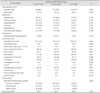
The demographic data of study subjects are given in Supplementary Table 1 (in the online-only Data Supplement). The patients were aged 62±11 years, and 64.9% (161/248) were male. The LDL particle size was 25.9±0.9 nm and the median NIHSS score was 3 (IQR=2-5). A history of diabetes mellitus, hemoglobin A1C and fasting glucose stroke was more frequent or elevated in the ≥50% stenosis group than in the no-atherosclerosis group. Smoking was more frequent in the no-atherosclerosis and ≥50% stenosis groups than in the <50% stenosis group. Twenty-one patients had taken lipid-lowering agents prior to the index stroke (a statin only in 19 patients, and a statin and fenofibrate in 2). The LDL particles were smaller in the ≥50% stenosis group (24.5±0.8 nm) than in the no-atherosclerosis (25.9±0.8 nm) and <50% stenosis (25.2±0.7 nm) groups (Supplementary Table 1).
Association between clinical variables and LDL particle size
LDL particle size was inversely correlated with age (r=-0.171, p=0.013), serum TG concentration (r=-0.155, p=0.024), TG/HDL ratio (r=-0.139, p=0.044), high-sensitivity C-reactive protein [high-sensitivity C-reactive protein (hs-CRP; r=-0.139, p=0.044)], NIHSS score (r=-0.228, p=0.012), and mRS score (r=-0.190, p=0.005). Furthermore, LDL particle size had marginally significant negative associations with hemoglobin (r=-0.118, p=0.086), white blood cell count (r=-0.122, p=0.077), hemoglobin A1C (r=-0.115, p=0.095), and fasting glucose level (r=-0.122, p=0.075). There was no statistically significant correlation between LDL particle size and any of the other variables.
Association between LDL particle size and stroke outcomes (severity and functional outcome at 3 months)
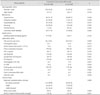
The relationship between LDL particle size and initial stroke severity was investigated after trichotomizing NIHSS scores. The LDL particles were smaller in the third tertile (NIHSS score ≥5) than in the second (NIHSS score 3 or 4) and first (NIHSS score 0-2) tertiles (24.8±0.7, 25.7±0.8, and 26.1±0.8 nm, respectively; p=0.004) (Table 1). Moreover, 194 (78.2%) of the 248 patients had a good functional outcome, and the LDL particle size in these patients was 26.1±0.8 nm. The LDL particles were smaller (25.5±0.7 nm, p=0.003) in the remaining 54 (21.8%) patients with a poor outcome than in patients with a good functional outcome (Table 2).
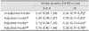
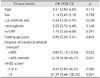
Regarding stroke severity, after adjusting for factors including age, sex, and variables with p<0.1 in univariate analysis (white blood cell count, total cholesterol, LDL cholesterol, hs-CRP, and fasting glucose), LDL particle size was independently and inversely associated with stroke severity [NIHSS score ≥5, reference NIHSS score 0-2; odds ratio (OR)=0.38, p=0.028] (Table 3). Furthermore, LDL particle size was independently and inversely associated with poor functional outcome at discharge (mRS score ≥3; OR=0.44, p=0.038) after adjusting for age, sex, and variables with p<0.1 in univariate analysis (hemoglobin, hs-CRP, fasting glucose, degree of cerebral arterial stenosis, and NIHSS score) (Table 4).
Discussion
The findings of this study show that small LDL particles were associated with stroke severity and poor functional outcome in the studied patients, even after adjusting for NIHSS score, which is a strong predictive factor for stroke outcome. There are a few previous reports on this relationship. One study of 200 patients with acute ischemic stroke found that small LDL particles were significantly associated with in-hospital mortality.14 Furthermore, in patients with coronary artery disease, and especially in those with acute myocardial infarction, the LDL particles were reportedly relatively small, and this tendency persisted during hospitalization.11 Consistent with this previous study in patients with acute myocardial infarction, the LDL particle size measured in the present study is also likely to have been affected by the acute-phase reaction of the lipolysis during acute ischemic stroke.
In addition, we found that small LDL particles were correlated with atherogenic molecules such as TG and hs-CRP. These data are relatively consistent with the results of a study that evaluated carotid artery stenosis using a hospital-based, cross-sectional design22 and of a study that measured the relative LDL particle sizes in patients with metabolic syndrome, insulin resistance, or coronary heart disease.3 Since both serum TG and hs-CRP are closely associated with atherosclerosis via the inflammatory pathway,1 the present findings suggest that small LDL particles play an important role in atherogenicity or the inflammatory reaction in atherosclerosis.
The presence of an association between LDL particle size and stroke severity and poor functional outcomes may be attributable to the following pleiotropic roles of small LDL particles. First, small LDL particles are more atherogenic than their larger counterparts.22 It is known that small LDL particles are more easily taken up by arterial tissue, suggesting greater transendothelial transport, and they exhibit increased binding with polyanionic proteoglycans, which play a deterministic role in atherosclerosis.2 Since progressive cerebral atherosclerosis is associated with a poor stroke outcome,23 the present results regarding the association between small LDL particles and stroke prognosis may be valid. Second, small LDL particles have a lower affinity for LDL receptors than do larger LDL particles.24 The lower affinity of small LDL particles for LDL receptors was reported to result in a longer retention time and consequently increased susceptibility to oxidative modifications.25 This altered balance in the antioxidant/oxidant mechanism may cause increased vascular thrombogenesis as well as severe atherogenesis.26 Moreover, increased thrombogenesis may contribute to atherothrombosis and poor stroke outcome. Finally, small LDL particles are involved in the inflammatory cytokine signaling pathways. The excess infiltration in the arterial intima and local enzymatic modification of small LDL particles may trigger the expression of inflammatory cytokines and vascular cell adhesion molecules. These cascades could ultimately lead to the formation of destabilizing lipid-dominant cores and rupture prone fibrous caps on plaque.27 This vulnerable atherosclerotic plaque is an important determinant for thrombosis-related stroke with poor outcome, thus explaining the present finding of a relationship between stroke outcome and LDL particle size. Small LDL particles have also been reported to be inversely correlated with tumor necrosis factor α and interleukin-1β.28 These inflammatory cytokines are associated with activation of nuclear factor-κB, which is triggered by hypoxia, reactive oxygen species, and several inflammatory mediators, and is responsible for neuronal cell death and neurovascular unit injuries.29 Therefore, the presence of small LDL particles could reflect the severity or outcome of ischemic brain injury.
This study was subject to some limitations. Blood samples were not drawn from a sample of the normal population for comparison. However, the objective of this study was to determine the relationship between LDL particle size and prognosis in stroke patients. Furthermore, such a comparative study between normal controls and stroke patients has already been performed,14 and it was found that LDL particles were smaller in stroke patients. Another limitation of the present study is that blood samples were obtained from acute stroke patients at admission, and LDL particle size was not serially assessed during the stroke time course. However, the patients' lipid and lipoprotein levels remained stable during the 4-week follow-up.30
In conclusion, small LDL particles are associated with initial stroke severity and are an independent predictor of poor functional outcome. Therefore, LDL particle size is a potential biomarker for the prognosis of atherothrombotic stroke.
 XML Download