

 PDF
PDF ePub
ePub Citation
 Print
Print


INTRODUCTION
Among the neurodegenerative parkinsonian disorders, progressive supranuclear palsy (PSP)1 is the second most common after idiopathic Parkinson's disease (IPD). Since levodopa responsiveness is poor and clinical deterioration is faster in PSP, it is important to be able to differentiate between IPD and PSP.2 It has been suggested that magnetic resonance (MR) measurements of several brainstem structures could overcome the limitations inherent in the clinical diagnostic criteria for PSP.3456789 The midbrain becomes atrophic in PSP patients, and thus it has been proposed that the decrease in the ratio of the midbrain area to the pons area (M/P ratio)67 and the characteristic shape of the upper brainstem (hummingbird sign)5 in the midsagittal plane could be used to discriminate between PSP and IPD. Quattrone et al.8 developed an index [the MR parkinsonism index (MRPI)] that reflects not only midbrain atrophy but also superior cerebellar peduncle (SCP) atrophy in PSP patients, and reported that MRPI is a better diagnostic tool for PSP.
The use of MR parameters in the diagnosis of parkinsonian disorders faces some obstacles. First, while the use of various structural parameters or their ratios or indexes [as measured using brain MR imaging (MRI) data] has been proposed for the differential diagnosis of parkinsonian disorders,3456789 the methods of measurement have varied between investigators and MRI protocols have not been standardized. Second, studies directly comparing diagnostic validity among different MR measurements are lacking. Third, although MRPI is reportedly superior to other diagnostic measurements, its use has been investigated by only two research groups, and these groups both used a unique, nonstandard MR protocol.81011 Furthermore, it remains to be determined whether MR images in standard MRI planes are as effective as MRPI in the diagnosis of PSP. Fourth, with the exception of a recent study by Morelli et al.,12 the influence of age has not been considered. Sung et al.9 reported that the diameter of the midbrain tegmentum (MBTegm) is decreased in PSP patients compared with normal controls. However, the diagnostic value of this parameter and its power to discriminate PSP from IPD have not been systematically examined.
The aim of this study was to determine whether the diameter of the MBTegm can aid in differentiating PSP from IPD, and which of the previously used MR measurement tools are the most useful for distinguishing between PSP and IPD.
METHODS
Subjects
Brain MR images of patients with IPD or PSP whose epidemiological and clinical data were collected based on a standardized registry protocol as well as three dimensional (3D) T1-weighted MRIs were analyzed retrospectively. This study was approved by Institutional Review Board in Hallym University Sacred Heart Hospital. IPD was diagnosed using the UK Parkinson's Disease Society Brain Bank criteria.2 Probable and possible PSP were diagnosed using the clinical diagnostic criteria of the Society for PSP of the National Institute of Neurological Disease and Stroke.1 In total, 125 patients with IPD or PSP were identified between March 2011 and February 2014. The distribution of the age at which the MRI was conducted among the IPD group did not conform to a normal distribution, and it was not practical to differentiate early-onset parkinsonian patients from PSP patients; therefore, 14 IPD patients who submitted to MRI at the age of ≤55 years were excluded. Thus, data from 111 patients were analyzed, consisting of 82 IPD patients and 29 PSP patients (21 probable and 8 possible).
MRI and measurements of brain structures
A multisequence 3-T MRI scanner (Philips Achieva 3.0T MRI System, release 3.2.1.1, Best, The Netherlands) was used to acquire standardized MR images consisting of T1-weighted 3D MP-RAGE axial images, T2-weighted axial and sagittal images, two dimensional gradient-echo T2*-weighted axial images, FLAIR-weighted axial images, and MR angiography images. The following parameters used to obtain T1-weighted 3D volumetric images: resolution, 1×1×1 mm3; repetition time/echo time, 800/26 ms; voxel size, 1.3×1.0×5.0 mm3; and interslice gap, 1.0 mm.
T1-weighted 3D axial MR images and images reconstructed therefrom were used to measure various brain structures. For the measurement of sagittal or coronal images, T1-weighted axial 3D MR images in DICOM format were reconstructed into sagittal and coronal images using OsiriX software (version 3 of the GNU General Public License, 2007, Free Software Foundation, Boston, MA, USA). All measurements were made using the same software. Any possible bias caused by considering patients with the same diagnosis together was avoided by shuffling MR images from PSP and IPD patients and analyzing them in a random order. All measurements were made twice with a 2-week interval by the same rater (Y.H.K.) who was blinded to the clinical diagnosis. The mean value of these two measurements was used for all statistical analyses except the intrarater reliability test.
MR references for measuring brain structures
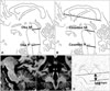
The brain structures that were measured are shown in Fig. 1. The length of the MBTegm was measured on an axial T1-weighted image using Sung's method.9 Briefly, an axial image encompassing the mid-mammillary body was chosen, and the distance from the interpeduncular fossa to the center of the aqueduct was measured (Fig. 1D). This distance was normalized by dividing it by the anterior commissure-posterior commissure (AC-PC) distance, which was measured in the reconstructed midsagittal plane, to generate the AC-PC-adjusted length of the MBTegm. To obtain the M/P ratio in the midsagittal plane, the midbrain and pons were measured using methods described in two previous publications, after magnifying the midsagittal images by a factor of eight.67 The M/P ratio was measured according to Oba's method (Oba M/P) by setting the lower margin of the midbrain area that corresponds to the upper margin of the pontine area to a line passing through the superior pontine notch and the inferior edge of the quadrigeminal plate; the lower margin of the pontine area was set to a line parallel to the lower margin of the midbrain area that passes through the inferior pontine notch (Fig. 1A).6 The M/P ratio was also measured using Cosottini's method (Cosottini M/P) by first drawing a line connecting the inferior borders of the genu and the splenium of the corpus callosum; the lower borders of the midbrain and pons were then set to lines parallel to that line and passing through the superior and inferior pontine notches (Fig. 1B).7 MRPI was measured using Quattrone's method8 with minor modifications (mMRPI). Briefly, although oblique coronal planes have been used previously81011 to measure the SCPs, reconstructed coronal images that were perpendicular to the AC-PC plane were used in the present study. The midbrain area and pontine area were measured on midsagittal MR images using Oba M/P.6 For measurements of the width of the middle cerebellar peduncle (MCP), a T1-weighted midsagittal volumetric MR image was chosen as the starting, reference view. The linear distance between the superior and inferior borders of the bilateral MCPs was measured in parasagittal images that best exposed the MCP between the pons and the cerebellum. The mean value of the measured widths of the left and right MCPs was calculated. The SCPs were measured as the linear distances between the medial and lateral borders of both SCPs at the middles of their extensions, as observed in reconstructed T1-weighted volumetric coronal MR images. The first image on which the inferior colliculi and SCPs were separated was used as the starting view for SCP measurements. SCP measurements were always performed on three consecutive sections. The SCP measurements were also averaged. The ratio of the pontine area to the midbrain area (P/M) and the ratio of the MCP width to the SCP width (MCP/SCP) were both calculated, and mMRPI was then calculated as (P/M)×(MCP/SCP)8 (Fig. 1C).
Statistical analysis
The difference in the sex distribution between IPD and PSP patients was analyzed using the chi-square test, and differences in the mean values of all continuous variables between groups were analyzed using independent t-tests. The intrarater reliability was assessed by calculating the intraclass correlation coefficient (ICC). Spearman correlation analysis was used to test correlations between MRI measurements and age at MRI. Receiver operating curve (ROC) analyses of the brain structure measurements were used to determine optimal cutoff values with the Youden index. The sensitivity, specificity, positive predictive value, and negative predictive value for differentiating PSP from IPD were determined for the cutoff values used for each measurement. ROCs were compared among the measurements of the four brain structures in a pairwise fashion. The threshold for statistical significance was set at p<0.05 for all tests. All statistical analyses were performed using MedCalc Statistical Software (version 13.2.2, MedCalc Software, Ostend, Belgium; http://www.medcalc.org; 2014).
RESULTS
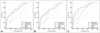
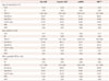
The demographic data for the IPD and PSP patients are given in Table 1. There was no difference in gender, disease duration, or Mini-Mental Status Examination score between the IPD and PSP patients. However, age at MRI (p<0.0005) and the total and motor scores on the Unified Parkinson Disease Rating Scale (p<0.01 and p<0.05, respectively) were higher in PSP patients than in IPD patients. The ICCs of all four MRI parameters (Oba M/P, Cosottini M/P, mMRPI, and MBTegm) were very high, ranging from 0.95 to 0.97 (Supplementary Table 1 in the online-only Data Supplement). Further analysis of the correlations revealed that in the IPD group, age at MRI was inversely correlated with Oba M/P, Cosottini M/P, and MBTegm, and positively correlated with mMRPI (Supplementary Table 2, Supplementary Fig. 1 in the online-only Data Supplement). There was no significant correlation between age at MRI and any of the MR measurements among the PSP patients.
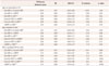
A comparison of the means of four MR measurements between IPD and PSP patients revealed that MBTegm, Oba M/P, and Cosottini M/P were higher and mMRPI was lower in IPD patients than in PSP patients (p<0.005-0.0001, independent t-test) (Supplementary Table 3 in the online-only Data Supplement). The mean AC-PC distance did not differ between the two patient groups (data not shown). ROC analysis of the four MR measurements used to discriminate PSP from IPD revealed that the AUC ranged from 0.69 to 0.76 (Table 2, first row). After matching the two patient groups for age at MRI, changes in the AUC ranged from 0.66 to 0.73 and were thus not remarkable (Table 2, second row). Restricting the diagnosis of PSP to probable PSP increased the AUCs slightly (range, 0.69-0.81); however, there was marked overlap of the 95% confidence intervals compared to ROC curve analyses between IPD and all PSP patients (Table 2, third row). Together these findings suggest that all of these measurements could be used to differentiate PSP from IPD, regardless of age at MRI or the diagnostic certainty of PSP. At the cutoff values with the highest Youden indices, mMRPI exhibited high sensitivity, while Oba M/P and Cosottini M/P exhibited higher specificity than the mMRPI (Table 2). These trends in sensitivity and specificity among Oba M/P, Cosottini M/P, and mMRPI were observed in age-matched cases and in comparisons between IPD and probable PSP. The sensitivity and specificity of MBTegm were comparable to those of other MR measurements, for which the sensitivity and specificity were high when differentiating between age-matched cases and between IPD and probable PSP, respectively.
To determine which of the four MR measurement methods was best for differentiating PSP from IPD, their ROC analysis results were compared (Table 3, Fig. 2). No difference in the AUC was found among MBTegm, Oba M/P, and Cosottini M/P. However, mMRPI was inferior to MBTegm for differentiating between probable PSP and IPD (p=0.049). In addition, although statistically not significant, mMRPI tended to be inferior to the two methods of measuring the M/P ratio (p=0.07 for Oba M/P vs. mMRPI; p=0.053 for Cosottini M/P vs. mMRPI).
DISCUSSION
The findings presented here suggest that MBTegm, two different methods of measuring the M/P ratio, and mMRPI are all useful for differentiating PSP from IPD, with high intrarater consistency and diagnostic accuracy, as demonstrated by the AUC values obtained in ROC analyses. Moreover, these findings were not affected by the age at MRI or the certainty of a diagnosis of PSP (probable PSP vs. both possible and probable PSP). A comparison of the AUCs among the four MR measurements suggests that mMRPI is less useful for discriminating PSP from IPD than the M/P ratio adjusted according to the AC-PC length.
One strength of this study is that it is the first to demonstrate the diagnostic value of MBTegm in differentiating PSP from IPD; the diagnostic value of this measurement is comparable to those of both Oba M/P and Cosottini M/P. The utility of measurements of the diameter of the whole midbrain on MR images in differentiating between IPD and PSP has been investigated previously.13 Although the mean diameter of the whole midbrain was found to differ significantly between PSP and IPD patients, this parameter exhibited poor diagnostic efficacy because of a large overlap in its range of values between the groups. The differences in the usefulness of the brain measurements found between the present study and that of Righini et al.13 may be due to differences in the techniques used to acquire MR images between them. The present study used 1-mm-thick, T1-weighted axial images, which most likely provided more consistent axial planes at the mid-mammillary-body level. Sung et al.9 reported that the diameter of the MBTegm was smaller in patients with PSP or subcortical ischemic vascular dementia compared to a normal control group. However, in the present study, although an IPD group was not included for comparison, the research aim was not to test diagnostic accuracy.9
The measurement of M/P ratio exhibited fair diagnostic validity. Massey et al.14 recently reported a new method of measuring the M/P ratio by applying an ellipse to the pons and midbrain, which demonstrated excellent discriminative power among pathologically confirmed cases of IPD, PSP, and multiple system atrophy (MSA). That method was not included in the present analyses because of possible inherent technical flaws, such as vagueness of the posterior margin of the ellipse in the pons and the unclear degree of tilting in the midbrain. Moreover, the data from Massey et al.14 exhibited excellent discrimination between PSP and MSA, but not between IPD and PSP.
Contrary to measurement of the M/P ratio, the results obtained using mMRPI in the present study were not completely in accordance with previous reports. To date, three reports from two research groups have focused on the diagnostic usefulness of MRPI. In contrast to a previous study in which the MRPI was found to be superior to other diagnostic parameters,11 the measurement of mMRPI in the present study exhibited high sensitivity but low specificity, and the diagnostic accuracy as determined by the AUC was smaller for this measurement than for MBTegm. Morelli et al.12 reported that MRPI was independent of age in both IPD and PSP patients, while the present research showed that mMRPI was influenced by the age at MRI in IPD patients but not in PSP patients. Given that the intrarater reliability in this study was high, the disagreement between these findings and those of previous studies may be due to differences in the MRI protocols. The SCP, MCP, and MR images were obtained in the previous studies in an oblique coronal plane parallel to the fourth ventricle floor, whereas reconstructed coronal images perpendicular to the AC-PC plane were used in the present study. This difference may have led to the disparate MRPI values observed between this study (mMRPI, 8.92) and that of Morelli et al.11 (MRPI, 13.63-13.7), although the measurements of the M/P ratio were very similar in the two studies (0.18-0.21 vs. 0.195-0.215). Since most clinical centers routinely study axial T1-weighted images, MBTegm is easier to measure and more practical to use.
This study was subject to several limitations. The study subjects were diagnosed clinically, and these diagnoses were not confirmed pathologically. In a recent comparative study of MRI and autopsy data, Whitwell et al.15 found that midbrain atrophy is more closely associated with clinical manifestations of PSP than with PSP pathology. As discussed above, differences between the present MRI protocol and those used in other studies may limit comparisons between (m)MRPI and other measurement tools. T1-weighted 3D volumetric images were used in this study, and the distance between MR slices was 1 mm, which may have resulted in overestimation of the diagnostic value of MBTegm.
In conclusion, simple measurements of MBTegm on an axial image at the mid-mammillary-body level appears to be comparable to the M/P ratio in the midsagittal plane with regard to discriminating PSP from IPD.

 XML Download