

 PDF
PDF ePub
ePub Citation
 Print
Print


Introduction
Neurocysticercosis (NCC) is caused by infection of the brain by the larval stages of Taenia solium. It is the most common helminthic infection of the central nervous system.1 Human cysticercosis is common in emerging countries such as Haiti.2,3 While intraventricular form of NCC is common, spinal involvement is rare, representing 1-2% of all cases.4,5 Asymptomatic forms are the most frequent. When symptomatic, NCC has a nonspecific presentation depending on the number, size, and location of the lesions. Hydrocephalus and epilepsy are the most frequent features.6
Hydrocephalus is caused by either cerebrospinal fluid (CSF) pathway blockade by ventricular cysts and/or inflammatory ependymal/arachnoidal changes.5,7 It can be treated by removal of the intraventricular cysts and/or CSF shunting, or endoscopic ventriculostomy.8,9 Although cysticidal drugs have proven effective, neurosurgical resection is necessary in cases of intraventricular cysts, giant subarachnoid cysts (usually located within the lateral fissure), or compressive cysts at the spinal level.10 The cases involving the three patients reported herein, all of whom originated from Haiti, illustrate various mechanisms of hydrocephalus. For two of the patients the hydrocephalus was related to intracranial NCC lesions, while in the third case it was attributable to cysticercosis of the cauda equina, which is a very uncommon pattern of presentation.
Case Report
Case 1
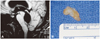
A previously healthy 19-year-old female sought medical attention due to headache, nausea, and balance disorders of 2 months duration. She was a student, and a daughter of a Haitian pig breeder. Clinical examination revealed a left ptosis, giddiness, and homolateral facial palsy. Computed tomography (CT) scan and magnetic resonance imaging (MRI) of the head disclosed obstructive hydrocephalus related to a multiloculated mass in the lower recess of the fourth ventricle (Fig. 1A). Surgery allowed en bloc removal from the lower part of the fourth ventricle of a cystic lesion containing a gelatinous mass that did not adhere to the surrounding nervous or vascular structures. On pathological examination, the cyst wall was found to comprise conjunctive tissue with very small numbers of lymphocytes. Parasitological examination revealed that the intracystic mass corresponded to the scolex of Taenia solium (Fig. 1B). Immunological blood tests for cysticercosis were positive. The patient was given albendazole (15 mg/kg/day for 3 weeks) immediately after surgery.
Case 2
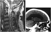
This 48-year-old Haitian male had been treated incompletely with albendazole in 1991 for NCC. He presented in May 2009 with headache, amnesia, cognitive impairment, right lower-limb weakness, and visual impairment with upward-gaze paresis, pupillary light-near dissociation, and convergence-retraction nystagmus (Parinaud syndrome). CT scan and MRI of the head disclosed multiple calcifications in the occipital lobe and major enlargement of the entire ventricular system, with leptomeningeal enhancement and septation of the subarachnoid space suggestive of extensive arachnoiditis, involving the posterior fossa, craniocervical junction, and upper cervical spine (Fig. 2). A ventriculoperitoneal shunt was inserted before initiation of a new treatment with albendazole associated with corticosteroids (60 mg/day). Ventricular CSF biochemical, cytological, and microbiological examinations produced negative findings. The patient recovered fully, with no recurrence during a 3-year follow-up period.
Case 3
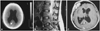
A previously healthy 25-year-old Haitian female, suffering only from headaches and back pain since delivery after an uneventful pregnancy 1 month previously, was admitted for sudden visual loss and impaired consciousness. She had no motor or sensory disturbance, but had complete visual loss with fixed, dilated pupils. Her body temperature was 38℃. CT scan and MRI of the head disclosed enlargement of the entire ventricular system and a calcification in the right occipital lobe (Fig. 3A). Insertion of external ventricular drainage resulted in dramatic improvement of consciousness and visual recovery. Her ventricular CSF biology was normal (protein, 0.4 g/L; leukocyte count, 12/mm3). Her body temperature rapidly rose to 40℃ and a lumbar puncture was performed. The CSF level of protein was 8.5 g/L, and the leukocyte count was 680/mm3, among which 61% were lymphocytes and 18% were eosinophils. These discrepancies between ventricular and lumbar CSF samples led to MRI of the spine, which disclosed a heterogeneous mass located in the cauda equina from L4 to S2. There was neither leptomeningeal enhancement nor subarachnoid cysts suggestive of arachnoiditis at the thoracic, cervical, or cranial levels (Fig. 3B). Since there was no other obvious explanation for the hydrocephalus (no obstructive lesion of the intracerebral CSF pathways nor at the craniocervical junction, and no abnormality in the spinal subarachnoid spaces at superior levels), this lumbosacral mass was considered to be responsible for the hydrocephalus, due to obstruction of the subarachnoid spaces by the mass itself and/or surrounding arachnoiditis. Surgery performed via a lumbosacral laminectomy allowed subtotal removal of an intradural, poorly limited, pearly white mass with small cystic formations surrounding the nerve roots, with thickening of the arachnoid layer. Histological examination revealed parasitic cysts. No scolex was found, but immunological blood and CSF tests for cysticercosis were positive. Although surgery led to disappearance of the symptoms, the patient was provided with adjunctive albendazole. There was no recurrence during a 15-year follow-up, with the patient leading a normal life.
Discussion
Cysticercosis usually affects the subcutaneous or muscular tissues, liver, lungs, or eyes. The nervous system is affected in nearly 4% of cases, and NCC constitutes the most frequent parasitic infection of the nervous system. Most lesions are intracranial. In most cases the spinal lesions are associated with other lesions of the nervous system.11 Hydrocephalus occurs in nearly 30% of NCC patients.2,6 Our three cases illustrate three distinct mechanisms.
Case 1 indicates that hydrocephalus can result from obstruction of the ventricles by a parasitic cyst located in the fourth (53%) or third (27%) ventricles. Cysts are less frequent in the lateral ventricles (11%) and aqueduct (9%). In this mechanism, hydrocephalus is revealed by signs of raised intracranial pressure such as headache, vomiting, and visual disturbances.5 Case 2 illustrates that hydrocephalus can also be caused by inflammatory/fibrotic changes in subarachnoid and ventricular spaces, without the finding of macroscopic parasitic cysts, leading to arachnoiditis and/or ventriculitis (granular ependymitis) and septation of the subarachnoid space. Parinaud syndrome is often observed in cases of ventriculitis/arachnoiditis-related hydrocephalus, as in case 2.5 Inflammatory and immunological markers of cysticercosis are positive in the CSF in cases related to widespread arachnoiditis and granular ependymitis due to multiple microscopic cysts. In contrast, in most cases of large obstructive cysts, inflammatory changes are localized and generally moderate, explaining the highly inconstant positivity of CSF markers. While a CT scan of the head can be sufficient to demonstrate hydrocephalus related to an intraventricular cyst, only MRI can reveal arachnoiditis in the posterior fossa or craniocervical junction.
The mean duration of signs and symptoms before diagnosis ranges from a few months for patients with ventricular cysts to few years for patients with ventriculitis/arachnoiditis. This discrepancy is explained by the symptoms worsening rapidly as a cyst reaches a critical size to cause obstruction of CSF pathways, while in "ventriculitis" cases the symptoms are due to a slowly evolving inflammatory process.
Our third patient potentially illustrates yet another mechanism. Hydrocephalus revealing tumors of the cauda equina, such as ependymomas, is well documented, but to the best of our knowledge there have been no cases of hydrocephalus revealing a parasitic cauda equina lesion reported in the literature so far.12
Intramedullary NCC forms are very rare, due to hematogenous dissemination. Location of the cauda equina is also infrequent and the most common clinical features are radicular pain, paraparesis, bowel and bladder incontinence, and sexual dysfunction.13 Hydrocephalus could be explained by a high CSF protein level (e.g., fibrinogen) caused by the inflammation and increased CSF viscosity, as in spinal tumors and Guillain-Barré syndrome.14 Another explanation is reduced obstruction of the spinal subarachnoid space with reduction of the total CSF compliance. Obstruction of CSF pathways could lead to exclusion of the lower compartment and reduction of CSF circulation volume. It could also prevent the normal compensation of CSF pressure variations.15 Recent flow-sensitive MRI studies support this theory.16
Therapeutic strategies depend upon the mechanism of hydrocephalus and the degree of responsibility of the parasitic lesion itself. Resection of the mass is the appropriate treatment when the ventricles or spinal canal are obstructed by the parasitic mass, and can be performed either by the endoscopic approach or by the direct approach. In all cases, medical treatment with a 3-week course of albendazole with steroids is mandatory.
In most cases, the role of biology can be limited to confirmation of the diagnosis (due to poor sensitivity). For patients with solitary intracranial or spinal lesions, as was the case for two of our three patients, the false-negative rate reaches 30% for biology diagnosis.17 However, recent advances have led to the development of assays with both high sensitivity and specificity, based on an enzyme-linked immunoelectrotransfer blot of either serum or CSF samples in patients with multiple NCC lesions.
In conclusion, NCC should be considered as a possible cause of hydrocephalus. In cases of tetraventricular hydrocephalus without intracranial explanation, MRI of the spine is mandatory to search for intraspinal lesions.
 XML Download