Abstract
Background
Opsoclonus-myoclonus syndrome (OMS) is a rare neurological disorder that is characterized by involuntary eye movements and myoclonus. OMS exhibits various etiologies, including paraneoplastic, parainfectious, toxic-metabolic, and idiopathic causes. The exact immunopathogenesis and pathophysiology of OMS are uncertain.
Case Report
We report the case of a 19-year-old male who developed opsoclonus and myoclonus several days after a flu-like illness. Serological tests revealed acute mumps infection. The findings of cerebrospinal fluid examinations and brain magnetic resonance imaging were normal. During the early phase of the illness, he suffered from opsoclonus and myoclonus that was so severe as to cause acute renal failure due to rhabdomyolysis. After therapies including intravenous immunoglobulin, the patient gradually improved and had fully recovered 2 months later.
Opsoclonus-myoclonus syndrome (OMS) is a rare neuro-ophthalmological disorder that occurs more commonly in children than in adults. Opsoclonus is characterized by involuntary, irregular, multidirectional, and conjugate saccadic eye movements without intersaccadic intervals. Myoclonus is characterized by brief, shock-like, involuntary movements caused by muscular contractions or inhibitions that predominantly affect the limbs, trunk, and head. Ataxia and encephalopathy may be additional features. Among the various etiologies of OMS, paraneoplastic, parainfectious, or idiopathic encephalitis are the most common causes, and an autoimmune-mediated brainstem dysfunction has been suggested as the underlying pathomechanism.1 The exact immunopathogenesis and pathophysiology of opsoclonus are uncertain. We report herein a case of OMS associated with acute mumps infection in an adult.
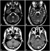
A previously healthy 19-year-old male was referred to a tertiary hospital with acute onset of involuntary eye movements, generalized myoclonus, and mildly confused mental status. He complained of fever, chills, myalgia, sore throat, and then dizziness that had developed 9 days previously. His initial vital signs were stable and he did not have a fever. Spontaneous eye movements were first observed 4 days previously by an otolaryngologist, and generalized myoclonus developed 2 days thereafter. There was no history of any medication or exposure to toxins. Neurological examinations revealed chaotic multidirectional saccadic eye movements with large amplitudes. While resting or moving he exhibited moderate-to-severe myoclonic jerks affecting the head, trunk, and all four limbs (Supplementary Video 1). He was disorientated with respect to both space and time. He was not able to stand or sit due to severe truncal ataxia. The results of initial brain magnetic resonance imaging (MRI) performed on the day of the admission, and follow-up images obtained on the 5th hospital day were normal (Fig. 1). Electroencephalography did not reveal any spikes or spike-and-wave components. The results of routine laboratory tests were normal except for elevated muscle enzymes [creatine kinase (CK) 1029 U/L, normal range 26-174 U/L]. Analysis of his cerebrospinal fluids (CSF) revealed normal cell counts and protein levels, and negative results for cytology and viral markers. Serological tests revealed no acute infections of rubella virus, cytomegalovirus, Epstein-Barr virus, syphilis, herpes simplex virus-1 and -2, human immunodeficiency virus (HIV), streptococcal infection, varicella-zoster infection, mycoplasma, or hepatitis A, B, or C. Tests for antinuclear antibodies were negative. Immunofluorescence analysis revealed that the patient's serum and CSF were negative for anti-Hu, anti-Ri, anti-Yo, and neuromyelitis optica antibodies. Contrast-enhanced chest computed tomography (CT) revealed a mediastinal mass detected, but biopsy proved this to be a benign lesion that had extensive necrosis and chronic inflammation without granuloma. Malignancy was not detected on positron-emission tomography-CT. On admission the patient had no symptoms or signs of parotitis, orchitis, or pancreatitis; however, his serum IgM antibody titer index for the mumps virus was elevated at 1.20 (negative <0.9), but he was negative for the IgG antibody. Enzyme immunoassays produced negative results for CSF IgM and IgG antibodies to the mumps virus. On the 26th hospital day his serum IgG antibody titer index to mumps virus was 1.40 (negative <0.9), and he was negative for the IgM antibody. There was no evidence of an alternative bacterial infection. He was thus diagnosed with OMS associated with mumps infection.
He was initially treated with clonazepam; however, his symptoms, and especially the myoclonus, became aggravated, and his muscle enzymes increased rapidly (CK=12835 U/L). Acute renal failure developed on the second hospital day due to rhabdomyolysis, requiring continuous infusion of midazolam and atracurium, and application of a mechanical ventilator. In addition, intravenous immunoglobulin (IVIg; 2 g/kg divided into 5 days) and valproate (initial loading dose, 1800 mg; maintenance dose, 1200 mg/day) was started. The patient subsequently gradually improved and so the midazolam and atracurium were tapered off, being discontinued on the 11th hospital day when the mechanical ventilator was removed. At that time small-amplitude opsoclonus was still observed intermittently, and myoclonus could be elicited only by stimuli such as being touched. The patient had fully recovered from his confused mental status, and had fully recovered from opsoclonus or myoclonus by 2 months.
The patient reported herein developed opsoclonus and myoclonus several days after a slight flu-like illness. During the early phase of the illness he suffered from opsoclonus and myoclonus that was sufficiently severe as to cause acute renal failure due to rhabdomyolysis. Serological tests revealed acute mumps infection. Intensive diagnostic assessment revealed no evidence of a remote neoplasm. We therefore attributed his illness to OMS associated with mumps virus infection.
Opsoclonus-myoclonus syndrome is associated with multiple etiologies (Table 1), including the paraneoplastic syndromes, parainfectious encephalitis, and toxic-metabolic states. However, in many cases no obvious cause can be found.1,2 The literature on OMS in adults is largely confined to single case reports and small case series. There are few collated data to inform physicians about the most common etiology among the diverse infections.2,3 It is clinically important that paraneoplastic OMS is differentiated from the parainfectious or idiopathic forms due to its poor prognosis3 and the necessity for treatment of the underlying tumor. A multi-institutional retrospective Spanish study showed that idiopathic OMS occurs mostly in younger patients, and is benign and more responsive to immunotherapy than paraneoplastic OMS.3 However, there were no clear clinical features specific to certain causes of OMS, especially in older patients. Based on the present case and our review of the literature, we suggest that searching for an acute viral infection such as mumps should be included when evaluating OMS.
The pathophysiology of opsoclonus remains uncertain. A disordered interaction between the omnipause and burst cells in the brainstem,4 or cerebellar dysfunction (especially of the fastigial nucleus)5 has been considered. However, there is as yet no histopathological or experimental evidence for this. Since saccades are controlled not only by the brainstem but also multiple brain structures such as the cerebellum, superior colliculus, thalamus, caudate nucleus, and frontal and parietal cortices, opsoclonus may be caused by multiple brain lesions or dysfunctions. In addition to opsoclonus, OMS presents with other signs including myoclonus, cerebellar ataxia, and encephalopathy. Therefore, diffuse brain lesions involving the brainstem and cerebellum may induce OMS.
One case report described a young boy who manifested with acute cerebellar ataxia together with opsoclonus and myoclonus following acute mumps infection with bilateral parotid swelling accompanied by fever.6 A lumbar puncture revealed pleocytosis (405/mm3) and an increased IgM titer against the mumps virus. Those authors suggested that CNS involvement in mumps infection reflected a direct invasion by the virus. Although treatment was not mentioned in the report, the patient's condition had resolved completely within 3 months. However, whether the pathomechanism of OMS is attributable to direct invasion or a toxic effect of infectious agents, or an autoimmune-mediated brain dysfunction has been a matter of debate in the previously reported parainfectious OMS cases. Humoral and cell-mediated immune mechanisms have both been implicated in paraneoplastic, idiopathic, and parainfectious OMS.1,7 Based on the detection of autoantibodies such as anti-Ri and anti-Yo, the frequent reversibility of symptoms, the good response to immunotherapy (IVIg and corticosteroid), and the paucity of findings on pathological examination, it has been suggested that the putative autoantigens reside on the neuronal tissue or in the synapse, and that the antibodies cause transient neuronal dysfunction rather than permanent neuronal degeneration.3 However, most patients with OMS are seronegative for all known antineuronal antibodies.2,3 In addition, there are no definitive links between various autoantibodies and neurological abnormalities.8 These observations suggest that a cell-mediated immune mechanism plays a role in the pathogenesis of OMS.
Several previous reports of OMS associated with HIV infection have documented that it may develop during the HIV seroconversion illness or during immune reconstitution after the initiation of antiretroviral therapy.9 The cause of OMS in HIV is probably related to autoimmunity, since this is common in HIV-positive patients, and OMS is also responsive to corticosteroid therapy. Our patient appears to have had an acute mumps infection before the onset of the symptoms. However, the results of brain MRI and CSF analyses carried out during the course of his illness were normal. His symptoms had completely resolved within 2 months after therapy, which included IVIg. These findings raise the possibility that autoimmune mechanisms are involved in the pathogenesis of OMS. This is the first report of OMS associated with mumps infection in Korea.
Figures and Tables
Acknowledgements
The present research was conducted by the research fund of Dankook University (BK21 Plus) in 2013.
References
2. Klaas JP, Ahlskog JE, Pittock SJ, Matsumoto JY, Aksamit AJ, Bartleson JD, et al. Adult-onset opsoclonus-myoclonus syndrome. Arch Neurol. 2012; 69:1598–1607.

3. Bataller L, Graus F, Saiz A, Vilchez JJ. Spanish Opsoclonus-Myoclonus Study Group. Clinical outcome in adult onset idiopathic or paraneoplastic opsoclonus-myoclonus. Brain. 2001; 124:437–443.

4. Ramat S, Leigh RJ, Zee DS, Optican LM. Ocular oscillations generated by coupling of brainstem excitatory and inhibitory saccadic burst neurons. Exp Brain Res. 2005; 160:89–106.

5. Wong AM, Musallam S, Tomlinson RD, Shannon P, Sharpe JA. Opsoclonus in three dimensions: oculographic, neuropathologic and modelling correlates. J Neurol Sci. 2001; 189:71–81.

6. Ichiba N, Miyake Y, Sato K, Oda M, Kimoto H. Mumps-induced opsoclonus-myoclonus and ataxia. Pediatr Neurol. 1988; 4:224–227.

7. Pranzatelli MR. The immunopharmacology of the opsoclonus-myoclonus syndrome. Clin Neuropharmacol. 1996; 19:1–47.


 PDF
PDF ePub
ePub Citation
 Print
Print


 XML Download