

 PDF
PDF ePub
ePub Citation
 Print
Print


Introduction
Obsessive-compulsive disorder (OCD) is a prevalent psychiatric disease that is characterized by disabling obsessions about intrusive unwanted thoughts and images, and/or compulsions expressed as ritualized repetitive behaviors.1 OCD has a lifetime prevalence of 2-3% in the general population,2 and typically 10-20% of affected people exhibit marked maladjustments in professional and social functioning even after treatments that include behavioral therapy and medications.3,4
Possible mechanisms underlying OCD are related to the functional dysregulation of the frontal-cingulate-thalamiclimbic circuit.5 Several psychiatric and neurologic disorders are thought to be associated with OCD or obsessive-compulsive symptoms (OCS) due to the involvement of this circuit. People with epilepsy (PWE) can also exhibit obsessional personalities linked to particular types of epilepsy.6 Uncontrolled temporal lobe epilepsy (TLE) was first reported with OCS and OCD.7 The frequency of OCD or OCS in controlled or uncontrolled TLE patients, which has ranged from 11% to 34.5%, was higher than that of healthy controls (0-3%).7,8,9,10,11 It has recently been reported that idiopathic frontal lobe epilepsy and idiopathic generalized epilepsy (GE) are also associated with a higher risk of developing OCS, with frequencies of 11.8% and 16.2%, respectively.11 Although these studies demonstrated a relationship between OCD or OCS and specific epilepsy syndromes, the participating subjects were not representative of the general population of PWE. Rather, the studies dealt with patients who were confined to a specific epilepsy syndrome7,8,9,10 or included a high proportion of subjects with uncontrolled epilepsy (UCE; nearly 50%) and who had normal neuroimaging results.11 Therefore, PWE with diverse etiologies and epileptic syndromes still need to be investigated in order to obtain an accurate understanding of the pathogenic mechanisms of OCS.
Observed risk factors for OCD or OCS in PWE were male gender, older age, longer duration of illness, TLE, the number of antiepileptic drugs (AEDs), and uncontrolled seizures with AEDs.9,10,11 However, one study found no relationship between OCD and age, gender, duration of disease, or seizure frequency.8 Depression and/or anxiety are commonly related psychopathologies with OCS.8,9,10,11 The side of the epileptic focus as a risk factor for OCS is controversial. A right-hemisphere focus in TLE had initially been suggested to predispose to OCD.12 However, other studies demonstrated an equal contribution between the right- and left-sided foci to OCD or OCS.7,8 Three recent studies found that a left-sided focus in TLE or other epilepsy types was a risk factor for OCD or OCS.9,10,11 A role of AEDs as a risk factor for OCS has not yet been reported. Patients with TLE taking carbamazepine (CBZ) were more likely to develop OCD than those with idiopathic GE taking valproate (VPA).8 PWE taking CBZ had a higher risk of developing OCS than healthy controls, but their risk did not differ from that of PWE taking VPA.11 However, these studies have a limitation that they did not discriminate between the impacts of AEDs and epilepsy syndrome on OCD or OCS. Although many risk factors for OCD or OCS have been suggested, their relative importance to determining OCS was not reported. Identifying decisive factors may help to minimize OCD or OCS and understand its prognosis.
Various psychiatric symptoms including depression and anxiety are the strongest predictors of the quality of life (QOL) in PWE.13,14 The QOL is higher in subjects with drug-refractory epilepsy but no depression or anxiety symptoms than in subjects with seizure freedom exhibiting depression or anxiety symptoms.13 Depression symptoms reportedly contribute more to the QOL than do adverse events associated with AEDs in seizure-free patients with monotherapy.14 Although OCS is commonly associated with depression, anxiety, and markedly impairment of professional and social functioning, the impact of OCS on QOL in PWE is poorly understood.
The above-described situation prompted us to measure the severity of OCS in PWE who visited our epilepsy clinic with diverse etiologies and epileptic syndromes. We also evaluated predictors of OCS among various demographic and clinical characteristics and investigated the impact of OCS on QOL.
Methods
Subjects
We included consecutive PWE who took AEDs and attended our epilepsy clinic between May 1, 2011 and March 31, 2012. Our clinic takes care of patients aged 15 years or older with various types of seizure control, etiologies, and epileptic syndromes; this population is similar to the overall community-based epilepsy population. Subjects were invited to participate in this study if they were aged 17 years or older, had a current diagnosis of epilepsy, had been taking one or more AEDs for at least 1 year, and had the ability to provide informed consent and to agree with the study protocol. Subjects who had insufficient information in their medical records, who had mental retardation or serious medical, neurologic, or psychiatric disorders that prevented them from understanding the questionnaire and cooperating with the study, or who refused to complete questionnaires were excluded. We recruited age-, gender- and education-matched healthy controls to compare the severity of OCS with PWE. These healthy controls were patients' caregivers, parents, children, relatives, or friends who visited our clinic and had no medical, neurologic, or psychiatric diseases.
Study design
This case-control study was approved by the institutional review board of Kyungpook National University Hospital, and all subjects gave written informed consent before participating in the study. Each patient was interviewed by a trained epileptologist (S.P.P.) who also reviewed the medical charts to collate demographic and clinical information in a computerized database. The information collected included age, gender, education, socioeconomic status, seizure type, etiology, epilepsy syndrome, age at onset, duration of epilepsy, side of the epileptic focus, EEG abnormality, MRI abnormality, concurrent medical diseases, family history of epilepsy, history of febrile convulsion, duration of AED intake, number of AEDs, AED action mechanism, seizure control, and depression symptoms as measured by the Beck Depression Inventory (BDI). Epilepsy was diagnosed according to the International League Against Epilepsy classification of seizures and epileptic syndromes.15,16 We divided the etiology into idiopathic and symptomatic epilepsy. We divided epileptic syndromes into three groups: TLE, extraTLE, and GE; where extraTLE included epilepsy syndromes with epileptic attacks originating from the frontal, parietal, or occipital lobe. We also divided seizure control into two groups: well-controlled epilepsy (WCE) and UCE. WCE was defined as freedom from seizures during the preceding year, while UCE was defined as experiencing a seizure during the preceding year despite an adequate intake of tolerated AEDs. We divided AEDs according to their action mechanisms into sodium-channel blockers and other types, because CBZ was reported to be an OCS-inducing drug. AEDs such as CBZ, phenytoin (PHT), oxcarbazepine (OXC), lamotrigine (LTG), topiramate, and zonisamide were included as sodium-channel blockers according to the review of Perucca and Mula.17 EEG abnormality was considered to be present when there were interictal epileptiform discharges (IEDs) on EEG recordings. The IED location was categorized into temporal or frontal IEDs, other focal IEDs, and generalized IEDs. According to the occurrence of OCS by the involvement of the frontal-cingulate-thalamic-limbic circuit, we examined the degree of OCS between temporal or frontal IEDs and other focal or generalized IEDs. The side of the epileptic focus was determined based on EEG and MRI findings. If the epileptic focus was generalized or ambiguous, we classified it as being generalized or unknown, respectively. Socioeconomic factors were classified as follows: having or not having a job; earning at least one million Korean won (KRW) per month (equivalent to US$ 900 per month) or less than one million KRW per month; having or not having a driving license; and being married, or divorced, bereaved, and unmarried.
Questionnaires
The MOCI is a 30-item questionnaire that evaluates OCS.18 It employed a dichotomous response format, and the total scores range from 0 to 30. It had good internal consistency (Cronbach's α=0.8) and was consistently correlated with the BDI score (r=0.37). This version of the MOCI employed factor analysis with orthogonal rotation (a linear transformation used to facilitate interpretation of results) to test whether individual MOCI items segregate into separate subscales of OCS. Only subscales with eigenvalues ≥1.0 were retained in the analysis, which yielded 22 items classified into 4 subscales: checking (6 items), tidiness (6 items), doubting (6 items, including 1 item that was duplicated in checking), and fear of contamination (4 items). Checking and doubting subscales represented compulsive behaviors, and tidiness and fear of contamination represented obsessive behaviors. We measured the MOCI total score and subscale scores. We determined that the cutoff of the MOCI total score for indicating the presence of OCS was 12, based on two standard deviations from the mean total score in healthy controls recruited from our study, because the original study did not suggest it.
The BDI is the most commonly used self-rating scale for depression.19 Participants rate 21 items on a scale from 0 to 3 according to how they feel at the time. The following cutoff scores are used: 0-16, normal; 17-20, mild depression; 21-23, moderate depression; and 24-63, severe depression. Subjects who score more than 16 points are considered to have depression. Cronbach's α value is 0.8.
The QOLIE-31 is a 31-item, self-administered questionnaire specifically designed to measure QOL in PWE.20 It consists of subscales addressing seizure worry, overall QOL, emotional well-being, energy-fatigue, cognitive functioning, medication effects, and social functioning. An overall score for the seven subscales is also calculated. Higher QOLIE-31 scores are indicative of better QOL. Cronbach's α values range from 0.69 to 0.86.
Statistical analysis
Data for continuous variables are expressed as mean±SD values, and those for categorical variables are expressed as frequencies. The independent-samples t test and Fisher's exact test were applied to compare variables between PWE and healthy controls. We used the Mann-Whitney U test for categorical independent variables and Spearman's correlation analysis for continuous independent variables to determine the relationship between demographic and clinical variables and the MOCI total score. Variables that were found to be significantly correlated with the MOCI total score were then included in a multiple linear regression analyses with stepwise selection. The probabilities of entry and exit were 0.05 and 0.1, respectively. Collinearity was addressed by performing collinearity statistical analysis. Predictors of the QOLIE-31 overall score were obtained as the same manner, and the MOCI total score were added as an independent variable. Findings from the linear regression analyses were used to construct a structural equation model to test the interrelations between variables and the QOLIE-31 overall score. The model fit was evaluated using path analysis, a method that estimates the relative importance of different paths of the independent variables onto the dependent variables. An acceptable model fit was defined as having a nonsignificant chi-square (χ2) value, Normed Fit Index (NFI) of ≥0.9, Comparative Fit Index (CFI) of ≥0.9, Goodness of Fit Index (GFI) of ≥0.9, and Root Mean-square Residual (RMR) of ≤0.05. Structural equation modeling was used to estimate the total effect of each predictor, in order to establish a linear model to predict the QOLIE-31 overall score with these interrelations accounted for. Except for the structural equation model, all statistical analyses were conducted with SPSS (version 19.0, IBM Inc.). Linear structural relationship 8.8 for Windows (Scientific Software International, Inc., Lincolnwood, IL, USA) for the path and structural equation modeling components of the analysis. The level of statistical significance was set at 0.05.
Results
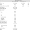
In total, 386 PWE were initially enrolled in the study. Among them, 86 were excluded because of their refusal to complete the questionnaires (n=22), inability to complete questionnaires due to mental retardation (n=20) or serious diseases (n=10), insufficient information about epileptic attacks in their medical records (n=5), young age (n=23), and having received less than 1 year of AED treatment (n=6). Therefore, 300 PWE (age: 35.3±10.9 years; 61.7% males) and 80 healthy controls (age: 34.5±13.7 years; 60% males) were included. Demographic and clinical characteristics of eligible subjects are summarized in Table 1. Age, gender, education level, and socioeconomic status (including job, income, having a driving license, and marital status) did not differ between PWE and healthy controls. Among 300 PWE, 217 patients (72.3%) had partial seizures. Symptomatic epilepsy was present in 117 patients (39%). The most common epilepsy syndrome was TLE (43.7%). The frequencies of left and right epileptic focuses were similar. EEG abnormality was present in 76 patients (25.3%), and the frequency of temporal or frontal IEDs was higher than those of other focal or generalized IEDs. MRI abnormality was found in 110 patients (36.7%), with etiologies of hippocampal sclerosis (n=36), trauma (n=25), brain anomaly (n=19), vascular injury (n=17), infection (n=10), tumor (n=2), and unknown (n=9). UCE was present in 122 patients (40.7%). Concurrent medical diseases were present in 38 patients (12.7%), comprising diabetes and other endocrinologic disorders (n=12), cerebrovascular disease and other neurologic disorders (n=11), hypertension and other cardiovascular disorders (n=10), brain and systemic tumors (n=4), gastrointestinal disorders (n=4), autoimmune disorders (n=3), renal diseases (n=2), and other diseases (n=3). The duration of AED intake and number of AEDs were 10.1±9.1 years (range: 1-52 years) and 1.5±0.7 (range: 1-4), respectively. The QOLIE-31 overall score and BDI score were 67.3±17.8 (range: 13.2-97.9) and 12.1±10.3 (range: 0-58), respectively.
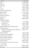
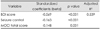
The severities of OCS in PWE and healthy controls are compared in Table 2. The MOCI total score was significantly higher in PWE than in healthy controls (p=0.002). Subscale scores of checking and doubting were also significantly higher in PWE than in healthy controls (p=0.01 and p<0.001, respectively). The presence of OCS, defined by a MOCI total score of 13 or above, was found in 60 patients (20%). Checking and doubting scores did not vary with the epileptic syndrome.
The correlations between demographic and clinical variables and the MOCI total score are summarized in Table 3. The variables found to be significantly associated with the MOCI total score were gender (p=0.011), job (p=0.001), income (p=0.001), having a driving license (p=0.001), seizure type (p=0.04), etiology (p<0.001), EEG abnormality (p<0.001), MRI abnormality (p=0.001), number of AEDs (p<0.001), seizure control (p<0.001), and the BDI score (p<0.001). That is, subjects with female gender, no job, lower income, no driving license, partial seizures, symptomatic etiology, abnormal EEG and MRI findings, larger number of AEDs, UCE, and depression were more likely to develop OCS. Epilepsy syndrome, the side of the epileptic focus, the location of IEDs, and the AED action mechanism did not affect OCS.
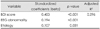
Predictors of the MOCI total score by stepwise linear regression analyses are listed in Table 4. The strongest predictor was the BDI score (β=0.453, p<0.001), followed by EEG abnormality (β=0.194, p<0.001) and etiology (β=0.107, p=0.031). Stepwise regression produced a three-variable model that explained 29.6% of the variance in the MOCI total score. According to the standardized β, the contribution of the BDI score to OCS was 2.34 times greater than that of EEG abnormality and 4.23 times greater than that of etiology. Tolerance was greater than (1-adjusted R2) and variance inflation factors were greater than 10 for all four variables, suggesting that they exerted independent effects without redundancy.
Predictors of the QOLIE-31 overall score are listed in Table 5. The strongest predictor was the BDI score (β=-0.569, p<0.001), followed by seizure control (β=-0.163, p<0.001) and the MOCI total score (β=-0.148, p=0.001). Stepwise regression produced a three-variable model that explained 53.9% of the variance in the QOLIE-31 overall score. According to the standardized β, the contribution of the BDI score to QOL was 3.49 times greater than that of seizure control and 3.84 times greater than that of the MOCI total score. Collinearity statistical analysis indicated that these variables exerted independent effects without redundancy.
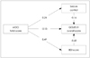
Complex interrelations between predictors and the QOLIE-31 overall score are illustrated by the refined path analysis model in Fig. 1. According to predefined criteria, the final model provided an acceptable fit to the data (χ2=23.91, p<0.001; NFI=0.950, CFI=0.950, GFI=0.970, and RMR=0.080). The MOCI total score, seizure control, and BDI score were found to exert direct effects on the QOLIE-31 overall score. The MOCI total score also exerted indirect effects on the QOLIE-31 overall score through seizure control and the BDI score.
Discussion
This study found that the severity of OCS was significantly higher in PWE than in healthy controls. One fifth of PWE were found to have OCS. Greater depression symptoms, IEDs on EEG recordings, and symptomatic etiology were closely related to the development of OCS, whereas the epileptic syndrome, side of the epileptic focus, location of IEDs, and AED action mechanism were not predictors of the development of OCS. The development of OCS appears to elicit psychosocial problems directly or indirectly by provoking depression or uncontrolled seizures.
We included patients whose clinical constituents were slightly different from those of the general epilepsy population,21,22 but they had diverse etiologies and epileptic syndromes, as indicated by the clinical history and EEG and MRI findings. Therefore, the results of the present study might lead to a better understanding of the pathogenic mechanisms of OCS. The frequency of OCS in our patients resembled the data obtained in other studies.7,8,9,10,11 Among subscales, checking and doubting were significantly increased, as also found by Isaacs et al.7 The OCS of PWE appear to be oriented around compulsive behaviors rather than obsessive behaviors.
Male gender, older age, longer duration of illness, TLE, larger number of AEDs, and uncontrolled seizures with AEDs as well as depression were previously proposed as risk factors for OCS in PWE.9,10,11 However, the previous studies did not measure the major predictors of OCS among these variables. We found that female gender, no job, lower income, not having a driving license, partial seizures, symptomatic etiology, abnormal EEG and MRI findings, larger number of AEDs, UCE, and depression as risk factors for OCS, with depression, IEDs on EEG recordings, and symptomatic etiology being major predictors. This is the first study to find that EEG abnormality is a strong predictor of OCS. As there are no studies to elucidate the short-term or long-term effect of IEDs on psychopathology, we hypothesize abnormal electrical discharges in the frontal-cingulate-thalamic-limbic circuit are related to the occurrence of OCS. We also found that the specific epilepsy syndrome, the side of the epileptic focus, and the location of IEDs were not risk factors for OCS. Differences in study design may produce different results, and future studies based on the video-EEG monitoring should attempt to clarify the exact epilepsy syndrome or epileptic focus. We also reported that AEDs that block sodium channels did not increase OCS more than AEDs having other action mechanisms. CBZ, PHT, OXC, and LTG have a mood-stabilizing effect,17 and hence these agents are not thought to provoke OCS.
The final goal in the management of PWE is to improve psychosocial functioning. The strongest predictors of QOL are depression and anxiety, followed by seizure control.13 We found OCS to be another important risk factor for QOL. The contribution of OCS to QOL was comparable to that of seizure control. Furthermore, the development of OCS appears to elicit psychosocial problems directly or indirectly by provoking depression or uncontrolled seizures. It is therefore suggested that clinicians should determine whether OCS are present when patients visit an epilepsy clinic, especially when they have depression, IEDs on EEG recordings, or symptomatic etiology. Unfortunately, brief and self-administered screening tools for detecting OCS-specifically designed for PWE in a busy clinical setting-have not yet been developed. A validated screening tool for OCS in PWE should be developed as soon as possible in order to improve their QOL and to minimize AED intractability and depression.
This study was subject to some limitations. First, since the study did not employ structured interviews for the diagnosis of OCS, such as the Structured Clinical Interview for DSM-IV axis I disorders23 and the Mini-International Neuropsychiatric Interview,24 we could not estimate the frequency of OCD. The Yale Brown Obsessive Compulsive Scale25 is a popular questionnaire for measuring OCS. However, since that questionnaire takes a long time to complete, we instead used MOCI as a screening test. Second, we did not elucidate the relationship between OCS and anxiety in PWE. Anxiety is a frequent psychiatric disorder accompanying PWE,26 and OCS frequently manifest in people who have anxiety disorders.27 Therefore, future studies should clarify the impact of OCS on QOL in relation to anxiety disorders. Third, since we determined the presence of epileptic syndrome based on the clinical history, EEG findings, and MRI findings, some of our diagnoses of epileptic syndrome may have been incorrect. To solve this problem, further studies that include PWE who complete video-EEG monitoring or who experience seizure freedom after epilepsy surgery are needed to clarify the exact location of the epileptic focus. Fourth, we did not investigate the impact of OCS on AED compliance. We found that OCS were likely to elicit uncontrolled seizures. Although OCS and uncontrolled TLE are reported linked to each other, OCS as a risk factor for uncontrolled seizures has not been proven, and patients with OCS are actually likely to have better seizure control than those without OCS due to higher AED compliance associated with their own frequent checking or doubting behavior. Therefore, a longitudinal study involving patients with newly diagnosed epilepsy should clarify whether OCS are likely to produce a favorable outcome in seizure control or to be a predictor of uncontrolled seizures.

 XML Download