

 PDF
PDF ePub
ePub Citation
 Print
Print


INTRODUCTION
Blepharospasm (BSP) is a focal dystonia characterized by repetitive involuntary sustained contractions of the orbicularis oculi muscle, causing spasmodic closure of the eyelids.1-3 It is frequently found in association with apraxia of eyelid opening (AEO).4 AEO has been described as a non-paralytic motor abnormality characterized by difficulty in opening the eyes at will in the absence of visible contraction of the orbicularis oculi muscle.4-6 When BSP occurs only in response to certain stimuli, such as a sudden visual threat or auditory or tactile stimulus, it is defined as reflex blepharospasm (R-BSP).3,7
Most of these ocular symptoms, such as BSP, are not associated with another identifiable associated disease. In this case, they are referred to as primary or idiopathic.4,5,7 However, secondary BSP has occasionally been reported in patients with parkinsonism and in those with focal lesions in the basal ganglia, diencephalon, or brainstem, in addition to those with an ophthalmologic disorder.8-12 AEO has been occasionally reported in patients with focal cerebral lesions and parkinsonism, particularly in those with progressive supranuclear palsy (PSP).6,13-15 Moreover, BSP and AEO have been reported as an coexisting dystonia in atypical parkinsonism, and are more commonly present in PSP.16 However, their clinical relationships are unclear, neither has it rarely been reported which disorder is related to several types of BSP.
Because there are no large clinical studies on several types of BSP such as reflex BSP (R-BSP) or preceding BSP prior to parkinsonism (Pre-BSP) in the literatures, we prospectively surveyed these symptoms in patients with parkinsonism. In the case of already existing BSP associated with parkinsonism, we retrospectively analyzed the characteristics and time intervals of BSP. We analyzed clinical features of BSP and AEO in patients with idiopathic Parkinson's disease (IPD), multiple system atrophy (MSA), and PSP. We subdivided MSA into MSA-p (predominantly parkinsonism) and MSA-c (predominantly cerebellar) satisfying diagnostic criteria proposed by Quinn.17 Similarly, BSP was subclassified into R-BSP, Pre-BSP, and BSP combined with AEO, and these subtypes were then analyzed individually.
Our aims were to investigate the clinical characteristics of BSP and AEO associated with various types of parkinsonism and to find out if there is any clinical significance of BSP to differentiate parkinsonism including subtypes of MSA.
MATERIALS AND METHODS
1. Patient Selection
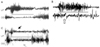
We enrolled 35 patients with BSP with or without AEO among 1113 patients with parkinsonism registered at the Movement Disorder Clinic of Samsung Medical Center from January 2002 to December 2003. Using the established diagnostic criteria for IPD,18 MSA17,19 and PSP,20 the 1113 patients with parkinsonism were subclassified as having; IPD (913 patients), probable MSA (190 patients; 134 MSA-p and 56 MSA-c), or probable PSP (10 patients). We subdivided MSA into MSA-p (predominantly parkinsonism) and MSA-c (predominantly cerebellar) satisfying diagnostic criteria proposed by Quinn.17 We clinically diagnosed BSP using the established definition.3,7 In the case of those with coexisting BSP and parkinsonism, we retrospectively analyzed the characteristics and time intervals of BSP. For diagnosis of AEO, we used diagnostic criteria proposed by Lepore and Duvoisin.6 These included (i) no sign of ongoing orbicularis oculi contractions such as lowering of the brows beneath the superior orbital margin; (ii) marked frontalis muscle overaction during period of inability to raise eyelids; (iii) no ocular motor/ocular sympathetic nerve dysfunction and ocular myopathy. To confirm the presence of AEO with BSP, we performed electromyography (EMG) on the orbicularis oculi (OO) and levator palpebrae (LP) muscles (Fig. 1) in 13 patients who were diagnosed by above diagnostic criteria. We included patients with long-term follow up for maximum 24 months, full evaluations for parkinsonism, and a clinical diagnosis confirmed by at least two special neurologists based at Movement Disorder Clinic.
2. Periocular Electromyography
After getting the patient's informed consent, muscle activities from the orbicularis oculi and levator palpebrae were recorded by means of 2 channel- EMG (Nicolet Biomedical Inc., Medicine, Wisconsin, USA), using monopolar needle electrodes inserted into the left upper lid. Proper placement of the needles was confirmed by muscle activities at the opening and closing of the lid. Reference surface electrodes were spaced about 1cm apart from the active electrodes on the orbital bony prominence. A ground electrode was fixed in the middle of the forehead. Trials of opening and closing of eyes were repeated several times to acquire optimal signals. The EMG signals were amplified in a conventional manner, filtered (10-1,000 Hz), displayed on a memory oscilloscope, and analyzed offline.
3. Clinical analysis
We analyzed the clinical features of BSP and parkinsonism including demography, onset age, onset interval to BSP, clinical diagnosis of parkinsonism, severity of parkinsonism, and relationship with levodopa treatment. Severity of motor symptoms of parkinsonism was evaluated by using motor UPDRS (part III of Unified Parkinson's Disease Rating Scale). To analyze the characteristics of BSP, we subclassified reflex BSP (R-BSP), BSP preceding parkinsonism (Pre-BSP), or BSP combined with AEO. R-BSP was defined when BSP occurred only in response to certain stimuli, such as a sudden visual threat or auditory or tactile stimulus.3,7 Pre-BSP was defined when BSP existed before presenting parkinsonism. Figure 1 showed EMG findings of EMG of BSP with or without AEO. Comparing with normal finding (Fig. 1-A), EMG showed a marked contraction of OO during eye closure (Fig. 1-B, 1-C). During eye opening, OO inhibition was incomplete and LP contraction was not sustained in BSP with AEO (Fig. 1-B). We also evaluated the coexistence of dystonia on other body parts.
4. Statistical analysis
All data are presented as means±standard error of mean (SEM). SPSS (version 12.0) program for windows (SPSS, Inc., Chicago, IL) was used for the statistical analysis. Sex differences and clinical characteristics of BSP data were analyzed using the Chi-square test. Onset age, onset interval to by parkinsonism, and severity of parkinsonism data were analyzed by using one-way ANOVA. Scheffe's test was used for post hoc comparisons. A p value of <0.05 was used as the criterion for statistical significance.
RESULTS
1. Clinical characteristics of BSP
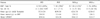
Thirty-five BSP patients with parkinsonism (mean age, 61.2±7.2 years), 19 men and 16 women, were enrolled in the study. Of these 35 BSP patients, 8 (22.8%) were diagnosed as having IPD, 15 (42.9%) as MSA-p, 5 (14.3%) as MSA-c and 7 (20%) as probable PSP. Of the 913 patients with IPD 8 (0.9%) had BSP, 15 of 134 (11.2%) patients with MSA-p, 5 of 56 patients (8.9%) with MSA-c, and 7 of 10 patients (70%) with PSP had BSP. BSP was more frequent in PSP and MSA than in IPD (p<0.05). In terms of sex differences, in contrast to the lack of gender preference in MSA-p, BSP was more frequent in men than women in IPD and in PSP. BSP with MSA-c only affected women. With except of these findings, no other significant clinical demographic difference was observed among various types of parkinsonism (Table 1).
2. Clinical significance of subtypes of BSP for differentiating parkinsonism
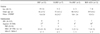
According to BSP subtype, i.e., R-BSP, Pre-BSP, and BSP with AEO, no significant differences were found between the parkinsonism types with respect to sex, age of onset, or severity of parkinsonism (Table 2). Seven of 35 BSP patients (20%) showed R-BSP. All R-BSP patients had been clinically diagnosed as having atypical parkinsonism (4 MSA-p, 1 MSA-c, and 2 PSP) (Table 2). Pre-BSP was observed in 5 of 35 BSP patients (14.3%: 1 IPD, 2 MSA-p, 1 MSA-c and 1 PSP), and was more frequently observed in atypical parkinsonism than in IPD (p<0.05). In our study, 13 of 35 patients with BSP (37.1%: 3 IPD, 4 MSA-p, 3 MSA-c and 3 PSP) showed combined AEO. The presence of AEO was more frequent in atypical parkinsonism than in IPD (p<0.05), but isolated AEO was not detected (Table 2).
In terms of the relationship between BSP and levodopa treatment, BSP occurred as an 'off' symptom in two IPD patients and as a "peak-dose" effect in one IPD patient. In 3 IPD patients and in 1 MSA patient, BSP was improved by levodopa medication. However, one PSP patient presented aggravation of BSP after levodopa medication.
3. BSP combined with other types of dystonia in parkinsonism
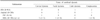
Nine (25.7%) of 35 BSP patients also had other types of dystonia. Types of dystonia and the clinical diagnoses of these patients were as follows; 4 with cervical dystonia (1 MSA-p, 2 MSA-c and 1 PSP), 3 with camptocormia - meaning forward bending of truncal muscles (2 IPD and 1 MSA-p), 1 with limb dystonia (1 IPD), and 1 with facial dystonia (1 MSA-p) (Table 3).
DISCUSSION
In this study, we prospectively surveyed BSP in patients with parkinsonism. In cases with coexisting BSP and parkinsonism, we retrospectively analyzed the characteristics and time intervals of BSP.
BSP is considered to be one of the more common forms of focal dystonia in patients with IPD and atypical parkinsonism.21 In our clinical series, by noting the frequencies of BSP among each group with parkinsonism, BSP was found to occur more frequently in atypical parkinsonism, MSA, and PSP than in IPD. However, no significant difference was found between the frequencies of BSP between subtypes of MSA, MSA-p and MSA-c.
In a recent report about BSP in IPD, gender was not found to be associated with the presence of BSP in IPD.12 According to another case study about dystonia in MSA, dystonia was observed twice as frequently in women.22 In our study, BSP with MSA-c only affected women. Our study also showed that BSP patients were mainly men in IPD and PSP, but than the gender were similarly represented in MSA-p.
According to previous studies,4-6,23 AEO either combined with or without BSP may occur in parkinsonism, especially in PSP. In the present study, we found that 13 of 35 patients (37.1%) with BSP showed AEO, and that BSP with AEO was more frequently detected in atypical parkinsonism. Although AEO might occur in many neurological disorders, we did not observe isolated AEO associated with parkinsonism.
The subclassification of BSP into R-BSP, Pre-BSP, and BSP with AEO was found to be more clinically helpful for the diagnosis of atypical parkinsonism, such as MSA and PSP. The present study suggests that R-BSP is a unique feature of atypical parkinsonism, because it was observed only in atypical parkinsonism. Although one study reported that idiopathic BSP does not lead to a parkinsonian syndrome,24 patients with Pre-BSP have a tendency to be diagnosed with atypical parkinsonism after long-term follow up. This finding contrasts with previous studies that presented patients with BSP only at advanced stage disease.11,25,26
No differences were observed in the clinical features of BSP between MSA-p and MSA-c despite a previous report that dystonia is common in untreated MSA-p.22 These findings support that BSP can be associated with any subtype of MSA.
The majority of previous studies have suggested that various types of dystonia can be observed in parkinsonism.16,21,22,27 We observed that other types of dystonia coexisted with BSP and AEO in parkinsonism. According to our study, even though we were unable to perform a statistical analysis due to small numbers, we presume that BSP combined with cervical dystonia or facial dystonia is more frequently related to atypical parkinsonism, whereas camptocormia or limb dystonia is more frequently related to IPD.
In terms of the relationship between BSP and levodopa treatment, we found that BSP might have occurred as an 'off' symptom or as a 'peak dose' effect of levodopa treatment in IPD. BSP that was improved by levodopa treatment was more frequently related to IPD, whereas BSP aggravated by levodopa treatment was more related to atypical parkinsonism. However, these suggestions were not statistically confirmable because of small patient numbers. Nevertheless, these relations are likely to be encountered in clinical medicine, and present a good clinical study topic for differential diagnosis in parkinsonism.
In conclusion, our study shows that BSP either with or without AEO is more frequently observed in atypical parkinsonism than in IPD, and suggests that R-BSP is a unique feature of atypical parkinsonism. In addition, our findings suggest that patients with Pre-BSP have a tendency to be diagnosed with atypical parkinsonism later, and that the presence of AEO indicates atypical parkinsonism. However, levodopa related BSP, including 'off' symptom, 'peak-dose' effect, or levodopa responsiveness were found to represent IPD rather than atypical parkinsonism. No differences were found between the clinical features of BSP in MSA-p and MSA-c.
 XML Download