

 PDF
PDF ePub
ePub Citation
 Print
Print


INTRODUCTION
Clinical symptoms and signs of cerebral venous thrombosis (CVT) are extremely variable, and the diagnosis of CVT is thought to be under-recognized because of its nonspecific symptoms.1 In the early angiographic era, CVT had a high (30-50%) mortality. However, CVT related deaths has been reduced considerably (6.5-15%) with the advance of diagnostic tools such as computed tomography (CT) and magnetic resonance imaging (MRI), as well as early application of anticoagulants following diagnosis.2
Unfractionated heparin has been the most common first-line drug for CVT,3,4 but its use has been the subject of much controversy because of reported hemorrhagic complications.2,5,6 The hemorrhagic conversion in the infarcted lesion is a major disincentive against the use of heparin. Low molecular weight heparin (LMWH) exerts an anticoagulant effect by inactivating factor Xa.7 LMWH also has a better antithrombotic-to-hemorrhagic ratio due to its low antithrombin effect.7 Perioperative LMWH use has been shown to have no increase in bleeding risk during operation and post-operative drainage.8,9 Additionally, several studies have equated the efficacy of LMWH with that of unfractionated heparin in the management of deep vein thrombosis,10 venous thromboembolism,11 and unstable angina.12,13 Furthermore, LMWH has much better bioavailability, that is, a longer plasma half-life and less variable anticoagulation effect with a fixed dose than unfractionated heparin.7
The numerous publications reporting the benefits of LMWH, particularly the antithrombotic effects and better bioavailability, suggest that LMWH can replace unfractionated heparin in the early treatment of CVT. The present study evaluates whether LMWH is safe, feasible, and perhaps a better first-line drug for CVT.
MATERIALS AND METHODS
1. Diagnosis of CVT
CVT was diagnosed using pre- and post-enhanced T1-weighted MRI (T1WI), CT, magnetic resonance venography, and/or cerebral angiography. The imaging study was performed at the time of hospital admission for initial diagnosis of CVT. To evaluate temporal changes to brain lesions, each patient was subjected to follow-up MRI and/or CT studies at discharge. Both direct and indirect signs of thrombosis in the intracranial venous system were used for the diagnosis of CVT. Direct evidence of CVT included thrombi in the cerebral vein and sinuses on any imaging modality.2 Intensified venous collaterals, the presence of cork screw veins, broken bridging veins, venous dilatation, and delayed venous emptying were considered as indirect evidence for CVT.2 Combined parenchymal lesions with hemorrhagic conversion were also confirmed by density or signal changes on CT, T1WI, and/or gradient echo images.2 An increase in the initial hemorrhage size and/or newly developed bleeding detected follow-up imaging was categorized as an LMWH-related hemorrhagic complication.
2. Causes of CVT
To evaluate the causes of CVT in patients, we assessed a history of risk factors that have been reported to be associated with CVT, such as oral contraceptive, parturition, infectious disease, ear disease, carcinoma, dehydration, marasmus, sepsis, acute lymphoblastic leukemia, chemotherapy, systemic lupus erythematosus, Behäet's disease, osteoporosis, diabetes mellitus, homocystinuria, and iron deficiency anemia.14 Laboratory blood tests were also performed to determine the presence of coagulopathy, such as complete blood count (CBC), liver function test (LFT), prothrombin time (PT), activated partial thromboplastin time (aPTT), levels of protein C and S, antithrombin III, lupus anticoagulant, and anticardiolipin antibody. If the above historical and laboratory evaluation for CVT revealed no abnormality, we classified the case as not-determined etiology.14
3. Management and clinical courses of CVT
Twice daily subcutaneous injection of 7,500 IU nadroparine (Fraxiparine®, Sanoffi-Aventis, France) immediately followed the initial CVT diagnosis and was maintained for 2 weeks. For secondary prevention after the termination of the drug, oral anticoagulant (warfarin) or antiplatelet agent (aspirin) was prescribed depending on the individual risk factors.
Initial manifestations were evaluated at CVT diagnosis and closely monitored for changes during nadroparine treatment until the initial neurological symptoms were normalized or stabilized. We recorded the onset of symptoms and/or signs prior to CVT diagnosis. We also recorded the interval from initial nadroparine treatment to initial evidence of clinical improvement. This interval time was used to estimate the drug's efficacy at managing CVT. Initial improvement was defined as the first signs of recovery from subjective symptoms observed at admission, such as headache or nausea, and/or objective neurological deficits, such as motor deficit, aphasia, and mental deterioration. Finally, the interval from initial nadroparin treatment to normalization or stabilization of initial neurological alterations was evaluated. Exacerbations of initial neurological deficits and/or newly developed neurological deficits after treatment were classified as, "ineffective" or as, "side effect".
RESULTS
1. Diagnosis of CVT
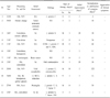
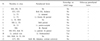
A total of 12 patients (male : female=5 : 7, age 21-76 years) were included in the present study (Table 1). Thrombi were noted in the cerebral venous sinus of all patients, serving as a direct evidence of CVT (Table 2). The thrombi were located in multiple sinuses in five patients. The venous thrombi were most commonly in the transverse sinus, followed by the superior sagittal and sigmoid sinuses. Associated parenchymal venous infarction was observed in nine patients. The parenchymal lesions were distributed in variable hemispheric regions, including the temporal, parietal, and occipital lobes. In patients 2 and 12, venous infarctions in deep structures, such as basal ganglia, thalami, and/or the centrum semiovale, were associated with the venous thrombi located in inferior cerebral veins (Table 2).
2. Causes of CVT
Common risk factors responsible for CVT were found in all but two patients (cases 6 and 7) (Table 1). Protein C and/or S deficiency were determined to be the most frequent cause, affecting 7 out of 12 patients. Deficiencies of both proteins were detected in three patients. Two patients had not only protein S and/or C deficiency, but also elevated levels of blood factor V (case 11) and blood factor VIII (case 12). Postpartum and oral contraceptives were checked in cases 4 and 8, respectively. Additionally, chronic otitis media in case 5 and osteomyelitis in the left lower tibia in case 9 were associated with protein C and/or S deficiency. Iron deficiency anemia (Hb=7.3 gm/dl) was detected in case 5. Case 2 had been diagnosed with stomach cancer 6 months previously but had no abnormality on the evaluated history and laboratory work-up. In all patients, warfarin was maintained at levels 2 to 3 of the international normal range of prothrombine time for secondary prevention of CVT.
3. Clinical manifestations and courses of CVT
Headache was the most frequent presenting symptom, and associated with neurological deficits, such as convulsive movement, sensory or global aphasia, hemiparesis, and hemianopsia. However, convulsive movement was the dominant manifestation without the headache symptoms in three patients. A mental status of deep drowsiness was the presenting manifestation in case 2. Interestingly, before the availability of imaging diagnosis for CVT, the likely presumptive diagnosis for case 2 was a seizure disorder. Other patients were initially determined as having variable diagnoses, with headaches in two patients, metastasis of stomach cancer, brain tumor, encephalitis, cerebral infarction, and meningitis (Table 1).
CVT was diagnosed rapidly (within 1 to 3 days) when the patients showed characteristic neurological deterioration at the hospital. However, the diagnosis was usually delayed (11 to 30 days from the initial onset of symptoms), when the patient first suffered from non-specific neurological manifestations, such as headache and nausea. Improvement of the initial symptoms was usually detected within 2 to 8 days after taking nadroparine, irrespective of a delay in the diagnosis of CVT. In two patients (case 3 and 8), improvement was detected after 20 days of nadroparine because clinical evaluations were impossible due to the inability to communicate. One patient showed sensory aphasia at admission, while another patient presented with a deep drowsiness during the management with nadroparine. Normalization to the previous neurological condition occurred rapidly in two patients, within 2 and 4 days in cases 1 and 12, respectively. Most patients normalized their initial neurological state within 11 to 60 days. Patient 9, who had marked thrombi and hemorrhagic conversion in both cerebral cortices, showed stabilization of right side hemiplegia by 222 days of nadroparine treatment, improving to Grade III.
4. Safety of LMWH for hemorrhagic-converted venous infarction
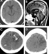
Hemorrhagic conversion was detected in five of the nine patients wth venous infarctions: cases 3, 7, 8, 9, and 10. In case 9, severe thrombi in the superior sagittal sinus, inferior sagittal sinus, and sigmoid sinus (Fig. 1-B) and parenchymal lesion without hemorrhagic conversion were detected in the initial imaging study (Fig. 1-A), Unfractionated heparin was administered immediately following the CVT diagnosis, and 2 days later the mental state had decreased from alert to deep drowsiness. Motor weakness on the right extremities had also aggravated to hemiplegia. On the follow-up brain CT study, hemorrhagic conversion had developed in the previous infarcted lesions (Fig. 1-C). The mental state markedly improved 2 days after replacing heparin with nadroparin and no aggravation of clinical symptoms was detected during the maintenance with nadroparin.
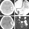
Associated hemorrhage was observed in the initial brain image of four patients. Again, nadroparine did not cause clinical or radiological aggravation. In case 3, a marked temporal lobe hemorrhagic infarction was observed on the initial CT, (Fig. 2-A) and the left lateral sinus was evident in cerebral venography performed at admission. (Fig. 2-B) The initiation of nadroparine treatment resulted in no aggravation of mental status, eventhough the surrounding edema increased in the follow-up study (Fig. 2-C). The patient recovered to an alert and cooperative status without neurological findings after termination of the nadroparine schedule. Encephalomalatic change at the hemorrhagic converted parenchymal lesion remained in case 9 (Fig. 1-D) and 4. (Fig. 2-D) Convulsive movement developed after hospital discharge in both of these patients, and an anti-epileptic drug was prescribed to prevent recurrence. Hemorrhagic-converted cerebral lesions were observed in the initial imaging study in three other patients. These patients showed clinical improvement after taking nadroparine, and no aggravation of the initial manifestations as well as radiological problems were observed. However, hemorrhagic converted lesions remained as encephalomalatic changes in the follow-up imaging studies.
DISCUSSION
The patients included in the present study showed improvement after taking LMWH without clinical aggravation suggesting its general superiority over unfractionated heparin at managing CVT. Unfractionated heparin had been the preferred choice for anticoagulation of CVT, with many series demonstrating good clinical prognosis.3,15,16 Recently, however, LMWH has been widely speculated as an alternative for managing and preventing venous thromboembolism with equal effectiveness.17-20
Although unfractionated heparin may prevent the progression of thrombosis and further infarction, it required very careful administration because of its hemorrhagic side effects.5,6 A previous randomized trial has shown that unfractionated heparin was both safe and effective at managing CVT.3 However, the results had been debated due to the small sample size, questionable outcome criteria, and a long treatment delay.21,22 In the present study, we also experienced clinical aggravation after initial heparin treatment in case 9, and newly developed hemorrhagic conversion was confirmed in the follow-up imaging study. Therefore, unfractionated heparin should be used with caution as a first-line drug for CVT patients.
LMWH presents a lower risk of bleeding than unfractionated heparin in the management of venous thromboembolism.10 The probability of bleeding is not increased during the perioperative period of major surgeries when LMWH was used.8,9 Therefore, LMWH would be expected to be a safe anticoagulant for managing CVT, especially for treating hemorrhage-converted venous infarctions. A previous randomized trial determined that LMWH was sufficiently safe for managing hemorrhagic converted CVT.20 CVT patients included in the present study showed no evidence of LMWH-associated hemorrhage. Even if they present hemorrhagic conversion in the initial imaging studies, the initial manifestations improved rapidly after LMWH was administered without clinical or radiological aggravations. Patient 9 exhibited heparin-induced clinical and radiological aggravations, but the clinial manifestations rapidly stabilized after an anticoagulation switch to LMWH. Therefore, the present study provides further supports for the safety of LMWH in managing CVT and venous thromboembolism.
Unfractionated heparin presents other practical limitations, such as a narrow window of therapeutic time, less predictable kinetics, platelet activation, and the inability to inhibit clot-bound thrombin.23 Consequently, unfractionated heparin requires frequent laboratory monitoring and close observation to prevent heparin-induced complications.24 On the other hand, LMHW has better bioavailability, limited non-specific binding, and non-dose-dependent half-life, as well as the ease of subcutaneous injections without laboratory monitoring.7 In several other studies, LMWH performed similarly, if not better, at preventing and managing both venous thromboembolism and CVT.17-20 Furthermore, the convenient subcutaneous injection without laboratory monitoring makes LMWH a better first-line CVT drug than unfractionated heparin.25,26
The ideal LMWH regimen for CVT has not yet been established, even though its efficacy and safety have been widely reported for venous thromboembolism and CVT. Long-term LMWH treatments (up to 3 months) are reportedly effective at managing symptomatic venous thromboembolism.27 Other studies have shown that short term LMWH treatments (5 to 7 days) was effective at treating thromboprophylaxis.28,29 Two weeks of LMWH maintenance was recently recommended for the prevention of venous thromboembolism.19 For CVT, however, LMWH was administered twice a day for 3 weeks in a randomized trial.20 In the present study, administration of LMWH twice a day for 2 weeks produced clinically acceptable results. Therefore, 2 weeks might be sufficient for CVT or thromboembolism management, although additional trials are required to verify the appropriate daily dosage and duration of LMWH maintenance.
In conclusion, LMWH was found to be safe and effective for managing CVT. The small number of tested patients and the lack of a control group receiving unfractionated heparin limit the applicability of our data. However, the rapid improvements without clinical or radiological aggravations strongly support previous studies showing the safety and efficacy associated with LMWH, and hence its potential as a better first-line agent for treating hemorrhagic converted CVT patients.
 XML Download