

 PDF
PDF ePub
ePub Citation
 Print
Print


Dear Editor:
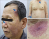
A 56-year-old Korean male presented with a 5-month history of multiple lesions on the face, trunk, and upper extremities. Physical examination revealed multiple asymptomatic, variable-sized red non-blanchable papules and nodules with peripheral erythema and telangiectasia (Fig. 1). The central component showed intense pulsation under digital exploration. The patient was an alcoholic and had suffered from liver cirrhosis for 10 years.
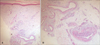
Blood tests showed an abnormal liver function which are as follows (normal range in parentheses): total bilirubin 9.2 mg/dl (0.2~1.4 mg/dl), aspartate aminotransferase 54 U/dl (9~40 U/dl), alanine aminotransferase 34 mg/dl (0~40 U/dl), lactate dehydrogenase 583 IU/L (208~405 IU/L), and alkalinephosphatase 218 U/dL (38~110 U/dl). Liver ultrasound and abdominal computed tomography revealed nodules on the liver surface and moderate amount of ascites in the peritoneal cavity. There was with no definite sign of hepatocellular carcinoma. Histopathological examination of right arm showed multiple dilated blood vessels within in the dermis (Fig. 2A). There were close approximation between the thick-walled arterioles and venules. Inflammatory cells including lymphocytes and histiocytes but not glomus cells were detected in the dermis (Fig. 2B). Based on the clinical and histopathological findings, a diagnosis of arteriovenous hemangioma (AVH) was made.
AVH is a rare, benign, acquired, cutanenous vascular tumor. The pathogenesis of AVH is unclear1. A number of hypotheses have been proposed. Some have suggested that it may represent a harmatomatous proliferation of the suprapapillary vascular plexus23. The pathogenic mechanism of AVH in relation to chronic liver disease is still unclear. Patients with liver failure have relatively high blood estrogen levels4 and some think that estrogens may be involved in the occurrence of AVH in such patients. However, the association is still unclear4.
Clinically, AVH usually presents as an asymptomatic, solitary, 0.5~1.0-cm-sized bluish to erythematous papule or nodule on the head or extremities1 and only 7% of cases have been reported to be multiple4. Histopathologic findings of AVH are well-circumscribed proliferation of blood vessels with thick and thin walls, lined by a single layer of endothelial cells in the upper and mid-reticular dermis1. AVH can be divided into superficial and deep type1. The deep type of AVH is a vascular malformation associated with shunting1. Cases of AVH associated with chronic liver diseases have been rarely reported45. Spider angioma is the most representative and classic vascular lesion of chronic liver disease5. AVH are caused by by proliferation of blood vessels, whereas spider angioma represent dilatation of pre-existing vessels5. This case was consistent with histopathologic features of the deep type of AVH.
The association between severity of liver disease and treatment outcome of AVH with chronic liver disease has not been reported. Our patient had been treated with a combination of vascular lasers (pulse dye laser and long pulsed Nd: YAG) but was not present for follow-up. He died of liver failure shortly after treatment.
We feel that our case is unique in that it presented as 1) multiple cutaneous AVH which is rare- to our knowledge, such case has never been reported in Korea; and that 2) our case of cutaneous AVH was associated with liver cirrhosis.
 XML Download