

 PDF
PDF ePub
ePub Citation
 Print
Print


Dear Editor:
Juvenile xanthogranuloma (JXG) is a benign, self-healing, non-Langerhans cell histiocytosis predominantly affecting infants and children. Usually, the clinical presentation is characterized by solitary or multiple yellowish or red-brown firm papules or nodules on the head, neck, and trunk12. Herein, we report the case of a solitary JXG with an unusual clinical presentation.
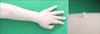
A 4-year-old boy presented with an asymptomatic nodule on the left forearm since 2 months. The lesion was a corn-shaped, erythematous to yellowish nodule measuring 0.5 cm in diameter and 0.7 cm in height. The apical part of the nodule showed marked hyperkeratosis (Fig. 1). The patient's parents reported that it had spontaneously developed without trauma. Clinically, the lesion was believed to be a cutaneous horn due to molluscum contagiosum or viral wart, and a shave biopsy was performed.
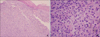
Histopathologic examination revealed hyperkeratosis and parakeratosis in the epidermis and dense intradermal histiocytic infiltrates, some of which contained foamy cells and Touton giant cells (Fig. 2). Histopathological findings were consistent with a diagnosis of JXG.
JXG was first described by Adamson in 1905. Histological examination revealed an ill-defined, unencapsulated, dense histiocytic infiltration in the dermis. In mature lesions, histiocytes have a foamy appearance, and Touton giant cells, which are characteristic of JXG, are observed. The clinical course tends to be benign, and lesions spontaneously regress over a period of months to years. The shape, site, distribution, and size of JXG lesions vary widely, often resulting in atypical clinical forms. A mixed form of small and large nodules, giant form, clustered form, generalized lichenoid form, subcutaneous form, flat plaque-like form, and paired form of JXG have been reported previously23.
Cutaneous horn is the clinical term for a circumscribed, conical, markedly hyperkeratotic protrusion that appears similar to the horn of an animal. The term refers to a reaction pattern and not to a specific lesion. Accordingly, the important point is not the horn itself, which is only keratin, but rather the underlying disease. Cutaneous horns can be benign (seborrheic keratosis and filiform verruca), premalignant (actinic keratosis and Bowen's disease), or malignant lesions (squamous cell carcinoma) in adults. However, a few reports in the literature describe cutaneous horns in children4. Molluscum contagiosum, viral warts, cutaneous leishmaniasis, and primary cutaneous inoculation tuberculosis have been reported. Other possible causes of a cutaneous horn are pyogenic granuloma, epidermal nevus, verruciform xanthoma, pilomatricoma, and verrucous dyskeratoma5. Therefore, an exact diagnosis of the underlying disease is important, and an adequate amount of material must be obtained from the base of the lesion to confirm the diagnosis.
Atypical clinical forms of JXG are usually diagnosed only after histopathologic examination. We propose that physicians must consider skin biopsy of cutaneous horns for accurate diagnosis.
 XML Download