

 PDF
PDF ePub
ePub Citation
 Print
Print


Dear Editor:
Meyerson phenomenon, or halo dermatitis, is characterized by the development of a halo or eczematous patch over another skin lesion, such as nevus cell nevus or nevus flammeus1. We report the case of a 10-month-old female infant who showed eczema within a pre-existing capillary malformation.
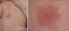
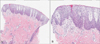
The infant presented with irregularly shaped, but relatively well-demarcated erythematous scaly patch on her back (Fig. 1). She showed an erythematous patch on her back at the time of birth. She had no past history and family history of atopic dermatitis or psoriasis of the infant. The current scaly eczematous lesion developed over the pre-existing congenital erythematous lesion 3 months ago without any triggering event. Biopsy of the skin sample obtained from the center of the lesion showed acanthosis with marked spongiosis in the epidermis and vascular proliferation with perivascular lymphocytic infiltration in the dermis (Fig. 2). She was eventually diagnosed with capillary malformation with overlying eczema. Mometasone furoate lotion was applied, but only slight and temporary improvement was observed after 2 weeks. Since then, the lesion waxed and waned without any other event like dye laser.
The first case report of an inflammatory reaction within a capillary malformation was published in 1996 and described dermatitis within nuchal-occipital port-wine stains2. Since then, only few cases of eczema over a capillary malformation corresponding to Meyerson phenomenon have been reported134. Most patients usually presented with sharply demarcated, scaly, and eczematous lesions confined within the borders of the capillary malformation1234. Histological examination showed epidermal hyperplasia and spongiosis overlying capillary ectasias1. Topical corticosteroid is usually successful for the treatment of eczema, but the lesions tend to recur after treatment discontinuation2. Treatment with pulsed dye laser alleviates both capillary malformation and the associated eczema13. However, in some patients, eczematous dermatitis was triggered by the laser treatment itself4. The pathogenesis of Meyerson phenomenon in the case of capillary malformation remains unclear despite several hypotheses. A hypothesis is that the skin within the capillary malformation shows abnormal dermal vasculature, and this ectatic capillary vessel possibly causes excessive production of proinflammatory cytokines, promoting the development of eczema3. Moreover, endothelial cells possibly play an important role in the inflammatory process3. This hypothesis is supported by the observation that eczema is alleviated by therapy with a dye laser13. Reversely, this proinflammatory hypothesis also can be explained in that eczema is triggered by post-laser inflammatory environment4. Another theory is that, in terms of nevus flammeus, which are oriented along the lines of Blaschko, this phenomenon is associated with genetic mosaicism. Thus, the microenvironment of the lesions may be different from the microenvironment of the surrounding skin4.
In Korean literature, some cases of Meyerson phenomenon were reported; however, the phenomenon was associated with seborrheic keratosis, compound nevus, and junctional nevus, but not capillary malformation5. Although Meyerson phenomenon with capillary malformation is not uncommon phenomenon in practice, it seems to be under-reported. Dermatologists should be aware that eczematous lesion could develop over the capillary malformation.
 XML Download