

 PDF
PDF ePub
ePub Citation
 Print
Print


Dear Editor:
Crohn's disease is an autoimmune condition involving the lower gastrointestinal tract that requires long term use of immunosuppressive agents due to its chronic and relapsing course. Diverse side effects of prolonged immunosuppression in Crohn's disease patients have been reported, including the development of eruptive benign melanocytic nevi and even malignant melanomas12. In this letter, we report the first Korean case of multiple melanocytic nevi formation in a patient with Crohn's disease undergoing immunosuppressive treatment with 6-mercaptopurine and intravenous (IV) adalimumab.
A 36-year-old Korean woman presented to the department of dermatology complaining of numerous widespread moles on her entire body except her face. She was diagnosed with Crohn's disease 12 years ago with an enterocutaneous and enteroenteric fistula complicated by intraabdominal abscess. Screening for tuberculosis was negative. Immunosuppressive therapy was initiated soon after the diagnosis, 6-mercaptopurine (50 mg per day) being the mainstay agent. The patient was occasionally given corticosteroid and started IV adalimumab ten weeks prior to her visit to our department. According to the patient, the number and the extent of moles increased rapidly only a few weeks after the initiation of adalimumab.
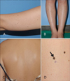
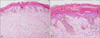
Skin examination revealed multiple characteristic melanocytic lesions of varying sizes (>30 in number, from 2 to 6 mm in diameter) (Fig. 1A~C). A skin biopsy was performed on three discrete lesions, which were the three largest ones (Fig. 1D). Common histologic findings among the three lesions included nevus cells arranged symmetrically and superficially in relatively well defined nests and lack of diffuse atypia within the lesions (Fig. 2). Accordingly the patient's cutaneous condition was compatible with eruptive benign melanocytic nevi. She is currently visiting our dermatologic clinic on a regular basis as surveillance for possible malignant transformation of the numerous nevi.
Developments of both inflammatory and melanocytic lesions such as eruptive nevi associated with immunosuppressive therapy, including the use of biologicals, have been reported previously134. Immunosuppressants such as azathioprine, 6-mercaptopurine and methotrexate are known to occasionally cause developments of eruptive nevi. Moreover, medical conditions including leukemia, pregnancy, erythema multiforme, epidermolysis bullosa and Stevens–Johnson syndrome have also been reported to induce the development of eruptive nevi even in the absence of immunosuppression3. This report adds the first Korean case of eruptive benign melanocytic nevi formation following IV adalimumab therapy for the treatment of Crohn's disease. The lesions developed newly and rapidly in previously normal skin. Previous cases suggest the tendency of melanocytic nevi to appear on specific locations (palms and soles); however, widespread distribution of nevi involving almost the entire body as in our case is a novel presentation1. Not only benign melanocytic lesions but also malignant melanomas have been reported to develop during or after anti-tumor necrosis factor (TNF)-α therapy235. Thus, a skin examination before and after the use of anti-TNF-α agents could be of value in identifying the development of melanocytic nevi, which often necessitates evaluation to rule out malignant melanoma.
Overall, this case adds clinical evidence that TNF-α plays a critical role in the differentiation and proliferation of melanocytes, inducing the development of melanocytic nevi.
 XML Download