

 PDF
PDF ePub
ePub Citation
 Print
Print


INTRODUCTION
Procalcitonin, consisting of 116 amino acids, is one of the precursor proteins of calcitonin, although its biological role is unknown12. When there is a bacterial infection, CALC1 gene expression is increased, and the expression of the precursor of calcitonin, procalcitonin, is subsequently increased in all of the cells in the body3. Synthesis of procalcitonin is stimulated by bacterial endotoxins and the proinflammatory cytokines, interleukin (IL)-1β, IL-6 and TNF, and suppressed by the interferon-γ secreted during viral infection4.
White blood cell (WBC) count, erythrocyte sedimentation rate (ESR) and C-reactive protein (CRP) are often used as markers of bacterial infection severity. However, in some cases, there are difficulties in using these markers as indicators of diagnosis and treatment due to their low sensitivity and specificity5. A recent study reported the value of procalcitonin in bullous impetigo, staphylococcal scaled skin syndrome, localized skin infection, diabetic foot infection, septic arthritis and osteomyelitis67. In addition, procalcitonin may be a useful tool for the initial diagnosis and monitoring of cutaneous infection or cutaneous soft tissue infection8. Hence, we conducted this study on patients with cellulitis to identify whether serum procalcitonin, in addition to the WBC, ESR, and CRP, as used in conventional measurements, was useful for the evaluation of disease severity and prognosis.
MATERIALS AND METHODS
Patients
We enrolled 160 patients who were diagnosed with cellulitis and erysipelas at the Department of Dermatology of Wonkwang University Hospital, from March 2012 to February 2015. Patient's medical records were reviewed retrospectively. Age, sex, involved site(s), infectious route, complications, systemic manifestations, and number of hospitalized days were evaluated. The study was approved by the ethics committee of Wonkwang University Hospital (IRB no. WKUH 201505-HRE-037).
Clinical and biomarker assessment
The diagnosis of cellulitis and erysipelas was based on the physician's clinical diagnosis and histopathologic findings. We considered erysipelas a synonym for cellulitis because it is difficult to clinically distinguish cellulitis and erysipelas. In order to assess the severity of the disease, we measured body temperature for systemic symptoms. Fever was defined as a body temperature of more than 38.0℃, according to the systemic inflammatory response syndrome criteria.
WBC (XN-9000; Sysmex Co., Kobe, Japan), ESR (ALI FAX; Alere Inc., Waltham, MA, USA), CRP (Modular p800; Roche diagnostics, Basel, Switzerland) and Procalcitonin (Vidas; BioMerieux Co., Lyon, France) levels were measured at the first day of admission as part of the biomarker assessment. Procalcitonin was classified into 5 groups according to the reference levels: (1) group 1, normal limit (<0.05 ng/ml); (2) group 2, possible local bacterial infection (<0.5 ng/ml); (3) group 3, minor systemic inflammatory response (<2 ng/ml); (4) group 4, sever sepsis (<10 ng/ml); and (5) group 5, septic shock (≥10 ng/ml). Patients were discharged when their CRP level was reduced to below 10 mg/L, or the only symptom that remained was mild swelling.
Statistical analysis
To evaluate differences between groups, independent t-test analysis and ANOVA were used to analyze variables. We analyzed cross-sectional correlations between WBC, ESR, CRP, and procalcitonin, and body temperature and number of hospitalized days. Correlations between the biomarkers and variables were examined both graphically and using the Pearson correlation coefficient. All analyses were performed using statistical software IBM SPSS Statistics ver. 21.0 (IBM Co., Armonk, NY, USA). p<0.05 was considered significant.
RESULTS
Patient distribution
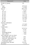
Patients' ages ranged from 3 to 84 years (mean, 48.0). Of the 160 subjects, 87 (54.4%) were male and 73 (45.6%) were female. The male-to-female ratio of the total patients was 1.2:1. Among the systemic symptoms, fever occurred in 27 (16.9%) cases. Complications were experienced in 13 (8.1%) cases; the complications included abscess, bursitis, necrotizing fasciitis and septic arthritis. In addition, recurrence occurred in 2 (1.25%) patients (Table 1).
Anatomic location of the disease
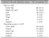
The most common site of disease was the lower leg (n=66, 41.3%) followed by the head and neck (n=39, 24.4%), foot (n=36, 22.5%), hand (n=8, 5.0%), thigh (n=4, 2.5%), arm (n=4, 2.5%), and forearm (n=3, 1.9%) (Table 2). When viewed as a whole, 106 (66.3%) cases arose from the lower limbs, while 39 (24.4%) cases were from the head and neck and 15 (9.4%) cases were from the upper limbs.
Infectious route
The route of infection could be inferred in 107 (66.9%) cases and 53 (33.1%) cases had cellulitis of unknown infectious origin. The most common possible route of infection was considered to be tinea pedis (n=49, 45.8%), followed by trauma (n=41, 38.3%), insect bites n=11, 10.3%), furuncle (n=4, 3.7%), and eczema (n=2, 1.9%) (Table 2).
Laboratory findings and relationships between body temperature and number of hospitalized days
WBC, ESR, and CRP levels were measured in a total of 160 patients, but procalcitonin level was measured in 144 (90.0%) patients, excluding 16 of the 160 patients. The average WBC level was 10.9×10³/µl, and 80 of 160 patients (50.0%) demonstrated leukocytosis. The average ESR and CRP levels were 26.0 mm/h and 67.5 mg/L, respectively. Elevated ESR and CRP were seen in 89 (55.6%) and 141 of 160 patients (88.1%), respectively. The average procalcitonin value was 0.35 ng/ml, and 65 of 144 patients (45.1%) demonstrated procalcitonin ≥0.05 ng/ml. The number of hospitalized days was between 3 and 38. The average was 9.3.
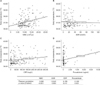
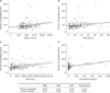
WBC, CRP, and procalcitonin showed a positive correlation with body temperature (p<0.05), but ESR had no relationship with body temperature (Fig. 1). WBC, ESR, CRP, and procalcitonin showed a positive correlation with number of hospitalized days (p<0.05; Fig. 2).
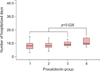
Procalcitonin could be divided into five groups according to its level: (1) group 1 (n=79, 54.9%), (2) group 2 (n=50, 34.7%), (3) group 3 (n=9, 6.2%), (4) group 4 (n=6, 4.2%), and (5) group 5 (n=0, 0%). Using Duncan post-hoc analysis, we found no differences in the first 3 groups and their relationship to the number of hospitalized days. However, there was a statistically significant difference between the first 3 groups and group 4 (p=0.028; Fig. 3).
Relationship between laboratory markers and severity of cutaneous symptoms
The most common cutaneous symptoms were erythema, tenderness, and swelling (100%) followed by pus drainage (n=21, 13.1%), bullae (n=12, 7.5%), and hemorrhagic swelling (n=8, 5.0%). There was a relationship between bulla formation and CRP and ESR (p=0.008 and 0.044, respectively). But differences between the others cutaneous symptoms and laboratory markers were not significant (Table 3).
DISCUSSION
In 1993, Assicot et al.9, first reported the clinical use of procalcitonin to differentiate between bacterial and viral meningitis. Recently, several studies have shown increased procalcitonin in patients with infectious disease10. An increased blood procalcitonin level reflects the severity of infection and can be helpful to predict the prognosis of patients1112. The procalcitonin level is also useful in the differential diagnosis of infections caused by bacteria and viruses, because it does not increase in viral infection13. Procalcitonin does not increase in patients with a severe inflammatory response unless the cause is infection9.
Procalcitonin is detected 2 hours after injection of Escherichia coli-derived endotoxin, reaches its highest concentration after 6 hours, and is maintained at the level for 9~24 hours. In contrast, CRP begins to increase after 12 hours and does not reach its peak level for 30 hours. Therefore, procalcitonin has the advantage of more rapid detection than CRP14.
Meanwhile, CRP, one of the acute phase proteins, is a useful measure of the inflammatory response. Even if there are no systemic symptoms, it is sensitive enough to still detect an inflammatory condition. This is useful in making diagnosis, but in some cases, it is difficult to interpret the results because an increased CRP level is non-specific, and cannot identify the cause of inflammation1516. ESR alone is also not sufficient for a diagnosis, although it too increases with inflammation1718.
In our study, analyses were performed with respect to the clinical usefulness of the procalcitonin level in cellulitis. Erysipelas is an inflammatory disease of the superficial dermis including the superficial lymphatics, and mainly occurs on the face and cellulitis is inflammation of the reticular dermis and subcutaneous fat, and presents with unclear margins. However, erysipelas is often considered a synonym for cellulitis affecting any skin area because of difficulties in accurately differentiating between the two diseases1920. Thus, patients with erysipelas are included in this study.
The procalcitonin level was shown to have a positive correlation with body temperature. Fever, body temperature above 38℃, is one of the criteria for systemic inflammatory response syndrome, and can be considered a factor indicating the severity of disease. Therefore, we thought that a higher procalcitonin level would correlate with higher disease severity. Pearson correlation coefficients calculated in this study between body temperature and parameters were WBC, 0.286; ESR, −0.042; CRP, 0.156); and procalcitonin, 0.180. WBC, CRP, and procalcitonin were all correlated with disease severity, and WBC appeared to have the highest Pearson correlation coefficient. These results suggest that the WBC level was the most valuable inflammatory indicator, but, procalcitonin was useful in evaluating the severity of disease.
Lazzarini et al.21 found that CRP and ESR levels were elevated in patients hospitalized for more than 10 days, but WBC had no relationship to duration of hospitalization. In this study, the Pearson correlation coefficients between the number of hospitalized days and parameters were WBC, 0.237; ESR, 0.241; CRP, 0.333; and procalcitonin, 0.179. Therefore, all parameters could be used as indicators to predict prognosis, especially, CRP level, which had the highest correlation coefficient and was the most useful predictor of prognosis.
Procalcitonin was divided into five groups depending on its level: group 1 had 79 patients (54.9%), group 2 had 50 (34.7%), group 3 had 9 (6.2%), group 4 had 6 (4.2%), and group 5 had 0 (0%). Interestingly, the number of hospitalized days in group 4 was significantly higher than that in the other groups. In a recent study, procalcitonin had a better diagnostic value when compared with CRP and WBC in erysipelas, and its concentration was relatively low in localized inflammatory state22. Our study was consistent with this previous study.
At a procalcitonin threshold of 0.05 ng/ml or more, the sensitivity and specificity for cellulitis were 45.1% (95% confidence interval [CI], 42.2%~45.1%) and 100% (95% CI, 97.0%~100%), respectively. The result revealed that a low procalcitonin level (<0.05 ng/ml) did not ultimately rule out cellulitis. And the procalcitonin level can be normal in cellulitis. However, clinicians should be highly cautious in the case of a procalcitonin level high enough to indicate poor prognosis.
In conclusion, procalcitonin reflected severity of disease in cellulitis and high levels suggested a poor prognosis. However, compared to procalcitonin, WBC and CRP are most closely associated with severity of disease and prognosis, respectively.

 XML Download