

 PDF
PDF ePub
ePub Citation
 Print
Print


Dear Editor:
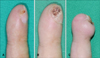
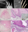
A 36-year-old man with a 2-month history of a hyperkeratotic nodule on the right little fingertip was transferred to our hospital (Fig. 1A). In our hospital, over 4 months, dye laser treatment was performed 4 times based on the initial diagnosis of subungual verruca. However, the lesion did not improve (Fig. 1B). Ultrasonography, radiography, magnetic resonance imaging (MRI), and three-dimensional (3D)-computed tomography (CT) showed osteolysis at the distal edge of the distal phalanx (Fig. 2A~D). Wide excision was performed based on the diagnosis of verrucous carcinoma (VC). The histopathological examination of the wide excision specimen revealed that the hypertrophic epidermis was large and bulbous, and proliferated downward, compressing the collagen bundles and pushing them aside; this resembled a "bulldozing" pattern. However, cellular anaplasia was absent, and mitotic figures were rare (Fig 2E, F). The pan-human papillomavirus was negative and Ki-67 was positive in the basal layer and suprabasal cells of the lesion (Fig. 2G). An erythematous swollen nodule developed 8 months after wide excision (Fig. 1C). Finally, amputation from the fifth proximal interphalangeal joint was performed. Histopathological examination of the amputation specimen revealed atypical squamous cell proliferation and bony invasion (Fig. 2H, I). At the 2-year follow-up, there were no signs of recurrence.
VC is a slow growing, well-differentiated, rare squamous carcinoma variant1. It tends to appear at 3 main sites: the oral and perioral regions (oral florid papillomatosis), the genital and anal regions (giant condylomata acuminata of Buschke-Lowenstein), and the sole or heel (epithelioma cuniculatum). VC can occur anywhere on the skin, and subungual VC is much rarer type.
The diagnosis of VC is difficult because of indolent growth and benign histologic features. The clinically differential diagnosis of VC may include verruca vulgaris, condyloma accuminata, keratoacanthoma, and squamous cell carcinoma2. In our case, verruca was the initial diagnosis. For the diagnosis of VC, a large, deep biopsy is essential. Histopathological findings of VC include hyperkeratosis, parakeratosis, and marked acanthosis. The proliferation of the epithelium usually has a pushing border that projects into the adjacent tissue and pushes it aside. However, there is absence of nuclear atypia, individual cell keratinization, and horn pearls. Immunohistochemical staining of Ki-67 provides the benefit to a differentiation among normal (limited to the basal cells), invasive squamous cell carcinoma (diffusely throughout the whole malignant epithelium), and VC (in the suprabasal cells and lower third of the epithelium)3. Imaging studies are helpful to make an early and accurate diagnosis of VC. In our case, ultrasonography, radiography, MRI, and 3D-CT showed bony invasion of the distal phalanx, implying malignancy. VC is characterized by local aggressiveness, and it has a low potential for metastasis4. Bony invasion appears to be more common in subungual VC because of delayed diagnosis and close relationship between the subungual area and the underlying distal phalanx2.
Subungual VC may be treated with wide local excision or Mohs micrographic surgery. For advanced cases, amputation is almost always necessary, and bony invasion requires amputation of the digit2. Other treatment modalities include radiotherapy, chemotherapy, CO2 laser, and imiquimod cream45. In our case, the patient underwent amputation of the digit.
We reported a rare case of subungual VC with bony invasion. For early and accurate diagnosis of subungual VC with bony invasion, it should be differentiated from subungual verruca using not only biopsy but also imaging tools.
 XML Download