

 PDF
PDF ePub
ePub Citation
 Print
Print


INTRODUCTION
Dermatofibrosarcoma protuberans (DFSP) usually occurs on the trunk or proximal extremities, and it typically appears as an indurated plaque. It is characterized by a low-grade malignancy and slow growth and is asymptomatic in most cases. It initially manifests as a firm papule or plaque that grows over several months or even years into a plaque or nodule. We report a case of DFSP in the inguinal area with multiple pedunculated nodules and a review of the literature.
CASE REPORT
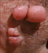
A 47-year-old man presented with multiple asymptomatic nodules in the inguinal area. The patient visited a local clinic 7 months ago for a single nodule in the right inguinal area. A skin biopsy was performed, and the biopsy results indicated that it was a benign tumor. However, he visited our dermatology clinic because the nodule grew larger and multiplied within 2 months. The skin lesion found in the right inguinal area had multiple, firm, protruding pedunculated nodules (Fig. 1). A skin biopsy specimen was obtained from the lesion. Histopathological examination results showed dense proliferation of the intradermal tumor cells, and the spindle cells were arranged in a storiform and cartwheel pattern (Fig. 2A). The spindle cells extended into the subcutis layer and showed a honeycomb-like appearance (Fig. 2B). Immunohistochemistry findings were positive for CD34 (Fig. 2C), confirming the diagnosis of DFSP.
The patient was referred to the department of plastic surgery to undergo wide excision and skin grafting. We have been following the patient's progress for 6 months, and to date, there has been no recurrence or metastasis.
DISCUSSION
DFSP is a rare malignant tumor (a dermal fibroblastoma), which accounts for less than 0.1% of all malignant tumors1. It generally occurs in adults in their 20s to 50s.
One study reported that it takes more than 10 years to diagnose most patients with DFSP after onset, because the tumor grows slowly over a long period and patients have no specific symptoms2. Several clinical cases of an unusual variant of DFSP have been reported previously such as atrophic, nonprotuberant, sclerosing, and fibrosarcomatous DFSP34. Two foreign cases of DFSP with pedunculated nodules had similar clinical manifestations to those in the present case. However, the case previously reported by Resnik et al.5 was that of a patient with a solitary lesion, and another case reported by Nakamura et al.6 involved a patient with multiple lesions with an indurated plaque and a pedunculated nodule. Therefore, the present case is distinct from the preceding cases in that this case of DFSP presented as multiple pedunculated nodules in the inguinal area.
Histologically, DFSP appears as dense spindle cell proliferation with a storiform and cartwheel arrangement, and in rare cases, atypical cells or mitotic figures are present7. Immunohistochemistry findings are positive for CD34 and vimentin antigens, but negative for S-100, actin, and desmin antigens1. The superficial stage of early DFSP may mimic neurofibroma, so a differential diagnosis is important. Therefore, histological examination of the dermis and subcutaneous fat layer is critical for making a definitive diagnosis.
DFSP rarely metastasizes, but local recurrence is common; one study reported a recurrence rate of 30%8. Treatment for DFSP ranges from wide excision and Mohs micrographic surgery to radiation therapy9. In the present case, wide excision was performed, and DFSP has not recurred during the last 6 months of follow-up.
The present case of DFSP demonstrated rare clinical manifestations, including rapid growth for 2 months and the presence of multiple, protruding, pedunculated nodules. This atypical presentation of DFSP could be misdiagnosed as another benign tumor such as neurofibroma or a skin tag. Therefore, it is important to be aware that DFSP could present as multiple pedunculated nodules and to treat it proper in a timely manner.

 XML Download