

 PDF
PDF ePub
ePub Citation
 Print
Print


INTRODUCTION
Psoriasis is a common chronic inflammatory skin disease, and incidence is reaching to 0.5%~3% of the population. It is a very debilitating disease, and its pathogenesis is still in a mystery. For the past thirty years, it has been thought that psoriasis is an adaptive immune-mediated disease, and that Th1 cells and their cytokines are importantly involved in the pathogenesis1. Recently, it has been demonstrated that unique type of interleukin-17 (IL-17) family cytokines are produced by Th17 cells, and these IL-17 family cytokines also have important roles in pathogenesis of psoriasis23. IL-17 family cytokines exert their profound effects on innate epithelial immune system, such as antibacterial responses and defense against parasitic infections4. Among IL-17 family cytokines, IL-17A is known to have important roles in skin inflammation5. Once activated by IL-17A, keratinocytes can produce abundant cytokines and inflammatory mediators, including CCL2, CCL20, CXCL1, CXCL2, CXCL3, and CXCL8, all of which are involved in the pathogenesis of psoriasis67. The IL-17A expression is highly associated with the severity of psoriasis8, and anti-IL-17A antibody is effective for the treatment of psoriasis9. These results suggest that IL-17A is a key pathogenic cytokine for psoriasis, and that inhibition of IL-17A axis is essential for disease resolution.
Psoriasis can be provoked and/or deteriorated by physical trauma on the skin, and this fact suggests that keratinocytes may be the important origin cells for psoriasis. Keratinocytes interact with other immune cells to expand the immune response. In addition, keratinocytes can contribute to immune surveillance through the expression of a variety of pattern recognition receptors including several Toll-like receptors (TLRs)10111213. It has been demonstrated that keratinocytes express almost all TLR family members including TLR7 and TLR9141516. Activation of TLRs by their corresponding ligands leads to upregulation of cytokine expression. For example, TLR9 ligand CpG treatment results in increase of IL-8 expression in HaCaT keratinocytes17. However, it has not been elucidated whether CpG induces IL-17A expression in keratinocytes.
In this study, we demonstrated that stimulation of HaCaT keratinocytes with CpG resulted in strong induction of IL-17A in a nuclear factor (NF)-κB-dependent way, and that keratinocyte-secreted IL-17A induces IL-22 production in the CD4+ T cells. Our data provide convincing evidence that keratinocytes play an important role in the pathogenesis of psoriasis.
MATERIALS AND METHODS
Cell culture
The human immortalized HaCaT keratinocytes were cultured in Dulbecco's Modified Eagle's Medium supplemented with 10% fetal calf serum (Life Technologies Corporation, Grand Island, NY, USA) and antibiotics, in a humidified incubator consisting of 5% CO2 and 95% air.
Real-time polymerase chain reaction
For determination of cytokines expression, small amount of reverse transcription mixture was subjected to quantitative real-time polymerase chain reaction (qPCR) using specific primer sets. The primer sequences were as follows: tumor necrosis factor-α (TNF-α), forward 5'-TG CTCCTCACCCACACCAT-3' and reverse 5'-GGAGGTTGA CCTTGGTCTGGTA-3', CCL20, forward 5'-CCACCTCTGC GGCGAAT-3' and reverse 5'-AGAATACGGTCTGTGTAT CCAAGACA-3', IL-17A, forward 5'-TCTGTGATCTGGGAG GCAAAG-3' and reverse 5'-CGTTCCCATCAGCGTTGAT-3', IL-22, forward 5'-CTGGCCAGGCTCAGCAA-3' and reverse 5'-GGATATGCAGGTCATCACCTTCA-3', GAPDH, forward 5'-ATGGAAATCCCATCACCATCTT-3' and reverse 5'-CGCCCCACTTGATTTTGG-3'. For qPCR, SYBR Green PCR master mix (Invitrogen, Carlsbad, CA, USA) was used.
Immunohistochemistry
All human skin samples were obtained under the written informed consent of donors. Paraffin section of skin specimen was dewaxed, and incubated with appropriate antibodies. After washing, section was incubated with secondary antibody conjugated to horseradish peroxidase and visualized with Chemmate envision detection kit (Dako, Carpinteria, CA, USA).
Western blotting
Psoriatic and normal tissues were lysed in a protein lysis buffer (Intron, Daejeon, Korea). The concentration of protein was measured by a BCA Protein Assay Reagent (Pierce Biotechnology, Rockford, IL, USA). Samples were separated using sodium dodecyl sulfate-polyacrylamide gels, then transferred to nitrocellulose membranes. After blocking with skim milk, membrane was incubated with anti-IL-17A antibody (Abcam, Cambridge, MA, USA) or anti-actin antibody (Sigma, St. Louis, MO, USA). Membrane was then incubated with peroxidase-conjugated secondary antibodies. Western bands were visualized using enhanced chemiluminescence (Intron).
Determination of cytokine and chemokine production by ELISA
The cell culture medium were harvested and frozen until used for ELISA assay. For detection of IL-17A and IL-22, commercial ELISA kits were used (R&D Systems, Minneapolis, MN, USA). All experiments were done at least three times, then presented as a mean±standard deviation.
Luciferase reporter assay
HaCaT cells were seeded in 6-well dishes and transduced with NF-κB reporter adenovirus for 6 hours16. After washing with PBS, fresh medium containing 10 µM CpG was added. Cells were harvested and luciferase activity was measured using commercial luciferase assay kit (Promega, Madison, WI, USA).
Isolation of CD4+ T cells
We isolated CD4+ T cells from human blood obtained from healthy volunteers, using MACSxpress CD4 T cell isolation kit (Miltenyi Biotec, Auburn, CA, USA). Briefly, whole blood was mixed with cocktail solution, then cells were separated in the magnetic field. Supernatant was collected and centrifuged, then resuspended in red blood cell lysis buffer. After centrifugation, cells were suspended in medium and stimulated with anti-CD3/CD28 antibodies (BD Biosciences, Franklin Lakes, NJ, USA).
RESULTS
CpG induces IL-17A expression in keratinocytes
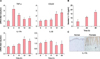
Because TLR9 ligand CpG has been known to increase the expression of IL-8 in HaCaT keratinocytes17, we questioned whether CpG can also induce the expression of other cytokines involved in the pathogenesis of psoriasis. To this end, we treated HaCaT keratinocytes with CpG, then performed qPCR. When treated with 10 µM CpG, expression of TNF-α, CCL20, and IL-17A was increased in a time-dependent manner (Fig. 1A). We chose one important cytokine IL-17A for a further study, as it is well known that IL-17A plays a pivotal role in pathogenesis of psoriasis. Consistent with the data on gene expression, CpG treatment resulted in increase of IL-17A secretion from HaCaT keratinocytes (Fig. 1B). Immunohistochemistry clearly showed that expression of IL-17A was increased in psoriatic lesional skin compared to normal skin (Fig. 1C).
NF-κB activation is required for CpG-induced IL-17A production
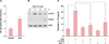
CpG activates NF-κB and results in increase of NF-κB-dependent gene expression in keratinocytes18. We determined whether CpG induces IL-17A expression via NF-κB-dependent way. First, we transduced HaCaT keratinocytes with NF-κB reporter adenovirus and then stimulated with 10 µM CpG. Consistent with previous data, CpG enhanced NF-κB luciferase activity (Fig. 2A). In parallel, phosphorylated-IκBα and CIAP2 was increased by CpG, further confirming that CpG activated NF-κB pathway (Fig. 2B). To determine the involvement of NF-κB activation in CpG-induced IL-17A production, we treated HaCaT keratinocytes with Bay11, a selective inhibitor for NF-κB pathway. As expected, pretreatment of Bay11 significantly blocked the CpG-induced IL-17A production. Pretreatment of CpG antagonist G-ODN also blocked CpG-induced IL-17A production (Fig. 2C). These data indicate that CpG induces IL-17A production in a NF-κB-dependent way.
IL-17A secreted from CpG-stimulated keratinocytes induces IL-22 production in CD4+ T cells
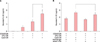
CD4+ T cells-derived IL-22 has been described as an important regulator of psoriatic inflammation19. We investigated whether IL-17A secreted from keratinocytes affects the IL-22 production in CD4+ T cells. We isolated CD4+ T cell from human blood, and then treated with conditioned medium of keratinocytes. As shown in Fig. 3A, conditioned medium obtained from CpG-treated cells (CpG-CM) clearly increased IL-22 production in CD4+ T cells, as compared with conditioned medium obtained from CpG-non-treated cells (CTL-CM). When the conditioned medium was neutralized with anti-IL17A antibody, IL-22 production in CD4+ T cells was significantly lowered (Fig. 3B), indicating that IL-17A secreted from keratinocytes was partly responsible for the production of IL-22 in CD4+ T cells.
DISCUSSION
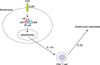
The role of keratinocytes in psoriatic inflammation has been increasingly noted202122. In this study, we demonstrated that CpG induced IL-17A expression in keratinocytes, and that this keratinocyte-derived IL-17A could affect IL-22 production in CD4+ T cells. Thus, our results provide additional evidence supporting the pivotal role of keratinocyte in pathogenesis of psoriasis, via the direct production of inflammatory cytokine IL-17A and boosting immune responses (Fig. 4).
In skin immune system, TLRs play as the key regulators that recognize pathogen-associated molecular patterns23. Different TLRs play different roles in various skin diseases in a context-dependent manner24. Keratinocytes, the main constituent of the epidermis, can contribute to innate immune surveillance through expression of various TLRs. TLRs can be categorized into two groups according to their locations. First is the surface-expressed TLRs that include TLRs 1, 2, 4, 5, 6, and 10. These TLRs act as the receptors for lipid-based ligands. Second is the intracellular TLRs that include TLRs 3, 7, 8, and 9. The nucleic-acid-based ligands bind to these TLRs13. Interaction of TLRs with different ligands activate various transcription factors such as nuclear factor NF-κB, which controls the expression of many genes involved in cell survival and immune responses2526. Implication for TLRs in the pathogenesis of psoriasis is supported by the fact that some effective drugs, such as vitamin D, can downregulate the expression of TLRs in keratinocytes27. In this experiment, we focused on the role of TLR9 in keratinocytes, since previous studies indicate that expression of TLR9 is increased in keratinocytes of psoriatic lesional skin, and that the nucleic acids play an important role in the pathogenesis of psoriasis2829. On the contrary, there is also a report that keratinocytes do not express TLR917. In this study, we showed that CpG, an TLR9 agonist, induced IL-17A expression through activation of NF-κB pathway in HaCaT keratinocytes. Our results somehow support the notion that keratinocytes express TLR9. However, whether other type of nucleic acid ligands including poly(I:C) and imiquimod can induce IL-17A production in keratinocytes remains to be determined.
Psoriasis is an innate immune-mediated disease, which is accompanied by some pathological alterations like immune cell infiltration and epidermal proliferation30. These alterations are mediated by several cytokines, among which IL-17A is regarded as the most important one. The IL-17A is mainly produced by Th17 cells which are activated by IL-23 produced by plasmacytoid myeloid cells. In addition to IL-17, IL-22 is known to be highly expressed in psoriatic lesional skin and to play a key role in the pathogenesis of psoriasis31. Like IL-17, IL-22 was originally produced by Th17 cells, however, it has been shown recently that IL-22 is produced not only by Th17 cells, but also by CD4+ T cells and CD8+ T cells32. Our data demonstrated that IL-17A secreted from keratinocytes can affect IL-22 production in CD4+ T cells, potentiating the possible involvement of keratinocytes in boosting epithelial innate immunity.
In summary, we demonstrated that CpG induced IL-17A production in keratinocytes, which in turn affected IL-22 production in CD4+ T cells, rsuggesting that keratinocytes modulate innate immune response thereby contributing to the pathogenesis of psoriasis.
 XML Download