

 PDF
PDF ePub
ePub Citation
 Print
Print


Dear Editor:
Dermatophytosis is common worldwide and is believed to affect more than 20%~25% of the world's population1. The epidemiology of dermatophyte infection is influenced by the changing patterns of migration, growth in tourism, immunocompetence of the host, pathogenicity of the infectious agents, availability of medical treatment, and changes in socioeconomic conditions23. We investigated the epidemiology of fungal skin infections through a retrospective analysis of patient's medical records between 1979 and 2013. Of total 4,275,715 patients, 415,526 patients with clinically suspicious fungal infection were collected at Catholic Skin Clinic in Daegu. Most of the patients were enrolled from Daegu and Gyeongsangbuk-do province. Microscopic examination with 15% KOH preparation and culture using potato dextrose agar corn meal Tween 80 media were performed for fungal examination. Culture media maintained at 24℃~26℃ were examined after 2 to 4 weeks. Of 415,526 patients, 131,440 KOH- and culture-proven patients were included in this study.
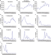
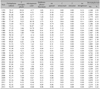
The annual number of patients with dermatophytosis ranged from 1,973 to 6,166 between 1979 and 2013 (Fig. 1B). The mean yearly isolation rate was reported to be 2,504 between 1979 and 1989. This yearly rate increased from 1990 onwards by an average of 4,329 patients per year. The ratio of patients with dermatophytosis among total patients was steady ranging from 2.18% to 5.21% (Table 1). Trichophyton rubrum was the most commonly identified dermatophyte in this study since the data collection began, followed by T. mentagrophytes, Microsporum canis, and Epidermophyton floccosum (Table 1). Of the 131,440 patients diagnosed with dermatophytosis during the study period, 116,164 patients (88.4%) had a T. rubrum infection. The ratio of T. rubrum infection among the dermatophytosis identified increased steadily during the study period (Table 1). The annual number of patients presenting with a T. mentagrophytes infection fluctuated between 120 and 505 during the period from 1979 to 2013 (Fig. 1D). However, the ratio of T. mentagrophytes infection remained stable after 1981 (Table 1). The number of M. canis infections showed a peak at 1986, however its ratio gradually decreased until 2013 (Fig. 1E, Table 1). The incidence of E. floccosum infection showed an abrupt decrease in the early 1980s and its low infection ratio remained constant until 2013 (Fig. 1F, Table 1). Although M. gypseum infection showed an isolated peak in 2004, its infection ratio was generally low throughout the study period (Fig. 1G, Table 1). The number of patients with T. verrucosum infection showed a peak in 1988, however its infection ratio remained steady until 2013 (Fig. 1H, Table 1). T. tonsurans infection became prevalent in late 1990s and it became the fourth most frequently isolated dermatophyte from 1995 onwards. However, its incidence showed a decreasing tendency until 2013 (Fig. 1I, Table 1). M. ferrugineum infection was no longer identified in this study after 1991 (Fig. 1J, Table 1). The prevalence of superficial fungal infections has changed significantly in the last century. The incidence of specific dermatophyte species has varied with time and place, due to factors including differences in hygiene levels, population migration, and the introduction of new treatment modalities4. The present study showed that the mean yearly incidence of dermatophytosis increased until 2003 but gradually decreased after 2004. This is assumed to be due to increasing urbanization, increasing fitness facilities, immunomodulatory therapy, the aging population, and the growing prevalence of obesity. Improvements in living conditions have generally been associated with a decline in zoophilic dermatophytes and an increase in anthropophilic dermatophyte infections3.
The evolution of prevailing T. rubrum infections in this study has been associated with the parallel increase in the prevalence of tinea pedis and onychomycosis5. A principal risk factor for developing a T. rubrum infection of the feet is the increased use of modern occlusive footwear. T. rubrum can be transmitted from infected to healthy persons via direct contact with infected skin or hair. Family history of tinea pedis and onychomycosis, advanced age, urbanization and obesity have also contributed to the increase in T. rubrum infections6. T. mentagrophytes, another main cause of tinea pedis, gradually decreased over the study period. This may be associated with public health education, improved hospital accessibility, environmental improvements, and increased lifestyle diversity7. The number of patients with M. canis markedly increased after 1979 and progressively decreased during the period from 1987 to 2013. The infection sources of M. canis include infected animals, particularly domestic cats and dogs. The major declining causes of these infections in Korea could be attributed to better public health education and improvements in personal hygiene8. In a previous study, E. floccosum, the main cause of tinea cruris, decreased rapidly after 19869. Our study confirmed a similar tendency. M. gypseum was the most common geophilic pathogen accounting for occasional epidemic spread under appropriate conditions1. T. verrucosum is a causative agent of tinea capitis associated with irreversible scarring and alopecia. Fortunately, the prevalence of T. verrucosum is decreasing. Since the first report in 1995, T. tonsurans infection was primarily observed in combat sports players in Korea, including wrestlers and judoists10. Infection due to T. tonsurans can occur by direct contact among humans or via contact with contaminated objects. A decreased incidence of T. tonsurans was observed in this study. M. ferrugineum, one of the most common causes of the non-inflammatory tinea capitis, was no longer identified in this study after 1991. T. rubrum is the most common cause worldwide for tinea pedis et unguium, which are becoming more common1. T. mentagrophytes is one of the most commonly isolated pathogens in Asia and America, causing tinea pedis et unguium1. However, T. rubrum and T. mentagrophytes are less common in Africa. Unusually, T. audouinii is the most prevalent pathogen in Africa1. M. canis is a prevalent agent of tinea capitis in the developed world1. A dramatic increase in T. tonsurans infections has been reported in the USA1.
Dermatophyte infections are still a common skin problem in Korea. This study will provide valuable information on current epidemiological trends for fungal infections in Korea. It will be helpful to predict coming fungal infections in Korea.
 XML Download