

 PDF
PDF ePub
ePub Citation
 Print
Print


Dear Editor:
We read with interest the article entitled "Effective Treatment of Congenital Melanocytic Nevus and Nevus Sebaceous Using the Pinhole Method with the Erbium-Doped Yttrium Aluminium Garnet Laser"1. We have used this method to successfully treat patients with an Er:YAG laser (Action; Lutronic, Goyang, Korea) in continuous-wave mode with a spot size of 1 mm and an output power of 0.2 mJ/cm2 2. We describe here a possible paradigm, a mimetic diagram, and a description of this donut ablation method with an Er:YAG laser.
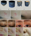
Fractionated ablation methods can include both space- and pulse duration-fractionation, yielding gentler and safer results. In addition to ablative and non-ablative resurfacing lasers, QS ruby and Nd:Yag lasers have been developed for fractionated methods3. The donut ablation method is a kind of space-fractionated ablation method. This method is an extension of the pinhole method and is based on the fractional photothermolysis theory, which posits that untreated skin is a source of rapid healing and intrinsic cooling of tissue4; however, whereas the pinhole method makes randomly or regularly distributed dot-shaped holes on the lesion, the donut ablation method makes line- or donut-shaped holes, or holes of various shapes, along the rim of the lesion (Fig. 1A~D). Complete removal of the lesion in the treated area requires the hole to be deep enough; that is, the appearance of normal tissue is the endpoint of treatment, resulting in a complete removal in the ablated fraction space. Any erythematous areas remaining after previous treatments were avoided to prevent scarring. Each lesion is treated in multiple sessions, with 1 to 2 month intervals between sessions.
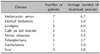
Using this method, we treated 26 patients with various skin lesions. Of these, 16 were almost completely treated, and all 26 showed at least a partial response (Table 1). One 31-year-old female with a nevus sebaceus on her forehead, which was a significant, cosmetically unacceptable size, was treated with this method. After six sessions, the lesion was completely resolved (Fig. 1E~G). A 53-year-old female with bilateral xanthelasma on the medial side of the eyelids was also treated with this method. After three sessions, the lesion was almost clearing without any side effects (Fig. 1H~K). Although xanthelasma can be treated with surgical excision, laser total ablation, chemical cauterization, or cryotherapy, these methods have significant risks of scarring and pigmentary changes after the treatment5. Another patient, a 40-year-old female with a deep dermal melanocytic lesion, was treated with five sessions of Er:YAG laser ablation that produced donut-shaped holes, resulting in almost complete elimination of the pigmented lesion (Fig. 1L~O).
The goal of donut ablation is maximally deep treatments to completely eliminate the lesion; however, the treatment area is confined to multiple dots, holes, lines, or segments to reduce adverse events such as scarring and recurrence. By contrast, the total ablation method utilizes limited penetration depths due to concerns about scarring. If skin lesions do not require removal in a single step, the results may be superior, with far fewer adverse events.
 XML Download