Journal List > Ann Dermatol > v.28(4) > 1046228

Hahn, Youn, Cha, Kim, An, and Ahn: Single Low-Dose Radiation Induced Regulation of Keratinocyte Differentiation in Calcium-Induced HaCaT Cells

Abstract

Background

We are continually exposed to low-dose radiation (LDR) in the range 0.1 Gy from natural sources, medical devices, nuclear energy plants, and other industrial sources of ionizing radiation. There are three models for the biological mechanism of LDR: the linear no-threshold model, the hormetic model, and the threshold model.

Objective

We used keratinocytes as a model system to investigate the molecular genetic effects of LDR on epidermal cell differentiation.

Methods

To identify keratinocyte differentiation, we performed western blots using a specific antibody for involucrin, which is a precursor protein of the keratinocyte cornified envelope and a marker for keratinocyte terminal differentiation. We also performed quantitative polymerase chain reaction. We examined whether LDR induces changes in involucrin messenger RNA (mRNA) and protein levels in calcium-induced keratinocyte differentiation.

Results

Exposure of HaCaT cells to LDR (0.1 Gy) induced p21 expression. p21 is a key regulator that induces growth arrest and represses stemness, which accelerates keratinocyte differentiation. We correlated involucrin expression with keratinocyte differentiation, and examined the effects of LDR on involucrin levels and keratinocyte development. LDR significantly increased involucrin mRNA and protein levels during calcium-induced keratinocyte differentiation.

Conclusion

These studies provide new evidence for the biological role of LDR, and identify the potential to utilize LDR to regulate or induce keratinocyte differentiation.

INTRODUCTION

Low-dose radiation (LDR; ≤100 cGy) is used for diagnostic imaging and interventional radiology1. We are continuously exposed to LDR from natural sources, medical devices, nuclear energy plants, and other industrial sources of ionizing radiation2. Therefore, it is important to understand the biological effects of LDR. The biological effects of LDR have been explained by the linear no-threshold (LNT) model, which considers that LDR effects are similar to those of high-dose ionizing radiation (HDR). The LNT model suggests that, similar as HDR, even very low exposure to LDR can cause harmful DNA lesions, such as single-strand breaks, double-strand breaks, apurinic/apyrimidinic site (either apyrimidinic or apurinic), DNA-DNA and DNA-protein cross-linking, and a plethora of base modifications3. However, there is some debate whether LDR is harmful to living organisms. Some studies have shown that LDR induces beneficial effects on DNA stability, growth rate, survival, life span, and immune activation, whereas other studies have shown that LDR has no biological effects 45678. Repeated exposure to LDR induces adaptation to ionizing radiation. Therefore, understanding the biological effects of LDR is crucial, particularly with the increasing use of LDR in medical and therapeutic protocols.
The epidermis is the outermost layer of the body, which forms a physical and immunological barrier. Keratinocytes are the most abundant cells in the epidermis9. Differentiating keratinocytes migrate from the stratum basale to the stratum corneum; this migration is implicated in forming the physical and immunological barrier and helping to form a multilayered epidermis. Mouse skin keratinocytes exposed to HDR exhibit accelerated differentiation due to the activation of protein kinase C (PKC) signaling pathways10. HDR-mediated activation of PKC induces the G2/M arrest and reduces keratinocyte stemness, which accelerates the initiation of keratinocyte differentiation11. In this study, we investigate whether LDR also accelerates keratinocyte differentiation.

MATERIALS AND METHODS

Cell culture and exposure to ionizing radiation

HaCaT cells were obtained from Cell Lines Service (Eppelheim, Germany) and cultured as described in a previous study12 in serum-free medium (GIBCO; Invitrogen, Carlsbad, CA, USA). Cells were cultured at 37℃ in a humidified atmosphere with 5% CO2. For exposure to ionizing radiation, cells were seeded in a 60 mm culture dish (SPL Life Sciences, Seoul, Korea) and exposed to a 137Cs γ-ray source (MDI-KIRAMS 137 irradiator; Korea Institute of Radiological and Medical Sciences, Seoul, Korea).

Cell viability analysis

Cell viability was determined by the tetrazolium salt (WST-1) assay (EZ-Cytox Cell Viability Assay kit; ITSbio, Seoul, Korea). HaCaT cells (1×10⁴ cells) were seeded into the wells of 96-well plates and cultured for 24 h. Then, cells were irradiated with 0, 0.01, 0.05, and 0.1 Gy gamma radiation (MDI-KIRAMS 137) and incubated for 28 h. Next, WST-1 solution was added to LDR-irradiated cells for 30 min. After incubation for 30 min, the absorbance of the samples at 450 nm was measured spectrophotometrically using a plate reader (iMark plate reader; Bio-Rad Laboratories Inc., Hercules, CA, USA). The optical density of each well was determined to quantify cell viability.

Western blot analysis

Western blot assays were performed on cell pellet lysates in radioimmunoprecipitation assay buffer with protease inhibitors (Roche, Basel, Switzerland), as previously described13. The antibodies used were anti-involucrin (Santa Cruz Biotechnology, Santa Cruz, CA, USA), and anti-β-actin (Sigma-Aldrich, St. Louis, MO, USA). Western blots were scanned and band intensities were quantified using ImageJ (National Institutes of Health, Bethesda, MD, USA).

Quantitative polymerase chain reaction

Quantitative polymerase chain reaction (PCR) was performed as described previously14. In brief, total RNA was isolated using TRIzol (Invitrogen; Life Technologies, Grand Island, NY, USA) according to the manufacturer's instructions. Then, cDNAs were synthesized from 1 µg total RNA using reverse transcriptase (Bioneer, Daejeon, Korea). PCR was performed using SYBR Green I in a PCR premix and a LineGene K cycler (BioER, Hangzhou, China). All results shown are representative of at least three independent experiments. All primers used for PCR have been reported in a previous study and include: involucrin forward primer, 5-CAAAGAACCTGGAGCAGG AG-3; involucrin reverse primer, 5-CAGGGCTGGTTGAA TGTCTT3; β-actin forward primer, 5-CGACAGGATGCA GAAGGAG-3; and β-actin reverse primer, 5-ACATCTG CTGGAAGGTGGA-3. β-actin was used as the internal loading control. Changes in relative levels of involucrin messenger RNA (mRNA) were obtained by relating each PCR product to its internal loading control.

Statistical analysis

Statistical significance was determined by t-test with Microsoft Excel 2016 (Microsoft, Redmond, WA, USA). A p-value 0.05 was considered statistically significant.

RESULTS AND DISCUSSION

LDR accelerates keratinocyte differentiation

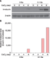
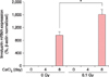
LDR increased involucrin protein levels in calcium-activated developing keratinocytes (Fig. 1). The relative involucrin expression level was elevated from 2,649.03 to 17,053.31 at day 8. LDR also induced increases in involucrin mRNA levels (Fig. 2). Involucrin is a representative marker of keratinocyte differentiation, and accumulates according to the level of differentiation15. Therefore, LDR-induced increases in involucrin mRNA and protein levels indicate that ionizing radiation facilitates calcium-mediated keratinocyte differentiation. In previous studies, gamma radiation has diverse effects on proliferation, DNA repair, release of cytokines and etc. in in vitro cellular as well as in vivo models1617181920. Especially, high gamma radiation increases calcium-induced keratinocyte differentiation through activating PKC signaling pathway in keratinocytes10. Thus, in our present study, we show that effects of LDR on keratinocyte differentiation.

LDR induces growth arrest by elevating p21 (Cip1/Waf1) expression

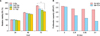
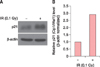
Song et al.10 show that HDR-induced acceleration of calcium-mediated keratinocyte differentiation is caused by a reduction in stemness and growth arrest due to PKC activation. Therefore, we tested whether LDR could induce keratinocyte growth arrest (Fig. 3). The results showed that LDR reduced keratinocyte viability in a time- and dose-dependent manner (Fig. 3), which is similar to the effects of HDR. Cell proliferation capacity was reduced by the exposure to 0.1 Gy LDR from 2.58 to 1.05 at 48~72 h, which suggested that LDR induced growth arrest. We also found that LDR elevated p21 (Cip1/Waf1) expression in the HaCaT cells (Fig. 4).
In general, increase of p21 (Cip1/Waf1) suppresses broad range of cyclin/CDK complexes, which leads to G1, G2 and S-phase arrest. Thus, p21 (Cip1/Waf1) represses stemness through the regulation of the cell cycle21, and is a well-characterized radiation marker that is activated by PKC1122. In keratinocytes, p21 (Cip1/Waf1) is increased by calcium-induced keratinocyte differentiation23. And overexpression of p21 (Cip1/Waf1) induces acceleration of keratinocyte differentiation23. Therefore, since keratinocytes initiates differentiation through halting cell proliferation and decreasing stemness, LDR-mediated elevation of p21 (Cip1/Waf1) facilitated calcium-mediated keratinocyte differentiation.
Ionizing radiation activates the PKC signaling pathway, which elevates the p21 expression levels. The PKC signaling pathway activation and elevation of p21 expression levels induces cell growth arrest and reduces stemness, which accelerates keratinocyte differentiation in HaCaT cells exposed to LDR. These results are consistent with previous results showing that HDR accelerates keratinocyte differentiation. Our results provide new evidence for the biological role of LDR, and suggest the potential utilization of LDR as an inducer of keratinocyte differentiation on in vitro model.

Figures and Tables

Fig. 1

Low-dose radiation stimulates involucrin accumulation in calcium-induced differentiating keratinocytes. (A) Western blot analysis of involucrin protein levels in control HaCaT cells (no exposure to ionizing radiation) and HaCaT cells exposed to 0.1 Gy gamma irradiation. In each experiment, equal loading was demonstrated by immunostaining for β-actin. (B) The band densities of interest were further analyzed by ImageJ. Involucrin levels were normalized against β-actin.

ad-28-433-g001
Fig. 2

Low-dose radiation elevates involucrin messenger RNA (mRNA) expression in calcium-induced differentiating keratinocytes. HaCaT cells were exposed to gamma radiation and control cells were untreated. After 4 h, keratinocyte differentiation was induced in HaCaT cells using 2 mM calcium for 4 and 8 days. Total RNA, isolated using TRIzol, was used to synthesize cDNA by reverse transcription, and was amplified by polymerase chain reaction. β-actin was amplified as a normalization control. Results are expressed as the mean percentage±standard error of the experiments performed in triplicate. *p<0.01.

ad-28-433-g002
Fig. 3

Low-dose radiation reduced cell viability and growth capacity in HaCaT cells. (A) HaCaT cells (1×10⁴ cells) were plated in 96-well plates and cultured for 24, 48, and 72 h after the exposure to the indicated dose of gamma radiation. Cell viability was measured by WST-1 as described in Materials and Methods. Results are expressed as the mean optical density±standard error, as a percentage of the control for the experiments performed in triplicate. *p<0.01. (B) Growth capacity was calculated by calculating the dRV/dT as derivative relative viability (%)/derivative time (h). IR: gamma irradiation.

ad-28-433-g003
Fig. 4

Low-dose radiation induced p21 (Cip1/Waf1) expression in HaCaT cells. (A) Western blot analysis of p21 (Cip1/Waf1) protein levels in control HaCaT cells and HaCaT cells exposed to 0.1 Gy gamma irradiation. Equal loading in each experiment was demonstrated by immunostaining for β-actin. (B) Band densities of interest were further analyzed by ImageJ; p21 (Cip1/Waf1) levels were normalized against β-actin. IR: gamma irradiation.

ad-28-433-g004

ACKNOWLEDGMENT

This study was supported by grants from the Ministry of Trade, Industry and Energy (grant no. 20131610101840) and the Ministry of Science, ICT and Future Planning (grant no. 20110028646) of the Republic of Korea.

References

1. Mobbs SF, Muirhead CR, Harrison JD. Risks from ionising radiation: an HPA viewpoint paper for Safegrounds. J Radiol Prot. 2011; 31:289–307.
crossref
2. Bae S, Kim K, Cha HJ, Choi Y, Shin SH, An IS, et al. Low-dose γ-irradiation induces dual radio-adaptive responses depending on the post-irradiation time by altering microRNA expression profiles in normal human dermal fibroblasts. Int J Mol Med. 2015; 35:227–237.
crossref
3. Bae S, Kim K, Cha HJ, Choi Y, Shin SH, An IS, et al. Altered microRNA expression profiles are involved in resistance to low-dose ionizing radiation in the absence of BMI1 in human dermal fibroblasts. Int J Oncol. 2014; 45:1618–1628.
crossref
4. Liang X, So YH, Cui J, Ma K, Xu X, Zhao Y, et al. The low-dose ionizing radiation stimulates cell proliferation via activation of the MAPK/ERK pathway in rat cultured mesenchymal stem cells. J Radiat Res. 2011; 52:380–386.
crossref
5. Ahmed KM, Nantajit D, Fan M, Murley JS, Grdina DJ, Li JJ. Coactivation of ATM/ERK/NF-kappaB in the low-dose radiation-induced radioadaptive response in human skin keratinocytes. Free Radic Biol Med. 2009; 46:1543–1550.
crossref
6. Moskalev AA, Plyusnina EN, Shaposhnikov MV. Radiation hormesis and radioadaptive response in Drosophila melanogaster flies with different genetic backgrounds: the role of cellular stress-resistance mechanisms. Biogerontology. 2011; 12:253–263.
crossref
7. Seong KM, Kim CS, Seo SW, Jeon HY, Lee BS, Nam SY, et al. Genome-wide analysis of low-dose irradiated male Drosophila melanogaster with extended longevity. Biogerontology. 2011; 12:93–107.
crossref
8. Ina Y, Sakai K. Activation of immunological network by chronic low-dose-rate irradiation in wild-type mouse strains: analysis of immune cell populations and surface molecules. Int J Radiat Biol. 2005; 81:721–729.
crossref
9. Eckert RL, Rorke EA. Molecular biology of keratinocyte differentiation. Environ Health Perspect. 1989; 80:109–116.
crossref
10. Song HJ, Cho CK, Yoo SY, Park KS, Lee YS. Increased induction of Ca2+-mediated differentiation by gamma ray is mediated by endogenous activation of the protein kinase C signaling pathways in mouse epidermal cells. Int J Radiat Oncol Biol Phys. 1998; 41:897–904.
crossref
11. Barboule N, Lafon C, Chadebech P, Vidal S, Valette A. Involvement of p21 in the PKC-induced regulation of the G2/M cell cycle transition. FEBS Lett. 1999; 444:32–37.
crossref
12. Cha HJ, Lee OK, Kim SY, Ko JM, Kim SY, Son JH, et al. MicroRNA expression profiling of p-phenylenediamine treatment in human keratinocyte cell line. Mol Cell Toxicol. 2015; 11:19–28.
crossref
13. Cha HJ, Bae S, Kim K, Kwon SB, An IS, Ahn KJ, et al. Overdosage of methylparaben induces cellular senescence in vitro and in vivo. J Invest Dermatol. 2015; 135:609–612.
crossref
14. Kwon KJ, Bae S, Kim K, An IS, Ahn KJ, An S, et al. Asiaticoside, a component of Centella asiatica, inhibits melanogenesis in B16F10 mouse melanoma. Mol Med Rep. 2014; 10:503–507.
crossref
15. Watt FM. Involucrin and other markers of keratinocyte terminal differentiation. J Invest Dermatol. 1983; 81:1 Suppl. 100s–103s.
crossref
16. Tsukimoto M, Homma T, Ohshima Y, Kojima S. Involvement of purinergic signaling in cellular response to gamma radiation. Radiat Res. 2010; 173:298–309.
17. Sudprasert W, Navasumrit P, Ruchirawat M. Effects of low-dose gamma radiation on DNA damage, chromosomal aberration and expression of repair genes in human blood cells. Int J Hyg Environ Health. 2006; 209:503–511.
crossref
18. Han SK, Song JY, Yun YS, Yi SY. Effect of gamma radiation on cytokine expression and cytokine-receptor mediated STAT activation. Int J Radiat Biol. 2006; 82:686–697.
crossref
19. Pourfathollah AA, Shaiegan M, Namiri M, Babae GR. Effect of gamma irradiation on lymphocyte proliferation and IL-8 production by lymphocytes isolated from platelet concentrates. Arch Med Res. 2008; 39:590–593.
crossref
20. Zhang Z, Zhang H, Liu F, Qiu M, Tong J. Effects of gamma radiation on bone-marrow stromal cells. J Toxicol Environ Health A. 2010; 73:514–519.
crossref
21. Yew TL, Chiu FY, Tsai CC, Chen HL, Lee WP, Chen YJ, et al. Knockdown of p21(Cip1/Waf1) enhances proliferation, the expression of stemness markers, and osteogenic potential in human mesenchymal stem cells. Aging Cell. 2011; 10:349–361.
crossref
22. Bluwstein A, Kumar N, Léger K, Traenkle J, Oostrum Jv, Rehrauer H, et al. PKC signaling prevents irradiation-induced apoptosis of primary human fibroblasts. Cell Death Dis. 2013; 4:e498.
crossref
23. Todd C, Reynolds NJ. Up-regulation of p21WAF1 by phorbol ester and calcium in human keratinocytes through a protein kinase C-dependent pathway. Am J Pathol. 1998; 153:39–45.
crossref
TOOLS
Similar articles