

 PDF
PDF ePub
ePub Citation
 Print
Print


Dear Editor:
Interstitial granulomatous dermatitis (IGD) is a rare and peculiar disorder with cutaneous and joint manifestations1. IGD may be associated with rheumatologic and hematologic disorders of underlying malignancies23. Herein, we report a case of IGD associated with rheumatoid arthritis (RA).
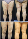
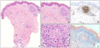
A 59-year-old Korean woman presented with multiple erythematous plaques on both extremities for 10 days (Fig. 1A). Upon physical examination, the plaques were linear shaped, non-tender, and palpable. She had been diagnosed with RA 17 years earlier. She had taken prednisolone, nonsteroidal anti-inflammatory drugs, and methotrexate to treat RA for 7 years. One month earlier, her arthritis symptoms had stabilized and the rheumatologist withdrew methotrexate. Laboratory test findings were non-specific except for an elevated C-reactive protein (CRP) level (7.12 mg/dl; normal range, 0.01~0.47 mg/dl) and erythrocyte sedimentation rate (ESR) (81 mm/h; normal range, 0~15 mm/h). Histopathologic findings indicated perivascular and interstitial lymphohistiocytic infiltration through the dermis and chronic granulomatous inflammation with collagen degeneration in the upper dermis. Immunohistochemically, most of the infiltrating inflammatory cells were CD 68-positive. Alcian blue staining revealed no mucin deposition in the areas of granulomatous inflammation (Fig. 2). Based on clinical and histological findings, she was diagnosed with IGD. A 7.5-mg methotrexate dose was given weekly and the daily prednisolone dose was increased from 5 mg to 15 mg. A topical 0.1% tacrolimus ointment and methylprednisolone cream were also applied daily for 2 months. The lesions were nearly cleared and ESR and CRP were found to be within normal limits after 2-month follow-up (Fig. 1B).
The most common clinical findings of IGD are asymptomatic multiple papules and plaques (70%~90% of cases)2. The "rope sign" was named for the linear prominent cutaneous cord-like lesions, considered pathognomic for IGD1. However, these lesions are not an essential feature, and are reported in only 9% of cases3. The histopathologic findings show interstitial CD 68-positive histiocyte infiltration around focally degenerated collagen24. Epidermal changes, mucin deposition, neutrophil and/or eosinophil infiltration, and vasculitis are usually absent 2. Differential diagnoses include interstitial granuloma annulare (IGA), interstitial granulomatous drug reaction (IGDR), and palisaded neutrophilic and granulomatous dermatitis (PNGD)124. In IGA, prominent mucin deposition and only focal interstitial infiltration could be found. Recent change of medication is present in IGDR, and IGDR presents with basal cell vacuolization and lichenoid changes with eosinophils2. Leukocytoclastic vasculitis and pandermal infiltration of neutrophil frequently present in early lesion of PNGD, and palisading granulomatous inflammation is the typical feature of fully developed one1.
The pathogenesis of IGD may be associated with immune complex deposition caused by immune reactants of systemic inflammatory diseases5. Interestingly, IGD lesions in our case began to occur in association with the flare-up of RA after methotrexate withdrawal, although the causal relationship remains unknown. From our case we suggest that IGD should be considered among the differential diagnoses for patients with rheumatologic disease and cord-like skin lesions.
 XML Download