

 PDF
PDF ePub
ePub Citation
 Print
Print


Dear Editor:
Cutaneous metastasis from osteosarcoma is exceedingly rare, with only 11 cases reported in the literature1. Giant cell–rich osteosarcoma is a rare variant of osteosarcoma, accounting for 1%~3% of conventional osteosarcomas2. Herein, we present the rare case of a patient with cutaneous metastasis of giant cell–rich osteosarcoma, which closely resembled a giant cell tumor. Owing to the different prognoses and treatment strategies for these tumors, it is important to ensure that the correct differential diagnosis is made.
A 43-year-old man presented with a 6-month history of nodules on his scalp. He had been diagnosed with giant cell-rich osteosarcoma in his right femur along with metastases to multiple organs, including the lungs and the mandible, 1 year ago. The metastatic lung nodules had increased in size despite 6 months of chemotherapy with ifosfamide, adriamycin, and high-dose methotrexate. Subsequently, oncologists changed the regimen to gemcitabine and docetaxel, but the tumor still progressed. Therefore, the regimen was changed once again, and he was admitted to receive a second cycle of cyclophosphamide and etoposide as a third-line regimen.
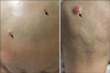
Upon physical examination, several firm skin-colored to erythematous nodules were palpated on his scalp, each measuring approximately 5~12 mm (Fig. 1). Histopathological findings showed many uniformly distributed, multinucleated giant cells containing pleomorphic nuclei. Mononuclear tumor cells had hyperchromatic nuclei with a variety of morphologies, such as round, oval, polygonal, and spindle-shaped (Fig. 2). Although atypical mitoses or dominant osteoid formations were hardly observed, these histological findings were consistent with those of giant cell-rich osteosarcoma, which was previously confirmed by right femur biopsy (Fig. 2). Three months later, he died of sepsis following bone marrow failure caused by the continued chemotherapy.
Several studies have reported that giant cell-rich osteosarcomas are sometimes misdiagnosed as giant cell tumors because of their similar clinicopathological features234. However, these tumors have extremely different clinical courses and prognoses. Giant cell tumors are usually benign with a metastatic rate of <2%, while the clinical course of osteosarcoma is extremely aggressive, with rapid hematogenous systemic dissemination and a tumor-related mortality of 75% within 5 years of diagnosis15.
For these reasons, Huang et al.3 suggested the use of some key factors to distinguish these two tumors, including osteoid formation, nuclear pleomorphism, and atypical mitotic figures3. However, osteoid formation cannot be found in small biopsy samples or samples from early lesions4. Tumor cell pleomorphism is not obvious in some cases2. Unless malignant osteoid formation is found, as in this case, it is difficult to confirm an osteosarcoma diagnosis without knowing a patient's cancer history. Unfortunately, not all cases present with features as informative as those in the case reported here.
Among 29 cases of giant cell-rich osteosarcoma, only 1 case of cutaneous metastasis had been reported in English literature234. With the histological similarity to giant cell tumors, this case emphasizes the value of careful examination of tumor cell pleomorphism and pathological mitoses when patients present with giant cell-rich tumors.

 XML Download