

 PDF
PDF ePub
ePub Citation
 Print
Print


INTRODUCTION
Papular elastorrhexis (PE), eruptive collagenoma (EC) and nevus anelasticus (NA) have been described as multiple small papules with decrease, fragmentation, or lack of dermal elastic fibers. The conditions were first described in 1987, 1966, and 1921, respectively123. The term nevus anelasticus was introduced by Staricco and Mehregan4 in 1961 as a more adequate expression for describing the entity named by Lewandowsky3 as naevus elasticus regionis mammariae in 1921.
It is suggested that PE, EC, and NA might be the same entity. Bordas et al.1 suggested that PE was a variant of NA in the first reported case of PE. In addition, it was suggested that EC is inseparable from PE5. Other authors have suggested that NA and EC should be considered variants of PE6.
Ryder and Antaya7 reviewed previous reports of the 3 entities and found remarkable similarities between them. All 3 begin to appear before the age of 20 years, and the sites of the lesions are mainly the trunk and upper extremities. A lack of history of trauma, inflammation, family history, or extracutaneous manifestations is also a common feature. All 3 showed a decreased amount of elastic tissue in biopsy specimens. Therefore, the authors concluded that PE, EC, and NA are the same entity.
Although changes in elastic fibers have been well evaluated in the 3 conditions, changes in collagen fibers have not attracted attention. There have been inconsistencies in previous reports' descriptions of collagen fibers. Some authors reported homogenized, thick collagen fibers. On the other hand, others reported that the collagen tissue was normal. We experienced an interesting case in 2011 and reported it as PE8. The patient had fine, compacted collagen fibers in the upper dermis, where elastic fibers were reduced and fragmented, which prompted our interest in changes in collagen fibers as an important histological feature in these diseases.
We reviewed previous reports of PE, EC, and NA, and changes in collagen and elastic fibers were investigated.
MATERIALS AND METHODS
We searched reports of PE, EC, and NA in PubMed and a Korean database. Demographic information and clinical manifestation of reported cases of the 3 diseases were summarized, and similarities and differences between the 3 conditions were analyzed.
We selected cases with histological figures from which we could evaluate the status of dermal collagen and elastic fibers. Changes in dermal collagen and elastic fibers of the select cases of the 3 diseases were summarized and compared.
According to the status of dermal collagen fibers, the selected cases were reclassified into 3 groups: (i) normal collagen group, (ii) fine, dense collagen group, and (iii) thick, dense collagen group. Demographic information and clinical manifestations for the 3 groups were summarized and compared.
RESULTS
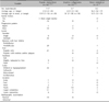
Twenty-four cases of PE1689101112131415161718192021, 12 cases of EC2223242526272829303132, and 2 cases of NA733 were found. One case of EC24 was reported in Korean in the Korean literature, and the others were in English. Demographic information and clinical manifestations for the cases are summarized in Table 1.
There were significant similarities between the 3 entities. All 3 diseases showed female predominance, and the average onset age was mid second decade. There was no family history except in 3 cases of PE from a single family20. The lesions progressed gradually in most cases. The lesional features of number, hardness, and location showed no significant difference between the 3 diseases. All cases had multiple lesions, and most lesions were slightly indurated to firm. The trunk and extremities were the major involved sites. All PE and NA cases in which relationships with hair follicles were described had nonfollicular lesions. Relationships with hair follicles were not mentioned in the EC cases. All lesions were papules of several millimeters in size in PE and NA, but 8 cases of EC had nodules and/or plaques along with papules. All PE lesions were whitish to hypopigmented, whereas 7 cases of EC had skin-colored lesions. There were no abnormal radiologic findings.
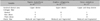
Among the reports, we selected only cases with histological figures in which we could evaluate the status of dermal collagen and elastic fibers. Six cases of PE689101618, 3 cases of EC242527, and 1 case of NA33 were selected. Changes in dermal collagen and elastic fibers in those cases are summarized in Table 2. The dermal area in which elastic fiber was reduced and fragmented was the upper dermis in 9 cases except in 1 case of NA, in which elastic fiber alteration was observed from the upper to deep dermis. Changes in collagen fibers in the elastic fiber-reduced area could be classified into 3 patterns: (i) normal collagen, (ii) fine, dense collagen, and (iii) thick, dense collagen. Four cases of PE8101618 and 2 cases of EC2427 had fine, dense collagen tissue. In 2 cases of PE 69 and 1 case of NA33, collagen fibers did not show any change; and only 1 case of EC25 had the thick, dense collagen fibers.
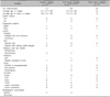
The 10 selected cases were reclassified into 3 groups according to the status of collagen fibers in the elastic fiber-reduced dermal area: (i) normal collagen group, (ii) fine, dense collagen group, and (iii) thick, dense collagen group. Demographic information and clinical manifestations for the 3 groups are summarized in Table 3. The fine, dense collagen group had all women, and the average onset age was 13.3 years (range, 3~25 years). Three cases progressed gradually, but 1 case progressed rapidly, with all lesions developing within 1 week. In all cases, multiple asymptomatic, less-than-1-cm-size, scattered papules developed on the trunk and/or extremities. Exceptionally, a few lesions also developed on the face and neck in 1 patient. The lesions were whitish to hypopigmented and slightly indurated to firm in most patients.
Three cases in the normal collagen group differed from cases in the fine, dense collagen group in their clinical manifestations. The first case9 reported as "eruptive PE" was the only male patient and had the latest onset age among the 10 selected cases. The involved sites of face and scalp also differed from the main involved areas. The lesions had developed rapidly, which was also unusual. The second patient33 was a 17-year-old female who had papules that were grouped and localized on the right areola. Elastic fiber alteration was observed from the upper to deep dermis in the biopsy specimen. Judging from the clinical figure, the last patient6 seemed to have numerous lesions.
One case with thick, dense collagen25 had a unique clinical manifestation: dozens of lesions were localized on the left trunk in a zosteriform distribution. Peculiarly, the color of the lesions was erythematous, and some lesions were confluent.
DISCUSSION
We searched reports of PE, EC, and NA in PubMed and the Korean database and found 24 cases of PE, 12 cases of EC, and 2 cases of NA. Demographic information and clinical manifestations of the 3 diseases had significant similarities on comparative evaluation. EC has been known to present with an acute onset2. However, 5 of the 7 cases of EC whose progression pattern was described progressed gradually, as in PE and NA. NA was first described as perifollicular lesions3. However, 1 of the 2 NA cases included had nonfollicular lesions, and a relationship with hair follicles was not described for the other patient.
We selected 10 cases with published histological figures in which we could evaluate the status of dermal collagen and elastic fibers. The cases were reclassified into 3 groups according to collagen tissue changes in the dermal area where elastic fibers were reduced and fragmented: (i) normal collagen group, (ii) fine, dense collagen group, and (iii) thick, dense collagen group.
Six cases in the fine, dense collagen group consisting of 4 cases of PE and 2 cases of EC showed similar clinical features. The condition developed in women between the 1st and 3rd decades, and the lesions progressed gradually. There were multiple asymptomatic, white to hypopigmented, slightly indurated to firm, less-than-1-cm-size, scattered nonfollicular papules on the trunk and/or extremities. Electron microscopic examination was performed in 1 case of that group27. Scanning electron microscopy of the dermis showed that collagen fibers did not form bundles. Collagen fibers were individualized, forming waved, compact masses resembling noodles, and hence, empty spaces normally seen between dermal collagen bundles disappeared. On the other hand, normal collagen bundles were easily observed in the normal skin, and empty spaces were seen among the bundles.
Three cases in the normal collagen group differed from the fine, dense collagen group in clinical manifestations. Each case had peculiar clinical features with few similarities. One case in the thick, dense collagen group also had clinical features that differed from other cases.
Ryder and Antaya7 suggested that PE, EC, and NA are the same entity. Our conclusions corroborated that suggestion. Furthermore, the fine, dense collagen group showed typical clinical and histological features of this condition. Until now, decrease in elastic tissue has been emphasized, but changes in collagen fibers also should be emphasized because collagen tissue mainly contributes to the formation of the lesions. It is possible that the decrease in elastic tissue is secondary to the change in collagen bundles—such as a dilutional effect.
Connective tissue nevi are acquired dermal connective tissue hamartomas characterized predominantly by an imbalance in the relative amount and distribution of collagen, elastin, or proteoglycans34. The nevi are classified according to histological aspects. When collagen predominates, they are called collagenomas. Lesions in which elastic tissue predominates are called elastomas3435. Therefore, we suggest papular collagenoma as a new name for the disease because it represents the disease's clinical and histological features.
Four cases with normal collagen or thick, dense collagen showed different clinical features as well as histological features from the fine, dense collagen group. We considered them to be conditions different from papular collagenoma or its variant.
 XML Download