

 PDF
PDF ePub
ePub Citation
 Print
Print


Dear Editor:
Spitz nevus is uncommon melanocytic nevus, and the head and neck are known to be the most common involved sites. To our knowledge, this is the first report of Spitz nevus arising on the perianal area.
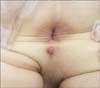
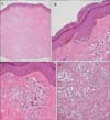
A 3-year-old boy presented with a pedunculated nodule on his perianal area (Fig. 1). The nodule was about 1 cm in diameter, with erythema and tiny black-pigmented spots anterior to the anal orifice. It had developed 1 year previously and had been gradually increasing in size. Shave excision was done for the diagnosis and treatment. Histopathological examination revealed numerous nests of cells in the entire dermis (Fig. 2A). In the epidermis, there were sparse basal pigmentations along the dermoepidermal junction and some nevus cells with large nuclei on the tip of the rete ridges (Fig. 2B). In the dermis, tumor cells were composed of large epithelioid nevocytes with abundant eosinophilic cytoplasm (Fig. 2C, D). Many tumor cells were uniformly atypical large epithelioid cells especially in the superficial dermis; however, there was no mitosis or Kamino body. Immunohistochemical staining revealed that the nest cells were superficially positive for HMB-45 and Ki-67. Because the lower margin contained tumor cells, we could not identify whether there was progressive dispersion of melanocytes to smaller aggregates or to single melanocytes in the deepest part of the lesion; therefore, we concluded that further excision with a clear margin or close follow-up for recurrence is needed. Since shave excision was accomplished, there had been no new lesions for about 1 year; however, he has not made a follow-up visit thereafter.
Traditionally, the sites of predilection of Spitz nevus are known to be the face, head, and neck in children, whereas in adults, the lower limbs have been recognized as the most common involved sites. However, in a study of 349 patients with Spitz nevus1, the most common location was the lower extremities in all age groups, except in the group older than 45 years, in whom the trunk was the most common site. There have been only three reports of penile solitary Spitz nevi in young adults234 and one report of Spitz nevus on the inner surface of the labium major5. There was no characteristic and common feature between all of these genital Spitz nevi. Although there are few reports of Spitz nevi of the genital area, to the best of our knowledge, this is the first reported case of Spitz nevus on the perianal area.
Unlike the typical Spitz nevus, the histopathologic findings of this case included epithelioid cells of large and pleomorphic morphology without a Kamino body. However, histologic findings that suggest the atypical Spitz tumor, such as higher cellular density and considerable mitotic rate, were not observed in this case.
In conclusion, this is the first case of Spitz nevus arising from the perianal area, and we suggest that Spitz nevus should be considered as a differential diagnosis in an unusual papular/nodular lesion on the perianal area in children.
 XML Download