

 PDF
PDF ePub
ePub Citation
 Print
Print


INTRODUCTION
The Q-switched 1064-nm neodymium-doped yttrium aluminum garnet (QS 1064-nm Nd:YAG) laser has gained popularity as a modality for the treatment of melasma and for nonablative skin rejuvenation. Traditionally, the QS 1064-nm Nd:YAG laser is used to treat pigmentation disorders such as lentigines, ephelides, nevus of Ota, and Hori's nevus1. Recently, it has been reported to be able to fragment melanin granules, dispersing them into the cytoplasm without causing cellular damage, resulting in the clinical improvement of melasma23. The QS 1064-nm Nd:YAG laser also has the ability to induce microdamage to the dermis, inducing dermal remodeling and neocollagenesis, resulting in nonablative skin rejuvenation, sometimes referred to as "laser toning." In laser toning, multiple and frequent low-fluence, large-spot-size treatments are used, typically with an 8-mm spot size and a fluence of 2.8 J/cm2. With the increasing demand for laser toning procedures, patients undergoing multiple sessions of laser treatment should be aware of the potential complications. A significant complication that has been increasingly reported in recent literature is guttate hypopigmentation and depigmentation after laser toning with QS 1064-nm Nd:YAG45. We present three cases of laser-induced hypopigmentation after laser toning that were referred to our tertiary dermatology referral center for treatment.
CASE REPORT
Case 1
A 51-year-old Chinese female patient presented with a complaint of facial dyspigmentation after multiple sessions of laser therapy performed by her general practitioner for the treatment of melasma on her face. She was treated with laser toning with QS Nd:YAG laser for her melasma and for skin rejuvenation. She was treated weekly, then daily, and subsequently several times a day with the laser toning procedure. She received 40~50 laser treatments during a 6-month period. She was otherwise systemically well and does not have any personal or family history of vitiligo. She started to notice guttate hypopigmentation 2~3 months into her laser toning treatment, which became more florid as the treatment frequency was increased.
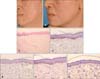
On examination, she had extensive speckled hypopigmented macules over the face. Some of the hypopigmented macules have coalesced to form larger patches over her cheeks. There was an observable hyperpigmentation in the background, which clinically looks like melasma (Fig. 1A). She did not have any hypopigmented areas elsewhere on her body. She was advised to stop the laser treatment immediately.
The skin biopsy done showed sections of skin that had focal loss of basal pigmentation as evidenced by Fontana-Masson staining. The Melanoma Triple Cocktail (human melanoma black 45 [HMB45]+melan-A+tyrosinase) from Ventana (Tucson, AZ, USA) was used to visualize basal melanocytes and their dendritic processes. In areas with preserved basal melanin pigmentation, the number of melanocytes was normal and the dendritic processes were prominent. In hypopigmented areas, the absolute number of melanocytes was decreased and the dendritic processes were markedly reduced. In addition, microphthalmia transcription factor (MITF) staining was performed, which confirmed that the number of melanocytes was preserved in the pigmented areas but were decreased in the hypopigmented areas. Solar elastosis was present. No pigmentary incontinence or ochronosis was seen (Fig. 1C~G).
Topical treatment was initiated with 0.1% tacrolimus ointment to the hypopigmented macules and hydroquinone-containing cream to the background melasma to decrease the contrast in pigmentation. On follow-up after a 2-year period of using only topical treatment and sunscreen, visible improvement of the hypopigmented macules was observed (Fig. 1B).
Case 2
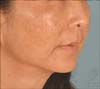
A 58-year-old Chinese female patient presented with hypopigmented macular spots on her face for the last 8 months after laser treatment. She had received weekly laser toning treatment with QS Nd:YAG for 1 year before the appearance of the hypopigmented macules on her face. The hypopigmented spots have increased in number and size during the months while she was receiving treatment. There were no preceding rashes or vitiligo. The hypopigmented spots were asymptomatic.
On examination, she had speckled hypopigmented macules on her cheeks and chin (Fig. 2).
She was treated with topical 0.1% tacrolimus ointment twice a day with no improvements.
Case 3
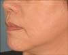
A 58-year-old Chinese female patient with a long-standing history of melasma presented with hypopigmented macules on her face that had been present for 1 year. She had noticed the hypopigmented macules appearing after starting laser toning treatment with the QS Nd:YAG laser for her melasma. She had undergone 2 years of laser treatment, sometimes at fortnightly intervals. She had no preceding rashes or a history of vitiligo. The hypopigmented macules were asymptomatic.
On examination, there were scattered guttate hypomelanotic macules on her face with a background of melasma (Fig. 3). She was treated with 0.1% topical tacrolimus with minimal improvements.
DISCUSSION
Laser toning with low-fluence, large-spot-size QS 1064-nm Nd:YAG laser has been reported to cause severe macular hypopigmentation as a treatment complication. Such acquired dyspigmentation can cause serious psychological distress to the patients. Recent studies that demonstrated patients with hypopigmentation induced by QS 1064-nm Nd:YAG laser treatment are summarized in Table 1121314.
The precise pathophysiology for this phenomenon has not been fully elucidated. It has been proposed that the 1064-nm QS Nd:YAG laser causes cumulative phototoxicity and cellular destruction of melanocytes12.
Previously, other forms of lasers, such as the CO2 laser and the ruby laser, have also been reported to cause hypopigmentation. Grimes et al.6 found normal or near-normal numbers of basal melanocytes in association with decreased melanin pigmentation in skin biopsy specimens taken from hypopigmented areas after CO2 laser resurfacing. Similar findings were observed by Liew et al.7 who studied patients with hypopigmentation induced by the ruby laser used for hair removal. In their study, the melanocyte numbers did not decrease on the S-100 stain; however, the dopa oxidase activity seemed to have been reduced. Both studies concluded that impairment of melanogenesis rather than the destruction of melanocytes was associated with laser-induced hypopigmentation. Hruza et al.8 investigated the effects of Q-switched ruby laser on the skin of normal human volunteers, and observed that low radiant exposures stimulated melanogenesis whereas high radiant exposures resulted in lethal injury to pigmented epidermal cells. With the advent of laser toning with the QS 1064-nm Nd:YAG laser, we are seeing another cause of laser-induced hypopigmentation as illustrated by our three cases. The histopathological features of hypopigmentation induced by the QS 1064-nm Nd:YAG laser have not been well delineated. A recent histopathological study by Kim et al.9 in patients with this type of hypopigmentation showed that the hypopigmented areas were melanopenic but not melanocytopenic, similar to the above-mentioned studies on other forms of laser-induced hypopigmentation. The authors suggested that laser toning results in decreased melanocyte function by downregulating factors affecting melanogenesis, including tyrosinase, tyrosinase-related protein-1 and -2, a-melanocyte-stimulating hormone, and nerve growth factors.
However, the histopathological findings in our first patient suggest that there was an absolute decrease in the number of basal melanocytes within the hypopigmented areas. The use of the Melanoma Triple Cocktail (HMB45+melan-A+tyrosinase) from Ventana increased the sensitivity of detecting melanocytes within the basal epidermis. In addition, the dendritic processes of these melanocytes could be visualized and compared as this antibody cocktail demonstrates cytoplasmic staining. It was observed that in areas showing preserved basal melanin pigmentation, the melanocyte numbers were normal and their dendritic processes were prominent. In comparison, hypopigmented areas showed a decrease in absolute melanocyte numbers and a marked reduction in their dendritic processes. Similarly, Kim et al.10 described a case of Nd:YAG laser-induced hypopigmentation that had decreased the number of functional melanocytes on histology, which concurred with the findings in patient 1. A recent study by Mun et al.11 analyzed the effects of QS Nd:YAG laser on melasma by using electron microscopy, and found that it resulted in decreased numbers of melanocytic dendrites and altered the ultrastructure of melanosomes, resulting in the lysis of melanin. Therefore, this led us to believe that hypopigmentation induced by the QS Nd:YAG laser could be due to lethal injury to melanocytes as a result of higher radiant exposures, coupled with decreased melanogenesis and shrinkage of melanocyte dendritic processes.
In our opinion, laser toning-induced hypopigmentation in melasma patients generally do not respond well to treatment. Such hypopigmentation often persists for many years despite a variety of topical and phototherapy treatments. Various treatment modalities have been proposed for such laser toning-induced hypopigmentation; however, none of them have been extensively studied. The aim of such treatment is to decrease the contrast between the hypopigmented areas with the background hyperpigmentation of melasma. In one study1, five patients with hypopigmentation induced by QS 1064-nm Nd:YAG laser treatment underwent narrow-band ultraviolet-B treatment. Repigmentation was achieved in three of the patients, and the results were maintained. The use of topical steroids and topical calcineurin inhibitors were also mentioned in previous studies.
Laser toning with Nd:YAG 1064-nm laser for the treatment of melasma should be used with caution and close monitoring. The development of guttate hypomelanotic macules may occur if treatment is done too frequently. Patients and physicians should be aware of such complications. Laser toning treatment should be limited to not more than once every fortnight, and the total number of treatment sessions should be limited to prevent the development of hypopigmented macules. The appearance of hypopigmented macules should alert the physician to stop the laser treatment. It would be beneficial if further studies could be done to determine the safety and efficacy of the various treatment modalities of laser-induced hypopigmentation.

 XML Download