

 PDF
PDF ePub
ePub Citation
 Print
Print


INTRODUCTION
Blastic plasmacytoid dendritic cell neoplasm (BPDCN) is a very rare hematopoietic precursor cell malignancy characterized by a striking predilection for cutaneous involvement1. BPDCN was first described in 1994 as a CD4+ lymphoma with a high expression of CD562. BPDCN was formerly called "blastic natural killer (NK) cell lymphoma" or "agranular CD4+ NK cell leukemia"34. In the past, the NK cell was thought to be the cell of origin, owing to the expression of CD56. However, it was confirmed in 2005 that BPDCN is derived from the precursor of the plasmacytoid dendritic cell (pDC); since then, the World Health Organization (WHO)-European Organization for Research and Treatment of Cancer has replaced the term "blastic NK cell lymphoma" with "agranular CD4+/ CD56+ hematodermic neoplasm"5. Currently, this neoplasm was renamed as BPDCN, and categorized under "acute myeloid leukemia (AML) and related precursor neoplasms" in the 2008 WHO Classification of Tumors of Haematopoietic and Lymphoid Tissues6.
Currently, there are no formal studies on the incidence of BPDCN in the general population7. A few available data indicate that its overall incidence is extremely low, accounting for 0.44% of all hematologic malignancies8 and 0.7% of cutaneous lymphomas9. BPDCN generally occurs in the elderly, with the mean age of affected patients ranging from 60 to 70 years4101112. It more often affects men than women7101112.
Most BPDCN cases involved the skin, and they present as solitary or multiple, bruise-like or erythematous papules/plaques or tumors4. However, it is not unusual that BPDCN is accompanied by extracutaneous involvement, including lymph node (LN), bone marrow (BM), and peripheral blood (PB) involvement. In addition, BPDCN is characterized by a highly aggressive behavior with a poor prognosis, and the potential to eventually progress to AML17. Since the first report of this entity, most of the reported cases have been in Western populations7101314; however, there was a comparatively small number of studies in Asians. Recently, 24 cases were reported in a Japanese nationwide study15, and seven cases in a single-center study in Korea16. Here, we report seven cases of BPDCN with skin manifestations at a single institution in Korea, and describe the clinical and pathological features, as well as the treatment outcomes.
MATERIALS AND METHODS
A retrospective analysis was conducted among patients with BPDCN, searched on the database of Samsung Medical Center by using the term "blastic plasmacytoid dendritic cell neoplasm," in the period between January 2010 and December 2014. The clinical data collected included sex, age at diagnosis, clinical pictures, complete blood count, blood sample microscopy, evaluations of the BM and LN, cytogenetic data, radiological studies (i.e., computed tomography and positron emission tomography), and pathologic findings of the skin lesion. In addition, treatment information concerning the first-line therapy, treatment response, relapse, and overall survival (OS) was collected. The WHO 2008 classification system6 was used for the diagnosis of BPDCN based on clinical, pathological, and immunophenotypical features. Pathological analysis was performed on skin, LN, and BM biopsy specimens. Immunophenotyping was performed by means of immunohistochemical staining. Formalin-fixed, paraffin-embedded tissue blocks were used for the immunohistochemical staining with the following agents: CD3, CD4, CD20, CD56, CD123, myeloperoxidase (MPO), and terminal deoxynucleotidyl transferase (TdT). The pathological diagnosis was made by a specialist at the department of pathology.
RESULTS
Clinical characteristics
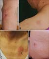
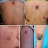
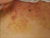
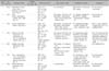
Seven patients were finally included in this analysis. The clinical data at diagnosis of the seven patients are summarized in Table 1. The median age of the patients was 52 years (range, 18~79 years), and six patients were male. At the initial presentation, five patients had skin manifestations. The remaining two patients had no skin lesions (patients 2 and 6) at baseline; however, they showed skin lesions after 4 and 12 months, respectively. Most of the skin lesions appeared as an erythematous nodule (Fig. 1), and/or brownish to bluish infiltrated bruise-like patch/plaque or tumor (Fig. 2). On the other hand, only one patient (patient 6) presented multiple erythematous firm papules (Fig. 3).
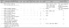
Complete staging investigations were applied to all of the patients. LN and BM involvements of BPDCN were found in six and four patients, respectively. PB involvement was found in two patients. Especially, patient 3 showed fever, general weakness, lymphadenopathy, hepatosplenomegaly, leukocytosis, and anemia. Furthermore, his BM evaluation revealed blast cells (up to 70%). These findings were consistent with features of acute leukemia. Conventional cytogenetic studies were performed in all patients. Three patients showed chromosomal abnormalities, including translocation, deletion, derivative chromosomes, and gain or loss of chromosomes in patients 3, 4, and 5, whereas the remaining four patients showed a normal karyotype.
Pathologic findings
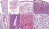
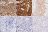
For pathological analysis and immunophenotyping, all patients underwent skin biopsies. The results of the skin biopsies are detailed in Table 2. As mentioned above, two patients (patients 2 and 6) had no skin lesion at the initial presentation. However, they showed skin involvement during the follow-up period, and underwent skin biopsies for the evaluation of disease progression. All of the biopsies from different patients, anatomic sites, and time points demonstrated similar histopathological features: diffuse or nodular dense neoplastic infiltration in the dermis, occasionally extending into the subcutaneous tissue, but sparing the epidermis with a Grenz zone (Fig. 4A~C). Some specimens (from patients 6 and 7) showed conspicuous extravasated red blood cells (RBCs) (Fig. 4D). Most of the specimens showed adnexal sparing (Fig. 4E); however, only one specimen (from patient 6) showed infiltrating tumor cells distributed along the adnexa (Fig. 4F). Notably, one specimen (from patient 5) revealed remarkable fibrotic changes in the dermis surrounded by tumor cells (Fig. 4G). At high magnification, there were monotonous populations of medium-sized cells containing a vesicular nucleus with irregular contour, fine chromatin, and indistinct nucleoli (Fig. 4H). Occasionally, they showed mitotic figures. The immunophenotyping revealed that the neoplastic cells were positive for CD4 (Fig. 5A), CD56 (Fig. 5B), and CD123 (Fig. 5C), but negative for CD3 (Fig. 5D), CD20 (Fig. 5E), and MPO (Fig. 5F). TdT staining, performed in three patients, was positive in patients 2 and 3 but negative in patient 1.
Treatment and outcomes
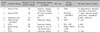
The treatment and outcomes of the seven patients are detailed in Table 3. Six patients received multiagent chemotherapy (CTx) as the first-line treatment. The CTx regimens are detailed in Table 3. After the first-line treatment, complete remission (CR) was achieved in four patients (patients 1, 2, 5, and 7); however, all of them showed relapse after 3~12 months. Most (patients 1, 2, 4, 5, and 7) of these patients showed cutaneous lesion as a sign of recurrence. Two (patients 1 and 2) of these patients received salvage CTx or autologous peripheral blood stem cell transplantation (PBSCT); nevertheless, they died because of disease progression and complications. Another one patient (patient 7) showed relapse only in the skin; however, the skin lesion disappeared after skin biopsy. This patient then received only close follow-up, and there has been no disease progression thus far. Patient 5 received palliative radiotherapy (RTx) with CTx, and is being followed to date. Patient 4, who received allogeneic PBSCT after partial remission, showed graft versus host disease (GVHD). Despite the GVHD, this patient maintained a stable disease state for 8 months. However, he had relapse and received salvage CTx. Patient 6 received consolidation RTx on the primary lesion (breast mass) and showed good response. However, after 2 months of RTx, the breast mass grew in size and was confirmed as recurrence on biopsy. Since then, she has received multiagent CTx; however, she showed involvement of the central nervous system and skin. Finally, she achieved a stable disease state and is being followed to date. PBSCT was considered as the primary therapy for her treatment. On the other hand, patient 3 refused any curative treatment at that time of diagnosis, and received only supportive care. He died after 6 months. The median progression-free survival (PFS) of the patients was 6 months (range, 2~12 months), and the OS was 17 months (range, 5~25 months). In contrast to those without skin lesion at diagnosis, patients with cutaneous involvement at the initial presentation had relatively long PFS. The median PFS was 8 months for those with skin involvement, and 3 months for those without skin involvement. However, the median OS was 15 and 18.5 months, respectively.
DISCUSSION
In this analysis, we describe the clinical and pathological features, and treatment and outcomes of seven cases of BPDCN in the Korean population. Clinically, their presentations were similar to those in previous reports. First, a male predominance was observed. Although a few studies did not show an increased prevalence in males1416, it is a universal finding that BPDCN affects male patients more, and the male-to-female ratio ranged from 2 to 7.2510111213. Secondly, although the mean age of our patients was slightly younger than in previous reports71013, they were middle-aged or elderly, except for one teenage patient. Third, all of our patients showed cutaneous involvement. Although two patients had no skin involvement at the initial diagnosis, they presented cutaneous lesions during the follow-up period. As a rule, BPDCN has a predilection to the skin; about 70%~85% of BPDCN cases had shown skin manifestations as the initial presentation in previous studies71417. Finally, our study revealed that the most common skin manifestation was bruise-like tumefaction or an erythematous nodule. These findings are similar to those of other previous studies1013. Notably, patient 6 in the present report had a breast mass as the initial presentation. It was similar to the case that Borchiellini et al.18 reported for the first time in 2013.
Pathological evaluation and immunophenotyping play an important role in the diagnosis of BPDCN1418. In cutaneous lesions, BPDCN characteristically infiltrates the dermis but spares the epidermis, and has a Grenz zone. As the disease progresses, it frequently extends into the subcutaneous layer but spares the adnexal structures1. In addition, the infiltration has a dense diffuse or nodular pattern. At high magnification, the tumor cells are characterized by a monomophic population of small to mediumsized cells with irregular nuclear contours, fine to evenly dispersed chromatin, various-sized or indistinct nucleoli, and scant to moderate amounts of cytoplasm without granules4. Characteristic extravasated RBCs responsible for bruise-like clinical manifestations are frequent, and mitoses are occasionally observed1. In our cases, all of the biopsies revealed typical features of BPDCN; however, the patients 5 and 6 additionally showed extraordinary findings such as infiltration along the adnexa or fibrotic changes in collagen.
On immunohistochemistry, BPDCN cells typically express a positive reaction to CD4, CD56, and CD123. Of note, staining for CD123 is typically strong, whereas that for CD4 and CD56 can be weak in some cases1920. The assessment of newer pDC-associated antigens, such as T-cell leukemia/lymphoma 1 (TCL1) and B-cell leukemia/lymphoma 11A (BCL11A) on immunohistochemical studies and blood dendritic cell antigen (BDCA)-2, BDCA-3, and BDCA-14 on flow cytometric analysis are potentially diagnostic for BPDCN1421. In contrast, tumor cells are usually negative for lineage-specific antigens of T-cells (CD3 and CD5), B-cells (CD19, CD20, and CD79), myeloid cells (MPO, CD13, and CD117), and NK cells (CD16 and CD57). However, a few markers of T-cells (CD2 and CD7) and myeloid cells (CD33) have been found frequently in tumor cells of BPDCN418. Moreover, CD68, a marker typically expressed by granulocytes and histiocytes, as well as by normal pDCs, is expressed in 50% of BPDCN cases4. TdT, a key antigen for precursor lymphoid cells is positive in one-third to one-half of cases4. Finally, BPDCN is negative for Epstein-Barr virus, unlike most true NK cell malignancies1. In the present report, immunophenotyping revealed that CD4, CD56, and CD123 were positive in all patients. These results were consistent with BPDCN, and there was no exceptional case.
Previous studies have described some karyotypic abnormalities. The representative chromosomal abnormalities are 5q21 or 5q34 (72%), 12p13 (64%), 13q13-21 (64%), 6q23 (50%), 15q (43%), and complete deletions of chromosome 9 (28%)22. In addition, the gene expression profile of BPDCN showed recurrent deletions of 4q34, 9p13-p11, 9q12-q34, and 13q12-q31, which contain tumor suppressor genes (Rb1 and LATS2) with diminished expression, as well as elevated expression of the oncogenes HES6, RUNX2, and FLT323. A recent gene expression study showed loss of the cell-cycle genes CDKN1B, CDKN2A, and TP5324. Among our cases, patient 4 had a deletion of 13q12-q22 included in the characteristic gene expression of BPDCN, as mentioned above. In contrast, the other patients did not show any major chromosomal abnormalities.
The clinical course of BPDCN is aggressive with a median survival of 12~14 months, regardless of the initial presentation4. However, owing to its rarity and only recent recognition as a distinct entity, there is no standardized therapeutic strategy for BPDCN. Most patients are treated with a variety of intensive combination CTx regimens, such as CHOP (cyclophosphamide, doxorubicin, vincristine, and prednisone) or hyper-CVAD (combination of course A [cyclophosphamide, vincristine, doxorubicin, dexamethasone] and course B [methotrexate and cytarabine], in an alternating fashion). Patients usually achieve resolution of the initial symptoms with the initial CTx1.
However, the disease often relapses, and the relapsed disease is generally resistant to the CTx agents previously used4. Roos-Weil et al.25 demonstrated that high-dose CTx followed by allogeneic stem cell transplantation (SCT) from related and unrelated donors could provide durable disease control in up to 50% of patients.
In contrast to that in adults, BPDCN in pediatric patients is clinically less aggressive, and show good response to acute lymphoblastic leukemia-type CTx14. Other authors suggested that the best long-term prognosis for relapse-free survival is expected in pediatric patients who ultimately underwent allogeneic BM transplantation after a CTx-induced remission2627. Furthermore, the outcomes were more favorable in cases that lacked cutaneous lesions at presentation14. However, the prognostic significance of cutaneous involvement is debatable in adult patients47102829.
In our cases, the median PFS revealed favorable outcomes in patients with cutaneous involvement; however, the median OS showed the opposite result. However, most of the patients are still alive until now, and these interpretations might be limited. Notably, patient 6, who lacked skin involvement at the initial presentation, showed a relatively long survival. In addition, patient 1, who had skin involvement exclusively, showed a longer survival than patient 2, who had blastic involvement, LN, and BM disease. Furthermore, patients 1, 2, 5, and 7 had achieved CR after the initial CTx. However, they showed relapse 3~12 months later. Especially, patient 2, who had no skin involvement at the initial presentation, showed early relapse and rapid progression despite receiving salvage CTx and autologous PBSCT.
Our study has some limitations. First, only a few patients were enrolled and analyzed. Owing to the small number of patients, it was difficult to carry out statistical analysis and a comparative study. Secondly, because of the retrospective design, the evaluation or follow-up of patients was not equivalent to each other. In addition, recently introduced diagnostic markers such as TCL1, BCL11A, BDCA-2, BDCA-4, and BDCA-14 were not examined. Especially, TdT staining was performed in only three patients. In fact, it was suggested that a high TdT expression (>50%) is associated with a favorable prognosis2930. However, we could not investigate the prognosis with only the three patients. Finally, four patients were still alive at the time of the last follow-up; therefore, careful attention is needed in interpreting the OS.
In conclusion, this report of seven cases of BPDCN in Korea, despite the small number of patients, might provide information about the clinical and histopathological characteristics of this neoplasm. As is well known, early diagnosis and appropriate treatment of BPDCN is crucial to improve the prognosis. Accordingly, both dermatologists and hemato-oncologists should be aware of this rare entity when confronted with unfamiliar skin lesions and hematological abnormalities. Notably, most of the patients in the present study showed skin manifestations as a sign of relapse. Therefore, it is important to carefully check the skin of the patients after treatment. In addition, in the treatment outcome, it was remarkable that all of the patients showed a high frequency of relapse after multiagent CTx without SCT, and underwent various courses regardless of the cutaneous involvement at the initial presentation. Further studies are needed to recognize the prognostic factors and establish the optimal treatment. Finally, we expect that this study would contribute to future studies such as meta-analyses, by introducing additional Asian cases.
 XML Download