

 PDF
PDF ePub
ePub Citation
 Print
Print


INTRODUCTION
In ancient times, when therapeutic options were much more limited than they are today, the use of systemic and external spa waters for the treatment of different physiological conditions was very popular. Even now, countless health resorts with specific spa waters continue this tradition. In dermatology, balneotherapy is mainly used for the treatment of psoriasis and atopic dermatitis. In particular, the water from the Dead Sea in combination with UV light is reported to alleviate the symptoms of psoriasis1. Spa waters from other sources are also reported to be useful in the treatment of inflammatory skin diseases23456. However, it is difficult to attribute the measured effects to specific parameters. There are data suggesting that the chemical and thermal properties of spa water have an impact on skin cell physiology2. Moreover, the mechanical effects of application (immersion, hydromassage, high-pressure showers) trigger physiological changes in the renal and cardiovascular systems with potentially beneficial effects on skin diseases7. Finally, the relaxing environment of a health spa can also aid in general health improvement with impacts on the skin.
The use of cell cultures is useful for determining the effects of spa water more precisely. The present study was aimed at testing two popular thermal spa waters from La Roche-Posay (LRP; L'Oréal, Clichy, France) and Avéne (ASW; Pierre Fabre, Paris, France) in direct comparison in a keratinocyte model (HaCaT). For this study, HaCaT cells were chosen, as they have been shown to respond similar to primary skin keratinocytes in previous assays89, but as a cell line, they lack interindividual variability. In this regard, proliferation, cytotoxicity, interleukin-6 (IL-6) expression, and formation of reactive oxygen species (ROS) after stimulation with ultraviolet B (UVB) were tested. Besides regular culture conditions, using double-distilled mineral-free water as a control, two natural mineral drinking waters (Adelholzener [St. Primus Heilwasser, Bad Adelholzen, Germany], Heppinger [Apollinaris, Bad Neuenahr-Ahrweiler, Germany]) were also included in the test.
MATERIALS AND METHODS
Cell culture
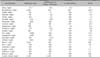
The spontaneously immortalized human keratinocyte cell line (HaCaT) (a generous gift from Norbert Fusenig, German Cancer Research Institute, Heidelberg, Germany) was cultured in carbonate-buffered Hank's medium with 5% fetal calf serum and 1% penicillin/streptomycin solution (Biochrom KG, Berlin, Germany) at 37℃ in a 5% CO2 atmosphere. Medium components in powdered form were solubilized using sterile double-distilled water (control) or spa water, for a concentration of 72% in the final medium. For spa water supplementation, (a) two thermal spring waters from LRP (L'Oréal) and ASW (Pierre Fabre) and (b) two natural mineral waters distributed as drinking waters from Heppinger and Adelholzener were used. The composition of the spa waters is given in Table 1. All experiments were performed in agreement with the local ethics commission.
DNA synthesis
HaCaT cells were cultivated in microwell plates at a density of 2×10⁴ cells/0.33 cm2. Cells were exposed for 24 h to spa water-supplemented medium. For the last 16 h, cells were pulsed with 5-bromo-2'-deoxyuridine (BrdU). Subsequently, the incorporation rate of BrdU was determined using a commercial enzyme-linked immunosorbent assay (ELISA) kit (Roche, Mannheim, Germany). Briefly, cells were fixed and immune complexes were formed using peroxidase-coupled BrdU-antibodies. A colorimetric reaction with tetramethylbenzidine (TMB) as a substrate gave rise to a reaction product measured at 450 nm in a scanning multiwell spectrophotometer (ELISA reader, MR 5000; Dynatech, Guernsey, UK).
Membrane integrity
Cell lysis was quantified using the cytotoxicity detection kit (Roche), which is based on the release of lactate dehydrogenase (LDH) from damaged cells. Briefly, HaCaT cells were seeded in microwell plates as described above and were treated with spa water-supplemented medium for 24 h. As a positive control (maximal cell damage), cells were treated with 1% Triton X-100 (Merck, Darmstadt, Germany). Subsequently, the cell-free supernatants were incubated with NAD+, which becomes reduced by LDH to NADH/H+. In a second step, NADH/H+ reduces a yellow tetrazolium salt to a red-colored formazan salt. The amount of red color is proportional to the number of lysed cells. For quantitation, the absorbance of the reaction product was measured at 490 nm using a multiwell spectrophotometer.
Enzyme-linked immunosorbent assay of IL-6
After preincubation for 1 h in the presence or absence of spa water-supplemented medium, HaCaT cells in phosphate-buffered saline, also reconstituted with the spa waters or double-distilled water, were irradiated with 150 mJ/cm2 UVB using a Psorilux-UVB-lamp (Heraeus, Hanau, Germany). Thereafter, cells were incubated with or without spa water-supplemented medium for 24 h. Then, cell-free supernatants were obtained and assayed for human IL-6 using commercial ELISA test kits (R&D Systems, Wiesbaden, Germany). Betamethasone-17-valerate (10 µg/ml) served as a positive control; dimethyl sulfoxide (DMSO) served as a solvent control. Briefly, supernatants were placed in microwell plates coated with antibodies against IL-6. After incubation with a horseradish-peroxidase conjugate, TMB (Sigma-Aldrich, Steinheim, Germany) was added, giving rise to a colored product measured at 450 nm in a scanning multiwell spectrophotometer (ELISA reader MR 5000).
IL-6 promoter transactivation assay
The human IL-6 promoter construct spanning 1,168 bp linked to a luciferase reporter gene in pGL3 basic (Promega, Mannheim, Germany) was a kind gift from Michèle Resche-Rignon10. Constructs were transfected into subconfluent HaCaT cells by lipofection (Lipofectamine reagent 2000; Invitrogen, Darmstadt, Germany). In order to standardize transfection efficacy, cells were co-transfected with a humanized Renilla luciferase vector (phRL; Promega). Transfected cells were treated with spa water-supplemented medium, betamethasone-17-valerate as positive control, or DMSO as a solvent control for 24 h. After irradiation with 150 mJ/cm2 UVB, cells were propagated for an additional 24 h in the above-mentioned media. Then, cells were lysed and activities of both luciferases were detected separately, using the Dual-Luciferase Reporter Assay System (Promega) and a luminometer (Berthold, Bad Wildbad, Germany).
Measurement of reactive oxygen species
HaCaT cells were incubated with spa water-supplemented medium in the presence of 100 µM 1,2,3-dihydrorhodamine (DHR 123; Sigma-Aldrich) or with 100 or 200 µM vitamin C, a known radical scavenger, as a positive control for 1 h at 37℃. Subsequently, the medium was substituted with PBS and cells were irradiated with 400 mJ/cm2 UVB. After incubation for 45 min, the fluorescence was quantitatively detected using a multiwell spectrofluorometer (Cytofluor; Applied Biosystems, Langen, Germany) equipped with 485 nm excitation and 560 nm emission filters.
Presentation of data and statistical analysis
All data are presented as mean values±standard deviations. Statistical significance in the data was evaluated by t-test (BIAS, Frankfurt, Germany). Each set of data relates to the untreated (asterisk) or irradiated (hash) control as indicated. Differences were considered significant at a level of p<0.05, represented by one icon, p<0.01, represented by two icons, and p<0.001, represented by three icons.
RESULTS AND DISCUSSION
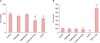
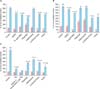
A general problem of studies dealing with spa waters is that most of these studies are initiated by the spa water manufacturers themselves. Independent scientific investigations are rare and necessary to providing a more solid basis to evaluate spa water-mediated effects. Here, we show that basic parameters such as proliferation and cytotoxicity were significantly decreased by LRP and ASW (Fig. 1). Moreover, both thermal waters reduced IL-6 levels in the medium after UVB irradiation to levels similar to that seen with betamethasone-17-valerate treatment (Fig. 2A). Interestingly, the drinking water from Heppinger also induced strong downregulation of IL-6. The IL-6 levels measured from non-irradiated cells were all at the detection limit of the test, making reliable statements infeasible. In the transactivation assay, both thermal waters showed a clear inhibitory effect on IL-6 after UVB stimulation, suggesting regulation at the promoter level (Fig. 2B). Finally, the formation of ROS after UVB was attenuated by LRP and ASW. Of note, both drinking waters from Heppinger and Adelholzener also showed distinct ROS suppressions (Fig. 2C).
Particularly in the last few decades, the French cosmetic industry has marketed thermal spa waters as cosmeceuticals, requiring some effort to prove cellular effects. The regulation of immunomodulatory parameters by spa water-supplemented media was observed in mast cells (ASW)11, Langerhans cells (LRP)12 and CD4+ T lymphocytes (Yong-gung oncheon, ASW)1314. In the latter, a partial shift from a Th2 to a Th1 cytokine profile was observed, offering a rationale for the treatment of atopic dermatitis14. Preliminary studies using cultured fibroblasts suggest enhanced plasma membrane fluidity by ASW15. Moreover, differentiation of skin keratinocytes as measured by the expression of involucrin and cytokeratins 1 and 10 was induced by ASW16.
Our results show that, although the thermal waters LRP and ASW differed in composition, they both efficiently suppressed the induction of a prototypical inflammatory cytokine and the formation of ROS after UVB. It seems likely that, in the case of LRP, the observed effects are mediated by the high selenium content (53 µg/L), which is a co-factor for glutathione peroxidase, a key-enzyme in the elimination of ROS17. Interestingly, ASW, which is almost free of selenium (<4 µg/L) showed anti-inflammatory properties. Here, the relatively high zinc content (20 µg/L) seems responsible for anti-inflammation18. Unfortunately, the composition list for the two mineral waters, as provided by the manufacturers, is incomplete. However, it could be speculated that boron, a trace element shown to act on keratinocytes19, is involved in the observed effects. Further studies should address these hypotheses.
In summary, our results give a scientific rationale for the application of spa waters in the treatment of chronic inflammatory skin diseases.
 XML Download