

 PDF
PDF ePub
ePub Citation
 Print
Print


INTRODUCTION
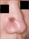
Nodular fasciitis (NF) is a benign proliferative myofibroblastic lesion that may regress spontaneously1. Although the etiology of NF is uncertain, histopathologically, it bears a close resemblance to organizing granulation tissue, and myofibroblastic proliferation may be initiated by a local injury or local inflammatory process, which supports a reactive proliferation theory triggered by trauma23. NF is frequently mistaken for sarcoma both clinically and histopathologically. Therefore, surgical excision is recommended for diagnosis and treatment to exclude malignancy4. However, for lesions occurring on the face, nonsurgical treatment and regular follow-up can be considered because surgical treatment may cause scarring on the face and the lesions may regress spontaneously.
For the nonsurgical treatment of NF on the face, triamcinolone intralesional injection (TA ILI) can be used on the basis of the fact that NF is a benign reactive or inflammatory condition of mesenchymal fibroblasts5. In addition, considering that early lesions of NF have a myxoid pattern of myofibroblast proliferation6, laser treatment can be effective in decreasing the size of the lesions through tissue contraction. Especially, NF on the face are usually 'intradermal type,' which extends beyond the fascia and subcutaneous tissue and invades into the skin, as facial muscles such as platysma, frontalis, and orbicularis oculi are located superficially underneath the skin7. Therefore, such intradermal type of NF can be affected by laser treatment from the skin surface. For example, multiple pinholes created by a carbon dioxide (CO2) laser on the skin surface can cause tissue contraction, resulting in size reduction and dermal collagen remodeling89.
In this study, the authors retrospectively reviewed cases of NF occurring on the face that were treated by using surgical and nonsurgical methods such as TA ILI and a pinhole method with a CO2 laser, and evaluated the outcomes of treatment.
MATERIALS AND METHODS
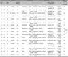
We performed a retrospective review of 16 patients (6 males, 10 females) with NF on the face clinically and histopathologically diagnosed from December 2010 to December 2013 at Severance Hospital in the Yonsei University Health System. Each patient's details from the medical record, including age, sex, onset location, trauma history, clinical photographs, histopathologic findings, treatment methods, clinical progress, and complication profile, were fully reviewed. The study protocol was approved by the institutional review board of Yonsei University Severance Hospital (IRB No. 4-2015-0928) and was conducted according to the Declaration of Helsinki Principles.
Histopathologic findings
To evaluate the possibility of nonsurgical treatments, we reexamined the histopathologic features. For this review, two dermatologists individually reexamined the H&E-stained sections, and evaluated the following features in a blind manner: depth of the lesion, type of cellular composition, and the presence of cellular atypia. Additionally, the presence of a loose myxoid pattern, which can provide a potential space for injectable agents, was also evaluated. If there were atypical cells in the H&E stained section, immunohistochemical staining for smooth muscle actin, desmin, pan-cytokeratin, CD34, S100 protein, and other markers, was performed to exclude malignancy.
Nonsurgical treatments
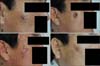
The nonsurgical approach was indicated when the diagnosis of NF is confirmed and the potential for malignancy is excluded with immunohistochemistry in the presence of atypical spindle cells. When recurrence occurred after the surgical treatment, TA ILI was used after the reevaluation of the excised specimen. The pinhole method with CO2 laser and TA ILI were repeatedly performed simultaneously every 4 weeks. Before the treatment, a topical eutectic mixture of 2.5% lidocaine and 2.5% prilocaine (EMLA cream; AstraZeneca AB, Södertälje, Sweden) was applied for local anesthesia. Treatment was performed with an output power of 1 to 4 W to make multiple small holes approximately 1 to 2 mm in diameter and spaced 2 to 5 mm apart (Fig. 1). Posttreatment pain was tolerable, and erythema spontaneously resolved. Immediately after the pinhole treatment, TA ILI was applied to the deep and firm part of the NF lesion.
Evaluation of the clinical outcomes
For the evaluation of the clinical outcomes, all photographic images of NF patients taken 3 months after the final treatment were individually evaluated by two dermatologists by using a visual analogue scale (VAS). The VAS is a 10-cm linear scale in which the left of the scale (score of 0) reflects the worst outcome and the right of the scale (score of 10) reflects the best outcome. At the end of the treatment, patients' satisfaction was evaluated on a 4-point grading scale (0=unsatisfied, 1=slightly satisfied, 2=satisfied, and 3=very satisfied).
Statistical analysis
To compare the outcomes of treatments between the surgical and nonsurgical methods, we performed statistical analysis by using the nonparametric Mann-Whitney U test, where p-values ≤0.05 were considered statistically significant. All data were analyzed by using SPSS 12.0 (SPSS Inc., Chicago, IL, USA).
RESULTS
The mean age of the 16 patients who had NF on the face was 36.6 years (range, 15~61 years). The average disease duration was 3.94 months (range, 3 weeks~12 months). In nine cases (56.3%), patients had histories of physical stimulation or trauma, such as facial massage, at least once before the development of NF lesions (Table 1).
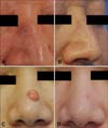
Histopathologic examination showed that NF consisted mainly of epithelioid and spindle cells. A loose myxoid structure was observed in all cases (Fig. 2). In five cases, a few atypical cells were noted in the H&E stain; however, they were all negative for desmin, pan-cytokeratin, CD34, S100 protein, or MART-1, and positive for smooth muscle actin in immunohistochemical stains. All lesions involved subcutaneous tissues, whereas seven cases (43.8%) showed the involvement of muscle layers, and in 10 cases (62.5%) dermal involvement was observed. Among the 16 cases, surgical excision was performed in 9 and recurrence developed in 7 of these 9 cases (77.8%), which were all histopathologically confirmed as NF from the excised specimens. However, the recurred lesions regressed after repetitive TA ILI (Fig. 3). In case no. 2, Mohs micrographic surgery was performed for the complete removal of the lesion because of some atypical cells with mitotic figures in the coin-sized lesion and the relatively older age of the patient (Fig. 4). The pinhole method with a CO2 laser and TA ILI, two to three times every 4 weeks, resulted in regression of the five cases of intradermal-type NF (Fig. 5). One of the two patients who did not receive any treatment showed spontaneous regression after 1 month (Table 2).
The final outcomes were evaluated on the basis of the VAS. The mean score of cases no. 12 to 16 who received pinhole method treatments (6.90±1.56) was slightly higher than that of cases no. 1 to 9 who received surgical excision (5.61±1.36); however, the difference was not statistically significant (p=0.163). Concerning patients' satisfaction, the group that received pinhole treatments scored 1 to 2; however, the group with recurrence after the surgical excision scored relatively lower (0~1) (Table 2). Neither the surgically nor the nonsurgically treated group reported adverse events such as severe pain or secondary infection.
DISCUSSION
Because malignant transformation seldom occurs in benign skin lesions, treatment is not always a requirement as long as patients do not mind the cosmetic problems resulting from the skin lesions. In cases of NF on the face, proactive efforts are needed to perform histopathologic evaluation and treatment because NF tends to grow rapidly on the face of adults between the age of 20 and 40 years (i.e., persons who are involved in many social activities)10.
Although wide excision is recommended for the treatment of NF on the extremities or trunk, it should be reconsidered in facial lesions as it presents several problems including scarring. First, surgical excision may not remove enough of the area surrounding the lesion because it is difficult to clearly determine the scope of the lesion because inflammation extends to the adjacent muscles and subcutaneous tissues. Particularly, the risk of insufficient excision can be higher in the face because surgeons try to reduce scarring as much as possible. Second, surgical procedures may act as another precipitating factor as NF is a myofibroblast proliferation that can be initiated by local injury. In this retrospective study, seven of nine patients (77.8%) who received surgical excision experienced local recurrence, which was a higher rate than those reported for other areas of the body1011. We assume that this result may be due to the difficulty in achieving complete surgical excision in facial lesions or the increased trauma caused by surgery. Therefore, when only considering scarring and the higher recurrence rate, surgical wide excision is not recommended for the first treatment of NF on the face.
Five patients who underwent nonsurgical treatments, such as a repeated pinhole treatments and TA ILI, in this study showed lesion regression and satisfaction. The prerequisite for nonsurgical treatment is a histopathologic confirmation to rule out sarcoma. It was reported that fibrosarcoma typically occurs in patients older than 50 years and in lesions >3 cm12. Although marked cellular atypia is rare in NF lesions, cells of fibrosarcoma show more nuclear pleomorphism with dense collagenous stroma and without inflammatory components. In immunohistochemical staining, lesional cells of NF can be reactive to vimentin, smooth muscle actin, muscle-specific actin, and KP-111. For the differential diagnosis, desmin, which is rarely expressed in NF, may be used to distinguish NF from leiomyoma and leiomyosarcoma13. In addition, it is possible to use CD34 to differentiate NF from dermatofibrosarcoma protuberans. However, the Ki67 index, which has been documented as useful for the diagnosis and prognosis of soft tissue sarcoma14, is not suitable for the assessment of NF because of the variation in NF lesions12.
Recently, along with the vast development of laser devices, various nonsurgical approaches have been applied in the treatment of benign skin tumors. For dermatofibroma (benign fibrous histiocytoma), one of the most common benign soft tissue tumors, a pulsed dye laser15, fractional laser with topical corticosteroid16, or traditional cryotherapy17 have been applied. As the cells of newly developed NF are embedded in a loose myxoid stroma6, intralesional delivery of medication can be done successfully. In our study, loose myxoid patterns were found in all cases, and TA ILI treatment of the lesions resulted in regression. However, the effects of intralesional injection may decrease for old lesions with increased cellular, fibrous components and decreased loose parts, because intralesional injection into fibrous and hard lesions is difficult to perform.
From our results, a pinhole method with a CO2 laser was an effective treatment option for NF on the face, especially those on anatomic locations that may involve significant aesthetic concern. CO2 laser treatment may induce tissue shrinkage and decrease in the size of NF lesion. The pinhole laser treatment has been reported to be an effective method in scar management by inducing tissue shrinkage and scar remodeling1819. When it is applied to myxoid lesions, its effects may become intensified because the CO2 laser wavelength is strongly absorbed by water. In addition, when the pinhole technique is combined with TA ILI, inhibition of fibroblast proliferation would also be achieved. As the scarring left by this procedure is much smaller than the size of the original lesion, scar revision can easily be performed, if necessary, after nonsurgical treatments. In this retrospective study, the superior cosmetic outcomes of the pinhole method resulted in a relatively higher patient satisfaction.
Nine of the 16 patients in this study (56.3%) had a history of receiving physical stimulation or trauma such as facial massages. Although the relation between NF occurrence and trauma has not been clearly defined, the possible occurrence of NF on the face should be closely monitored as persons become more interested in skin care that may give physical stimuli to skin.
Despite the limitation of being a retrospective study with a relatively small sample size, we experienced successful outcomes with a nonsurgical approach to NF on the face. However, potential malignancy should not be overlooked, and before any nonsurgical approach, histologic confirmation with a sufficiently deep skin biopsy including skin and facial muscle parts should be done first. For lesions showing recurrence or regrowth with different clinical features from the original lesion, additional skin biopsy would be required for further histologic analysis. In conclusion, considering the potential aesthetic disfigurement and recurrence after conventional wide excision, repetitive TA ILI and CO2 pinhole laser can be recommended as a less invasive treatment option.


 XML Download