

 PDF
PDF ePub
ePub Citation
 Print
Print


Dear Editor:
Acute pigmentation due to tanning is commonly understood as increased melanization of the epidermis observed in the skin after ultraviolet (UV) exposure, and the mechanisms underlying this condition are well understood now. Keratinocyte-derived gene products are upregulated by UV irradiation and act as paracrine factors in the skin to stimulate melanogenesis and melanin transfer by melanocytes1. Although acute pigmentation disappears over time, some types of hyperpigmentary disorders such as freckles, solar lentigines, and melasma, tend to persist if patients do not receive any treatments such as topical cosmetic products, medication, or laser.
Solar lentigines are dark brown spots that occur on sun-exposed areas2, typically on the face, upper back, and shoulders. Multiple solar lentigines are considered a hallmark of aged skin. It is thought that cumulative UV exposure causes these spots. Therefore, pigmented spots of solar lentigines can be considered as indications of photoaging. Melasma is a common acquired symmetrical hypermelanosis on sun-exposed areas of the skin and is very common among Oriental women3. The major etiological factors include genetic influences, exposure to UV radiation, and sex hormones. However, the mechanisms underlying the persistence of hyperpigmentation in solar lentigines and melasma are not yet fully understood.
Keratinocyte growth factor (KGF) or fibroblast growth factor-7 (FGF-7) is a member of the FGF family4. KGF is secreted from cultured stromal fibroblasts derived from the skin and gastrointestinal tract, and is expressed in vivo in dermal cells, but not in epidermal cells5. In addition, this paracrine growth factor also plays a role in the stimulation of melanogenesis56, proliferation of human melanoblasts, and differentiation of melanocytes7. A previous study reported higher levels of KGF in five patients with solar lentigines, suggesting the permeation of KGF from the dermis to the epidermis, which may result in the persistence of solar lentigines6. In this study, we quantitatively investigated the accumulation of KGF in the epidermis of patients with two major types of hyperpigmentary disorders, facial solar lentigines, and melasma to identify novel effective topical measures for their treatment.
We examined 24 Korean women with newly diagnosed facial solar lentigines and 13 others with newly diagnosed melasma, which were determined on physical examination and histological examination. This study was approved by the ethics committee of Ajou University Hospital (No. MED-KSP-12-171). Punch biopsies from lesions and perilesional normal skin were obtained from each patient. The perilesional normal skin was taken from the area within 1 cm away from the lesional border. Twenty-four pairs of facial solar lentigines and 13 pairs of melasma tissue were prepared for immunohistopathological examination.
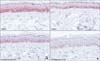
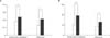
We examined KGF protein accumulation in the epidermis of both facial solar lentigines and melasma. Paraffin-embedded sections of both lesional and perilesional normal skin were processed with monoclonal antibodies against KGF (dilution, 1:100; Abcam, Boston, MA, USA) for 20 min at 48℃. The stained area per epidermal area (SA/EA) and SA per single rate ridge length (SA/1R) of lesional and perilesional skin were measured. In both facial solar lentigines and melasma lesions, distinctly positive immunoreactivity against KGF was noticed in the epidermis, whereas perilesional normal skin only showed weak immunoreactivity (Fig. 1). The SA/EA of perilesional normal skin samples was 0.184±0.139 for facial solar lentigines and 0.134±0.071 for melasma, and that of lesional skin samples was 0.237±0.107 for facial solar lentigines and 0.210±0.084 for melasma (Fig. 2A). The differences were statistically significant (p=0.014 for facial solar lentigines and p=0.016 for melasma). The SA/1R of perilesional normal skin samples was 10.216±7.194 for facial solar lentigines and 8.699±4.923 for melasma, and that of lesional skin samples was 19.350±8.744 for facial solar lentigines and 13.172±4.709 for melasma (Fig. 2B). The differences were statistically significant (p=0.000035 for facial solar lentigines and p=0.014 for melasma).
In the present study, we demonstrated that KGF protein accumulation in the epidermis of facial solar lentigines and melasma was significantly increased. We performed the following two analytical techniques: first, the SA for KGF was normalized to the EA; second, the SA for KGF was normalized with 1R, which is independent from epidermal thickness. In both analyses, we found a statistical difference between perilesional normal and lesional skin of both facial solar lentigines and melasma. In this study, we did not take the duration and stage of solar lentigines and melasma into consideration because of the small number of patients, although they could have affected the results. We expect that a study with a larger number of patients in varied stages and duration of solar lentigines and melasma will demonstrate the correlation between KGF protein accumulation level and the duration or stage of solar lentigines and melasma.
Iriyama et al.8 revealed that the degradation of heparan sulfate at the dermal-epidermal junction in photoaged skin impaired the function of the basement membrane, which regulates the transfer of several growth factors between the epidermis and dermis. Moreover, they demonstrated that heparan sulfate at the dermal-epidermal junction was specifically reduced in solar lentigines on the human back, and that the degradation of heparan sulfate enhanced melanogenesis in a skin equivalent model. In addition, recent histological studies on melasma described changes in the basement membrane in the lesional skin of melasma910. The basement membrane structure in lesional skin was not intact and appeared disrupted. Thus, the loss of heparan sulfate at the dermal-epidermal junction of facial solar lentigines and the loosening of the basement membrane in melasma may enhance the transfer of dermis-derived factors such as KGF. Our present study strongly supports the hypothesis that in hyperpigmentary disorders, such as facial solar lentigines and melasma, the fibroblast-derived KGF is transferred into the epidermis in which it accumulates, thereby leading to the persistence of hyperpigmentation. Thus, targeting the dermal-derived KGF itself and the degradation of heparan sulfate at the dermal-epidermal junction, which allow an excess amount of KGF to transfer into the epidermis, could be effective topical measures like cosmetic or medication use for treating hyperpigmentary disorders.
 XML Download