

 PDF
PDF ePub
ePub Citation
 Print
Print


Dear Editor:
Pyodermatitis-pyostomatitis vegetans (PD-PSV) is a rare, chronic, inflammatory dermatosis characterized by mucocutaneous vesiculopustular eruptions1. The cutaneous lesions present as papulopustules that coalesce to form large vegetating plaques. The oral lesions manifest friable and yellowish pustules that form a "snail-track". A strong correlation of PD-PSV with inflammatory bowel disease (IBD) is well documented. Ulcerative colitis (UC) is more commonly associated with PD-PSV than Crohn's disease2. In this study, we describe a case of PD-PSV associated with Crohn's disease.
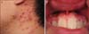
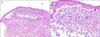
A 27-year-old male patient presented with a 2-month history of bullous eruption on the whole body. Physical examination revealed annular and polycyclic erythematous vesicopustules on the face, neck, trunk, and extremities (Fig. 1A). The gingiva was involved, exhibiting coalescing pustules with a characteristic "snail-track" shape (Fig. 1B). The patient also had a 2-year history of Crohn's disease that has been treated with mesalazine and azathioprine. Histology revealed epidermal hyperplasia and intraepidermal neutrophilic microabscess with some eosinophils and acantholytic keratinocytes (Fig. 2). The direct and indirect immunofluorescence tests were negative for immunoglobulin (Ig) G, IgA, and C3, and the routine laboratory findings were normal except for peripheral blood eosinophilia (1,040/µl). Analysis of the serum cytokine level revealed elevated tumor necrosis factor (TNF)-α (18.55 pg/ml), interleukin (IL)-8 (8.31 pg/ml), and IL-10 (100.10 pg/ml). However, the level of interferon (IFN)-γ, IL-17A, IL-4, and IL-6 were within normal limits. A diagnosis of PD-PSV was made, and the patient was treated with prednisolone (20 mg/day), dapsone, and colchicine. The skin and oral lesions were greatly improved within 2 weeks; however, a low dose of prednisolone and dapsone was required to control the disease.
PD-PSV is a rare inflammatory dermatosis of unknown cause. The association with IBD occurs in approximately 70% of cases2. Of the 61 cases of PD-PSV, 36 (59%) involved coexistent UC and 7 (11%) were associated with Crohn's disease. This suggests a much stronger association of PD-PSV with UC than with Crohn's disease.
In most cases, gastrointestinalsymptoms of IBD precede PD-PSV. However, cutaneous lesions may precede gastrointestinal symptoms in approximately 15% of patients3, indicating the need for the evaluation for IBD even if patients with PD-PSV have no gastrointestinal symptoms. Commonly, the clinical course of PD-PSV is parallel to that of IBD. Treatment of IBD with sulfasalazine or mesalazine or surgical treatment may lead to the resolution of PD-PSV. Also, the severity of coexisting IBD has an effect on the prognosis and treatment of PD-PSV.
PD-PSV should be differentiated from other blistering diseases presenting vegetating vesicopustular eruptions such as pemphigus vegetans, IgA pemphigus, subcorneal pustular dermatosis, dermatitis herpetiformis, and herpes simplex. The pustules with characteristic "snail-track" ulcers and the association with IBD distinguish PD-PSV from other blistering diseases. Moreover, intraepithelial and subepithelial splitting with neutrophilic and eosinophilic microabscess and negative immunofluorescence findings sug gest PD-PSV rather than other immunobullous disorders such as pemphigus.
The serum cytokine profile revealed enhanced TNF-α, IL-8, and IL-10. As far as we know, this is the first report of a serum cytokine analysis in PD-PSV. TNF-α is a prominent cytokine that is associated with most inflammatory skin diseases4. The high IL-8 concentrations imply neutrophil chemotaxis, as shown by the histological findings. IL-10 is an anti-inflammatory cytokine produced by Th2 cells and regulatory T cells. Thus, the elevation of IL-10 might be paradoxical; however, IL-10 is also a potent B-cell stimulator, and high serum levels of IL-10 and IL-8 were reported in patients with UC, suggesting the close relation between PD-PSV and UC5.
 XML Download