Journal List > Ann Dermatol > v.27(5) > 1046074

Kim, Kim, Lew, Lee, Hong, Jang, Park, Seo, Bae, Choi, Suhr, Lee, Ko, Park, Son, Seo, Lee, Cho, Park, and Roh: Consensus Guidelines for the Treatment of Atopic Dermatitis in Korea (Part I): General Management and Topical Treatment

Abstract

Background

Since the treatment guidelines for atopic dermatitis (AD) were released by the Korean Atopic Dermatitis Association (KADA) work group in 2006, there have been several advances in AD management.

Objective

We aimed to establish updated evidence- and experience-based treatment guidelines for Korean AD.

Methods

We collected a database of references from relevant systematic AD reviews and guidelines regarding general AD management such as bathing and skin care, avoidance of exacerbating factors, education and psychosocial support, and the use of moisturizers and topical anti-inflammatory and antipruritic drugs. Evidence for each statement was graded and the strength of the recommendation for each statement classified. Thirty-nine KADA council members participated in three rounds of voting to establish an expert consensus of recommendations.

Results

Basic AD treatment includes proper bathing and skin care, avoidance of exacerbating factors, proper education and psychosocial support, and use of moisturizers. The regular use of moisturizer has a steroid-sparing effect and reduces relapse episodes. The short- and long-term use of topical corticosteroids and calcineurin inhibitors improves AD symptoms and should be encouraged to use in an active and proactive treatment. Wet-wrap therapy can be used for rapid recovery of acute exacerbation. Topical antipruritic drugs cannot be recommended for the treatment of AD.

Conclusion

This report provides up-to-date evidence- and experience-based treatment guidelines for AD regarding general management and topical treatment. In addition, the average agreement scores obtained by a panel of experts based on the Korean healthcare system and patient adherence are presented.

INTRODUCTION

Atopic dermatitis (AD) is a chronic relapsing inflammatory skin disorder caused by an individual's combined genetic, environmental, and immunologic background1. Several therapeutic guidelines for AD treatment from work groups in various countries have been published2345. To provide individualized and long-term AD treatment, various factors must be considered, including age, provocative and socioeconomic factors, patient adherence, and psychological status. The health care system and cultural background of a country can also affect the treatment decisions of both physicians and patients.
The Korean Atopic Dermatitis Association (KADA) convened the Korean AD Treatment Guideline Task Force group to develop updated evidence-based and practical AD treatment guidelines based on the Korean health care system and patient adherence. These revised treatment guidelines provide up-to-date, evidence-based treatment recommendations and the average agreement scores of expert panel members are presented for each key statement.
Recommendations for AD treatment are divided into two parts: nonsystemic treatment, including AD skin management and topical treatment, and systemic treatment. This report is the first of two parts in a guidelines series and includes information on AD management, such as bathing and skin care, avoidance of exacerbating factors, education and psychosocial support, and the use of moisturizers and topical anti-inflammatory drugs, antibiotics, and antipruritic drugs. Clinical questions are focused on therapeutic effects, detailed plans of action, side effects, cost-effectiveness, and measures to enhance patient compliance for each treatment.

MATERIALS AND METHODS

In developing the Korean guidelines for managing AD, KADA convened a working group of 12 dermatologists representing AD experts nationwide. The panel followed the methodology for developing guidelines detailed in the 2011 Guidance for the Development of Clinical Practice Guidelines from the National Evidence-based Healthcare Collaborating Agency6.

Database and literature research

The 12 members of the working group each conducted separate comprehensive computerized database searches of MEDLINE (accessed by PubMed) and Embase from January 1, 2005, to December 31, 2014, using combinations of the search terms "atopic eczema", "atopic dermatitis", "bathing", "cleanser", "triggering factor", "environment", "avoidance", "clothing", "food", "education", "psychosocial support", "topical agents", "emollient", "moisturizer", "wet wrap treatment", "topical" "topical corticosteroid", "calcineurin inhibitor", "pimecrolimus", "tacrolimus", "antibiotics", "antimicrobials" "topical anesthetics", "topical antihistamine", "capsaicin", "tiacrilast", and "phosphodiesterase inhibitors". These searches were supplemented by hand-searching references from related systematic reviews and guidelines of other groups. The members collected all the relevant statements on managing AD from the identified references.

Evaluation of the literature

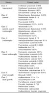
The members of the working group graded the evidence of each statement and then classified the strength of the recommendation for each statement. The evidence of each statement was graded as follows: level 1, systematic review of randomized controlled trials (RCTs) and individual RCTs; level 2, systematic review of cohort studies and individual cohort study (including low-quality RCTs); level 3, systematic review of case-control studies and individual case-control studies; level 4, case series (and poor-quality cohort and case-control studies); and level 5, expert opinion. The strength of each recommendation was classified as A (level 1), B (levels 2 and 3), C (level 4), or D (level 5) (Table 1)7.

Consensus process

A total of 39 of 54 KADA council members participated in the vote on the draft guideline statements. Each statement was given along with its grade of evidence and recommendation strength. Each participant had one vote per statement. Participants scored their level of agreement with each draft statement on a scale of 1~9, where 1 represented strong disagreement and 9 represented strong agreement. Each voting score was assigned to one of three groups: disagreement (1~3), neutrality (4~6), and agreement (7~9). Consensus was defined as ≥75% of participants scoring within the 7~9 range (agreement).
A second vote was performed after disclosing the additional information of previous voting results, including the mean score and proportion of agreement.

RESULTS

Bathing and basic skin care

Basic skin care, including bathing, represents a first strategy to maintain a good skin barrier and prevent and eliminate more severe infections, ultimately resulting in the normalization of immunologic responses and inflammation1. Bathing can provide hydration of the skin and removal of scale, crust, and irritants such as sweat and allergens. The application of moisturizer soon after bathing is crucial for the further maintenance of hydration, because water easily evaporates from the skin, resulting in a higher transepidermal water loss8.
The recommendations for bathing are summarized in Table 224910111213. Generally, a maximum bathing frequency of once per day is recommended2. Strongly scrubbing with a towel is not recommended9. The proper duration of bathing is a short period of time (e.g., 5~10 min)210, as some experimental studies have revealed that significantly longer exposure to water can disrupt the structural integrity of the skin barrier141516. Warm or lukewarm water (27℃ to 30℃) is recommendedas the avoidance of temperature extremes is established in the management of AD17. There are very limited data about the usefulness of bathwater additives in the treatment of AD.
AD skin is vulnerable to increased skin surface pH, which has various effects on barrier function, desquamation of corneocytes, induction of inflammation, secretion of lamellar bodies, and antimicrobial defense18192021. Nonsoap cleansers (e.g., syndets, aqueous solutions), which have a neutral or low pH and are less allergenic, nonirritating, and fragrance-free, are recommended. Soap-based cleansers, which have a high pH and contain surfactants, should be avoided, because they can cause dry skin, irritation, and contact dermatitis111213. Antiseptic-containing cleansers are not recommended due to the limited duration of action of antiseptics and limited clinical data regarding their effectiveness3. Although one RCT provided evidence for the effectiveness of a bleach bath to control moderate to severe AD infections (2b, B)2223242526, the use of a bleach bath in the treatment of AD failed to achieve a consensus among Korean experts. Elimination of the belief that natural ingredients in cleansers or emollients are better is necessary. "Natural" does not mean "safe"; sometimes, these natural ingredients may have a higher risk of producing contact dermatitis.
Gentle drying after bathing (a soft-pat dry with a towel and the avoidance of rubbing) is recommended27. After bathing and soft-pat drying, the application of moisturizer is recommended.

Avoidance of exacerbating factors

Recognition of exacerbating or triggering factors is an indispensable component of the personalized avoidance strategies in patients with AD, and these recommendations are summarized in Table 34528293031323334353637. Such factors may differ according to age, environment, and individual social and daily lifestyles. In addition to climate and pollution, other known AD-exacerbating factors include clothing, the presence of house dust mites (HDMs) in the home, cosmetics, certain foods, dietary changes, physiological stressors (infections, sweat, etc.), and emotional stress38. One recent multicenter, cross-sectional, epidemiologic study including 125 adults and 116 children with moderate to severe AD found that the factors most commonly perceived by patients as triggers of AD flares were perfumes and personal hygiene products (44% of adults, 34.5% of children), followed by articles of clothing, presence of HDMs, sudden changes in temperature, and stress. The foods most frequently perceived as triggers were sweets (3.88%), seafood (2.33%), and eggs (2.33%) among adults and eggs (7.63%) and fish (3.05%) among children.
In the second half of childhood and in adulthood, the most common airborne allergens exacerbating AD are derived from HDM species. In more than 90% of Korean homes, the level of exposure to HDMs is clinically remarkable. More than 40% of patients with AD are sensitized to HDMs39. The severity of AD is related to indoor concentrations of HDMs in children without sensitization to HDM, and this concentration likely acts as a nonspecific irritant as well as an allergen40. Avoidance of HDMs is an effective method that can be used to control AD exacerbation282930313241. At 25℃ and 75% relative humidity, HDMs can grow well and their antigen concentrations can accumulate3334. Controlling the environment and vacuuming mattresses daily can significantly decrease one's level of exposure to HDMs35.
Allergens should be determined by assessing clinical symptoms and laboratory test results. The proper diagnosis of contact dermatitis using patch testing and other tools to confirm true immunoglobulin E (IgE)-mediated allergies must be undertaken to avoid specific contact with the allergen34538.
To diagnose a food allergy, clinical symptoms or signs after suspected food allergen intake or exposure must be reproducible, as broad-panel allergy testing unrelated to a clinical history of a reaction to certain foods should be avoided. The National Institute of Allergy and Infectious Diseases Food Allergy Expert Panel advocates food allergy testing only in cases of children younger than 5 years of age with moderate to severe AD and treatment resistance despite proper management and topical therapy and/or consistent episodes of an immediate allergic reaction after eating a specific food3637. Food diaries are useful for this purpose. If IgE-mediated food allergy is suspected, a diet eliminating the candidate food allergen for 4~6 weeks is recommended. If patients responded to the elimination diet, an oral food challenge should be done to confirm the causal relationship of food intake and AD lesions. Consultation with a dietitian is recommended for advice on suitable avoidance and alternatives327.
Wearing soft-textured clothing and avoiding rough-textured fabrics and tight garments are preferred42. Although wearing silk fabric may be helpful, it is not clear whether silk is superior to cotton4344. One recent meta-analysis failed to show the effectiveness of the use of functional textiles in AD treatment45. Another study indicated that residual laundry detergent in clothes is an aggravating factor of winter xerosis46. Water softening using an ion-exchange method provided no additional benefit to usual care47. There is a paucity of well-controlled studies about the effects of environmental modification on AD. General consensus recommendations include the avoidance of mechanical and chemical irritants (such as wool, acids, and bleaches) and organic solvents (such as formaldehyde and toluene).

Education and psychosocial support

Aggravation due to mental stress can be easily identified in AD patients, and minimizing such stress can be helpful in controlling the disease28484950. Psychotherapeutic approaches and behavior therapy can be considered to manage individual emotional factors that trigger AD, such as viscous itch-scratch cycles, comorbidity with anxiety and depression, and low quality of life451.
Patient and parent education is effective in the management of AD52535455 and should aim to provide information about the clinical characteristics of AD (etiology, clinical manifestations, and disease course in layperson language), aggravating and relieving factors, self-management, and improving coping skills. Psychological and psychosomatic interventions are necessary and important tools of educational programs.
A multidisciplinary team (e.g., doctors, nurses, psychologists, and dieticians) approach is recommended (1a, A)56. There is evidence that a systematic educational program considering the factor of age substantially improves AD severity scores53. Workshops and educational sessions led by a doctor, nurse, or other health care professional57, as well as live video or practicing interventions, can be beneficial to patients by improving their knowledge of AD and the use of bathing, skin clearing, and moisturizers and other topical treatments (2b, B)4. Recommendations are summarized in Table 42848495052535455.

Moisturizer

AD patients have very dry skin and defective skin barrier function. Regular use of moisturizer is a basictherapy for the management of AD. Recommendations for moisturizer use are summarized in Table 545958596061626364656667. Mild AD can be improved by the appropriate use of moisturizer. Moisturizer application should also be maintained in patients with moderate to severe AD, which also requires topical or systemic anti-inflammatory treatment during acute flares to induce remission5859. Data from RCTs show that moisturizers have a long- and short-term steroid-sparing effect in mild to moderate AD and in preventing AD flares60616263. The antipruritic effect of certain moisturizers in the treatment of AD has also been proven in RCTs64. Some moisturizers have been found to restore a defective skin barrier while decreasing exposure to irritants and showing anti-inflammatory and antimicrobial effects626566. The Barrier Enhancement for Eczema Prevention trial, a large-scale RCT on the effectiveness of intensive moisturizer therapy in early life to prevent AD and/or allergic reactions or reduce disease severity, is underway with encouraging results in a pilot study6869.
Although no clinical trials have studied the proper amount or frequency of moisturizer use in AD patients, moisturizer should be used at least twice daily, and more frequently during acute flare-ups. Adult patients with AD should use approximately 250 g or more of moisturizer per week467 and apply it to their whole body, regardless of the presence of lesions, since barrier defects and subclinical inflammation may also be present on lesion-free skin. The recommendation is to use moisturizer after taking a bath while the skin is still moist (within 3 min). During acute flare-ups, moisturizer should be used more frequently in conjunction with anti-inflammatory treatment and continued as maintenance therapy459. Moisturizer treatment without the use of anti-inflammatory agents during acute flares may increase one's susceptibility to bacterial or viral infection70. Conventional moisturizers contain occlusives, humectants, and emulsions. Newer classes of emollients designed to restore skin barrier defects include distinct ratios of lipids that resemble physiological compositions, such as ceramides:cholesterol:essential fatty acids in the ratios of 3:1:1 or 1:1:1. Although these new moisturizers are used worldwide as prescribed devices for the treatment of AD, only a few studies have demonstrated the effects of moisturizer on skin barrier function recovery and symptoms and signs of AD in a randomized and controlled manner596265.
A few moisturizer-containing substances with nonsteroidal anti-inflammatory effects such as N-palmitoylethanolamine and sunflower seed oil have been developed, and some of them have been shown to lessen pruritus, xerosis, and inflammation and have steroid-sparing effects in a small number of controlled studies60616263646566.
Various types of moisturizer are available as creams, ointments, oils, gels, or lotions. Ointment or oily cream-type moisturizers, rather than lotions, are suitable for AD patients. However, some patients may not like these formulas because they feel greasy. At bedtime, lotion or aqueous cream-type moisturizers are desirable. In winter, higher lipid contents are preferable.
Because moisturizers including proteinaceous antigens or haptens may induce an allergic reaction, it is not desirable to use them in children younger than 2 years71.
An over-the-counter moisturizer with a petrolatum-base was proven to be as effective as prescribed moisturizers in the treatment of children with mild to moderate AD in an RCT72, which suggests the cost-effectiveness of moisturizer treatment is a factor in selecting a moisturizer. Because adherence to moisturizer usage is key to the successful treatment of AD, patient preferences and tolerances should be considered when selecting a moisturizer to enhance adherence to the therapy.

Topical corticosteroids

Topical corticosteroids (TCSs) are important anti-inflammatory drugs for managing AD, especially during acute flares. TCS recommendations are summarized in Table 6456473747576777879. TCSs significantly improve AD lesions compared to placebo7374. Data from a meta-analysis of RCTs showed that TCSs are effective in the relief of AD pruritus63 (relative risk=0.66, p<0.001) and have a significant effect in reducing Staphylococcus colonization75.
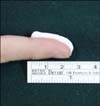
TCSs are classified according to their potency (Table 7)76. Most TCSs are enough strong to be used only once daily77. The fingertip unit (FTU) is commonly suggested, as it has been found that the amount of TCS from the distal skin crease to the tip of an adult patient's index finger is equivalent to approximately 0.5 g TCS. Using 1 FTU of TCS, applying the amount on the surface area of the two palms of the patient is appropriate (Fig. 1)78.
Application of TCSs should be tapered if signs of inflammation disappear79. Proactive treatment with TCS therapy (e.g., once- or twice-weekly application) to areas known to commonly relapse in the maintenance period may help to reduce acute flares737480. Schmitt et al.80 concluded in their systematic review that long-term (40-week) proactive therapy with TCS did not result in skin atrophy and telangiectasia or systemic side effects, such as adrenal suppression, and can be safely used during the AD maintenance period.
On skin with coexistent infections, TCSs have been widely used along with systemic or topical antibiotics. However, a review by Cochrane did not find any additional benefit from the concomitant use of antibiotics and TCS compared to the use of TCS alone for AD treatment81. Combination therapy with a topical calcineurin inhibitor (TCI) did not have a synergistic effect in one study82, but it did in others8384.
Local side effects of TCS include steroid acne, flushing, skin atrophy, hypertrichosis, striae, telangiectasia, and allergic contact dermatitis, which may occur occasionally in the treated area85; however, these side effects can be resolved with discontinuation or appropriate treatment. The risk of cataract or glaucoma development when TCS is applied to the periorbital area is uncertain. Systemic side effects are extremely rare, but they have been reported. Children have a greater chance of developing adrenal suppression, since they have a relatively greater body surface area-to-weight ratio and a higher systemic absorption. Routine screening tests for systemic side effects of TCSs are not required.
Potent TCSs can be used in the following amounts to avoid systemic and local side effects: 15 g/month in infants, 30 g/month in children, and 60~90 g/month in adults4. For infants and children, the elderly, or pregnant woman, mild to moderate TCSs can be used instead of more potent varieties of TCSs86.
For pregnant women, potent or very potent TCSs should be chosen as a second-line option for as short a time as possible, and relevant obstetric care is needed, as TCSs increase the possibility of fetal growth restriction. It has been proven that the use of ≤200 g TCSs during the pregnancy period is not associated with fetal growth restriction86.
A meta-analysis of RCTs recommended that superpotent TCSs be used only once daily, because this is as beneficial as twice-daily application87.
Steroid phobia is widespread and can cause treatment failure among AD patients. To increase treatment compliance, it is important to educate children and their parents about the side effects of TCSs and signs and symptoms of worsening eczema459.

Wet-wrap therapy

Wet-wrap therapy (WWT) can be helpful to quickly reduce AD severity, and it is often useful for acute flares and/or recalcitrant disease88899091929394959697. For better results and to reduce the risk of infection, WWT use must be based on proper education and can be administered on an outpatient or inpatient basis8894. A recent RCT demonstrated that a 4-week proactive schedule of WWT with diluted TCSs was superior to WWT with moisturizer in children with severe AD89. The first step of WWT is to apply topical agents to the lesion. Next, the skin is covered with a wet inner layer of tubular bandages followed by a dry outer layer. Gauze or a cotton suit can be used as an alternative. The WWT can be maintained from several hours to a day at a time (an illustration of how to use WWT can be found online at http://www.atopy.re.kr/academy/news_02.asp). Further RCTs and clinical studies are needed to determine the ideal topical agents to use as well as duration, covering agent, and frequency. WWT recommendations are summarized in Table 887889394.

Topical calcineurin inhibitors

TCIs exert anti-inflammatory effects by blocking calcineurin-dependent T-cell activation, which leads to the release of proinflammatory cytokines and mediators of AD2. TCIs used to treat AD include pimecrolimus cream (1%) and tacrolimus ointment (0.03% in children and 0.1% in adults), which have been proven to help improve and control AD in both children and adults59899. Recommendations for TCI use are summarized in Table 949899100101102103104105.
A meta-analysis showed that tacrolimus 0.1% and midpotency TCS hydrocortisone butyrate 0.1% are comparable in the efficacy, whereas the efficacy of tacrolimus 0.03% is inferior to hydrocortisone butyrate 0.1% but superior to low-potency TCS hydrocortisone acetate 1%98. Pimecrolimus cream has been considered less efficacious than midto high-potency TCS, although there are no studies comparing pimecrolimus and TCS3. Tacrolimus is approved for the treatment of moderate to severe AD, and pimecrolimus is approved for mild to moderate AD599.
Korean dermatologists strongly recommend the use of TCIs for both active and proactive treatment of AD. The 2012 European guidelines recommend TCIs as a first-line therapy in the short- and long-term treatment of AD5. Proactive therapy with tacrolimus ointment could reduce the frequency of relapse4. The 2013 Asian-Pacific guidelines recommend TCIs as a second-line therapy for both acute AD and long-term proactive treatment. The 2014 American guidelines also recommend the use of TCIs for the active and proactive treatment of AD2.
In a proactive regimen, TCI application 2 to 3 times a week on frequent recurrent sites can effectively reduce relapses4. After the acute flare is controlled, long-term management with topical tacrolimus significantly decreased the number of episodes compared with vehicle and increased the time to relapse and number of flare-free days100101. The efficacy and safety of topical tacrolimus 0.1% use for 6 month to 5 years is well documented in children with moderate to severe AD5. In adults with mild to moderate AD, pimecrolimus cream 1.0% also reduced the occurrence of flares102.
TCI has no risk of cutaneous atrophy, and it has few harmful effects on collagen synthesis103104. This favors the use of TCIs over TCS in thin-skinned areas such as the eyelid, perioral region, genitalia, axillary area, or inguinal fold, as well as for long-term management. TCI has steroid-sparing effects and that can be maintained during long-term use (up to 12 months)2106.
The most common local side effects of TCIs are stinging and burning sensations. Irritation is seen more frequently with TCIs than with TCSs but tends to decrease with repeated applications or when patients are shortly pretreated with TCSs107108. Physicians should provide this information to patients when TCIs are first prescribed to prevent premature discontinuation of TCIs. Infections with virus such as herpes simplex, eczema herpeticum, or molluscum contagiosum have been reported during TCI use109110111, but these studies failed to prove a causal relationship106112.
Topical tacrolimus and pimecrolimus have been approved for adults and children older than 2 years5109. However, topical tacrolimus 0.03% and pimecrolimus can be safely used in children younger than 2 years, even in infants105.
There are several case reports of lymphoma and skin cancer in patients treated with TCIs. However, there is currently no scientific evidence of an increased risk of malignancy due to TCI use113.
Routine blood monitoring of tacrolimus or pimecrolimus levels is not needed in AD patients, as systemic absorption after topical application of both TCIs is negligible114115.

Topical antipruritic agents

1) Topical anesthetics

Local anesthetics are sometimes used to achieve a temporary antipruritic effect. The antipruritic effect of local anaesthetics was reported in patients with AD116; however, controlled clinical studies are lacking (4, C).
The routine use of topical anesthetics in AD is not recommended as an antipruritic therapy4.

2) Topical cannabinoid receptor agonists

Topical cannabinoid receptor agonists have been reported to have antipruritic and analgesic effects (4, C)117. However, further studies are needed to recommend their routine use.

3) Topical capsaicin

Capsaicin is the major constituent of hot chili peppers and has an antipruritic effect in various dermatoses. Some previous studies reported a reduction of itching in AD patients after use of topical capsaicin (4, C)118119. However, no controlled study has been performed to date, and further studies are needed.

4) Topical doxepin

A controlled study documented that an antipruritic effect of doxepin 5% cream in AD patients (2b, B)120. However, RCTs on the use of doxepin in AD are lacking.

5) Topical mast cell stabilizers

Tryptase and histamine, released by mast cells, lead to the induction of pruritus in AD. Some studies have reported the usefulness of mast cell stabilizers in AD (2b, B)121. However, further controlled trials are needed.

Other topical anti-inflammatory agents

1) Phosphodiesterase inhibitors

Phosphodiesterase-4 (PDE4) inhibitors produce anti-inflammatory effects by inhibiting the secretion of various cytokines from inflammatory cells such as lymphocytes and monocytes. Roflumilast, an oral PDE4 inhibitor, is used to treat moderate chronic obstructive pulmonary disease, and apremilast, which has a relatively low incidence of adverse effects, is approved by the US Food and Drug Administration in the treatment of active psoriatic arthritis122. However, systemic side effects such as nausea and vomiting are relatively common with these drugs. Studies on the development of topical PDE4 inhibitors are underway, although thorough study and experimentation will be required before clinical use123124. Nevertheless, recent studies have demonstrated the clinical efficacy and safety of topical PDE4 inhibitors, which will likely be used as topical anti-inflammatory agents in the treatment of AD.

DISCUSSION

Because AD chronically relapses, the active participation of patients in basic skin care, avoidance of aggravating factors, and appropriate application of topical medicines is important to achieve an effective treatment outcome2345. Patient education and psychological intervention are closely related to the prevention of recurrence and shortening of disease duration4849505152535455.
This report provides the level of evidence, recommendation strength, and average agreement scores of an AD expert panel regarding the general management and topical treatment of AD. Most recommendations regarding environmental control are based on both expert consensus and personal experience. The assessment of aggravating factors and their avoidance should be personalized. Indiscriminate food restriction is unnecessary and can lead to malnutrition and growth problems in children with AD3459. Food allergies should be determined by elimination and challenge tests of suspected food while considering both medical history and results of laboratory tests.
Education can lead to greater improvement in ADmanagement by increasing patient adherence to therapy 5354. It is necessary to provide specific and individualized information, such as how to use moisturizer or anti-inflammatory agents and the adequate amount to apply, according to each patient's disease course. Moisturizer itself has steroid-sparing and antipruritic effects. The regular use of moisturizer lowers the number of relapse episodes60616263646566. However, moisturizer cannot replace topical anti-inflammatory treatment. During flare-ups, active topical anti-inflammatory treatment should be primarily considered. "1 FTU" is a useful tool that can be used to explain to AD patients the adequate amount of topical anti-inflammatory agent to apply78. Even after the AD lesions disappear, patients with frequent relapsing disease courses require proactive treatment with TCSs or TCIs3459.
Steroid phobia is widespread due to the inconsistency of information regarding its usage125. If a caregiver has mistrust about TCS use, this can lead to a missed opportunity of timely anti-inflammatory intervention and increased flare-ups that require more intensive treatment. Many patients tend stop using TCSs because they confuse the side effects of TCS use with signs and symptoms of worsening AD, which leads to treatment failure. Specific education is needed regarding effective and safe long-term TCS usage, TCS side effects, and signs and symptoms of worsening eczema to increase patient adherence to TCS therapy and achieve successful AD management459. Education and psychosocial support is essential, especially for AD patients with frequent relapse episodes451.
WWT can increase the efficacy of TCSs or moisturizer and facilitate a faster recovery of AD lesions88899091929394959697. To achieve better results and reduce the risk of infection, WWT use must be based on proper education on an ambulatory or inpatient basis.
Evidence supporting the routine use of topical antipruritic drugs is lacking in the treatment of AD. However, in patients with refractory pruritus, the use of topical antipruritic agents can be considered.
This report might require supplementary statements or a revision of recommendations based on upcoming publications and results of the many ongoing clinical studies of AD. These guidelines provided evidence- and experience-based recommendations and will be helpful for physicians and AD patients in improving treatment outcome and quality of life.

Figures and Tables

Fig. 1

The fingertip unit (FTU) method is used in to determine the amount of topical corticosteroids (TCS) or calcineurin inhibitors to apply. The amount from the distal skin crease to the tip of an adult patient's index finger is equivalent to approximately 0.5 g TCS. 1 FTU generally covers the surface area of two palms.

ad-27-563-g001
Table 1

Level of evidence and strength of the recommendations

ad-27-563-i001

RCT: randomized controlled trial.

Table 2

Expert consensus recommendations for bathing and basic skin care

ad-27-563-i002
Table 3

Expert consensus recommendations for avoidance of exacerbating factors

ad-27-563-i003
Table 4

Expert consensus recommendations for education and psychosocial support

ad-27-563-i004

AD: atopic dermatitis.

Table 5

Expert consensus recommendations for moisturizer use

ad-27-563-i005

AD: atopic dermatitis.

Table 6

Expert consensus recommendations for TCSs

ad-27-563-i006

TCS: topical corticosteroid, FTU: fingertip unit, AD: atopic dermatitis.

Table 7

The potency of topical corticosteroids

ad-27-563-i007

Modified from Ference and Last (Am Fam Physician 2009;79: 135-140)76.

Table 8

Expert consensus recommendations for WWT

ad-27-563-i008

WWT: wet wrap therapy, AD: atopic dermatitis.

Table 9

Expert consensus recommendations for TCIs

ad-27-563-i009

TCI: topical calcineurin inhibitor, AD: atopic dermatitis.

ACKNOWLEDGMENT

This work was supported by the Korean Atopic Dermatitis Association. We appreciate the active participation and advice of the 54 KADA council members in developing this expert opinion consensus.

Notes

Conflict of interest Young Roh is a clinical trials investigator for Astellas and Novartis. Hyun Chang Ko is a clinical trials investigator for Astellas.

References

1. Elias PM, Eichenfield LF, Fowler JF Jr, Horowitz P, McLeod RP. Update on the structure and function of the skin barrier: atopic dermatitis as an exemplar of clinical implications. Semin Cutan Med Surg. 2013; 32:2 Suppl 2. S21–S24.
crossref
2. Eichenfield LF, Tom WL, Berger TG, Krol A, Paller AS, Schwarzenberger K, et al. Guidelines of care for the management of atopic dermatitis: section 2. Management and treatment of atopic dermatitis with topical therapies. J Am Acad Dermatol. 2014; 71:116–132.
3. Sidbury R, Tom WL, Bergman JN, Cooper KD, Silverman RA, Berger TG, et al. Guidelines of care for the management of atopic dermatitis: Section 4. Prevention of disease flares and use of adjunctive therapies and approaches. J Am Acad Dermatol. 2014; 71:1218–1233.
4. Ring J, Alomar A, Bieber T, Deleuran M, Fink-Wagner A, Gelmetti C, et al. Guidelines for treatment of atopic eczema (atopic dermatitis) part I. J Eur Acad Dermatol Venereol. 2012; 26:1045–1060.
crossref
5. Rubel D, Thirumoorthy T, Soebaryo RW, Weng SC, Gabriel TM, Villafuerte LL, et al. Consensus guidelines for the management of atopic dermatitis: an Asia-Pacific perspective. J Dermatol. 2013; 40:160–171.
crossref
6. Kim SY, Jee SM, Lee SJ, Lee YJ, Park JE, Nam MH, et al. Guidance for development of clinical practice guidelines. 1st ed. Seoul: National Evidence-Based Healthcare Collaborating Agency;2011. p. 607–608.
7. Phillips B, Ball C, Sackett D, Badenoch D, Straus S, Haynes B, et al. Oxford Centre for Evidence-based Medicine-Levels of evidence [Internet]. Oxford: Centre for Evidence Based Medicine;2001. 05. updated 2009 March. cited 2014 March 4. Available from: http://www.cebm.net/oxford-centre-evidence-based-medicine-levels-evidence-march-2009/.
8. Simpson E, Trookman NS, Rizer RL, Preston N, Colón LE, Johnson LA, et al. Safety and tolerability of a body wash and moisturizer when applied to infants and toddlers with a history of atopic dermatitis: results from an open-label study. Pediatr Dermatol. 2012; 29:590–597.
crossref
9. Katayama I, Kohno Y, Akiyama K, Ikezawa Z, Kondo N, Tamaki K, et al. Japanese Society of Allergology. Japanese guideline for atopic dermatitis. Allergol Int. 2011; 60:205–220.
crossref
10. Baker M. NICE guidance points the way to tackling eczema in children. Community Pract. 2013; 86:40.
11. Shaughnessy CN, Malajian D, Belsito DV. Cutaneous delayed-type hypersensitivity in patients with atopic dermatitis: reactivity to surfactants. J Am Acad Dermatol. 2014; 70:704–708.
crossref
12. Cheong WK. Gentle cleansing and moisturizing for patients with atopic dermatitis and sensitive skin. Am J Clin Dermatol. 2009; 10:Suppl 1. 13–17.
crossref
13. Shukuwa T, Kligman AM. Disaggregation of corneocytes from surfactant-treated sheets of stratum corneum in hyperkeratosis on psoriasis, ichthyosis vulgaris and atopic dermatitis. J Dermatol. 1997; 24:361–369.
crossref
14. Bouwstra JA, de Graaff A, Gooris GS, Nijsse J, Wiechers JW, van Aelst AC. Water distribution and related morphology in human stratum corneum at different hydration levels. J Invest Dermatol. 2003; 120:750–758.
crossref
15. Warner RR, Stone KJ, Boissy YL. Hydration disrupts human stratum corneum ultrastructure. J Invest Dermatol. 2003; 120:275–284.
crossref
16. Fluhr JW, Lazzerini S, Distante F, Gloor M, Berardesca E. Effects of prolonged occlusion on stratum corneum barrier function and water holding capacity. Skin Pharmacol Appl Skin Physiol. 1999; 12:193–198.
crossref
17. Langan SM, Bourke JF, Silcocks P, Williams HC. An exploratory prospective observational study of environmental factors exacerbating atopic eczema in children. Br J Dermatol. 2006; 154:979–980.
crossref
18. Lee HJ, Lee SH. Epidermal permeability barrier defects and barrier repair therapy in atopic dermatitis. Allergy Asthma Immunol Res. 2014; 6:276–287.
crossref
19. Sakai T, Hatano Y, Zhang W, Fujiwara S. Defective maintenance of pH of stratum corneum is correlated with preferential emergence and exacerbation of atopic-dermatitis-like dermatitis in flaky-tail mice. J Dermatol Sci. 2014; 74:222–228.
crossref
20. Lee HJ, Yoon NY, Lee NR, Jung M, Kim DH, Choi EH. Topical acidic cream prevents the development of atopic dermatitis- and asthma-like lesions in murine model. Exp Dermatol. 2014; 23:736–741.
crossref
21. Angelova-Fischer I, Dapic I, Hoek AK, Jakasa I, Fischer TW, Zillikens D, et al. Skin barrier integrity and natural moisturising factor levels after cumulative dermal exposure to alkaline agents in atopic dermatitis. Acta Derm Venereol. 2014; 94:640–644.
crossref
22. Wong SM, Ng TG, Baba R. Efficacy and safety of sodium hypochlorite (bleach) baths in patients with moderate to severe atopic dermatitis in Malaysia. J Dermatol. 2013; 40:874–880.
crossref
23. Huang JT, Rademaker A, Paller AS. Dilute bleach baths for Staphylococcus aureus colonization in atopic dermatitis to decrease disease severity. Arch Dermatol. 2011; 147:246–247.
crossref
24. Craig FE, Smith EV, Williams HC. Bleach baths to reduce severity of atopic dermatitis colonized by Staphylococcus. Arch Dermatol. 2010; 146:541–543.
crossref
25. Barnes TM, Greive KA. Use of bleach baths for the treatment of infected atopic eczema. Australas J Dermatol. 2013; 54:251–258.
crossref
26. Huang JT, Abrams M, Tlougan B, Rademaker A, Paller AS. Treatment of Staphylococcus aureus colonization in atopic dermatitis decreases disease severity. Pediatrics. 2009; 123:e808–e814.
crossref
27. Leung TNH, Chow CM, Chow MPY, Luk DCK, Ho KM, Hon KL, et al. Clinical guidelines on management of atopic dermatitis in children. Hong Kong J Paediatr. 2013; 18:96–104.
28. Oosting AJ, de Bruin-Weller MS, Terreehorst I, Tempels-Pavlica Z, Aalberse RC, de Monchy JG, et al. Effect of mattress encasings on atopic dermatitis outcome measures in a double-blind, placebo-controlled study: the Dutch mite avoidance study. J Allergy Clin Immunol. 2002; 110:500–506.
crossref
29. Koopman LP, van Strien RT, Kerkhof M, Wijga A, Smit HA, de Jongste JC, et al. Prevention and Incidence of Asthma and Mite Allergy (PIAMA) Study. Placebo-controlled trial of house dust mite-impermeable mattress covers: effect on symptoms in early childhood. Am J Respir Crit Care Med. 2002; 166:307–313.
crossref
30. Gutgesell C, Heise S, Seubert S, Seubert A, Domhof S, Brunner E, et al. Double-blind placebo-controlled house dust mite control measures in adult patients with atopic dermatitis. Br J Dermatol. 2001; 145:70–74.
crossref
31. Ricci G, Patrizi A, Specchia F, Menna L, Bottau P, D'Angelo V, et al. Effect of house dust mite avoidance measures in children with atopic dermatitis. Br J Dermatol. 2000; 143:379–384.
crossref
32. Sanda T, Yasue T, Oohashi M, Yasue A. Effectiveness of house dust-mite allergen avoidance through clean room therapy in patients with atopic dermatitis. J Allergy Clin Immunol. 1992; 89:653–657.
crossref
33. Yella L, Morgan MS, Arlian LG. Population growth and allergen accumulation of Dermatophagoides farinae cultured at 20 and 25℃. Exp Appl Acarol. 2013; 60:117–126.
crossref
34. Yella L, Morgan MS, Arlian LG. Population growth and allergen accumulation of Dermatophagoides pteronyssinus cultured at 20 and 25℃. Exp Appl Acarol. 2011; 53:103–119.
35. Wu FF, Wu MW, Pierse N, Crane J, Siebers R. Daily vacuuming of mattresses significantly reduces house dust mite allergens, bacterial endotoxin, and fungal β-glucan. J Asthma. 2012; 49:139–143.
crossref
36. Boyce JA, Assa'ad A, Burks AW, Jones SM, Sampson HA, Wood RA, et al. Guidelines for the diagnosis and management of food allergy in the United States: summary of the NIAID-sponsored expert panel report. Nutr Res. 2011; 31:61–75.
crossref
37. NIAID-Sponsored Expert Panel. Boyce JA, Assa'ad A, Burks AW, Jones SM, Sampson HA, et al. Guidelines for the diagnosis and management of food allergy in the United States: report of the NIAID-sponsored expert panel. J Allergy Clin Immunol. 2010; 126:6 Suppl. S1–S58.
38. Katayama I, Kohno Y, Akiyama K, Aihara M, Kondo N, Saeki H, et al. Japanese Society of Allergology. Japanese guideline for atopic dermatitis 2014. Allergol Int. 2014; 63:377–398.
crossref
39. Jeong KY, Park JW, Hong CS. House dust mite allergy in Korea: the most important inhalant allergen in current and future. Allergy Asthma Immunol Res. 2012; 4:313–325.
crossref
40. Kim J, Lee S, Woo SY, Han Y, Lee JH, Lee IY, et al. The indoor level of house dust mite allergen is associated with severity of atopic dermatitis in children. J Korean Med Sci. 2013; 28:74–79.
crossref
41. Arshad SH, Bateman B, Sadeghnejad A, Gant C, Matthews SM. Prevention of allergic disease during childhood by allergen avoidance: the Isle of Wight prevention study. J Allergy Clin Immunol. 2007; 119:307–313.
crossref
42. Mason R. Fabrics for atopic dermatitis. J Fam Health Care. 2008; 18:63–65.
43. Ricci G, Neri I, Ricci L, Patrizi A. Silk fabrics in the management of atopic dermatitis. Skin Therapy Lett. 2012; 17:5–7.
44. Kurtz EJ, Yelverton CB, Camacho FT, Fleischer AB Jr. Use of a silklike bedding fabric in patients with atopic dermatitis. Pediatr Dermatol. 2008; 25:439–443.
crossref
45. Lopes C, Silva D, Delgado L, Correia O, Moreira A. Functional textiles for atopic dermatitis: a systematic review and meta-analysis. Pediatr Allergy Immunol. 2013; 24:603–613.
crossref
46. Kiriyama T, Sugiura H, Uehara M. Residual washing detergent in cotton clothes: a factor of winter deterioration of dry skin in atopic dermatitis. J Dermatol. 2003; 30:708–712.
crossref
47. Thomas KS, Koller K, Dean T, O'Leary CJ, Sach TH, Frost A, et al. A multicentre randomised controlled trial and economic evaluation of ion-exchange water softeners for the treatment of eczema in children: the Softened Water Eczema Trial (SWET). Health Technol Assess. 2011; 15:v–vi. 1–156.
crossref
48. Suárez AL, Feramisco JD, Koo J, Steinhoff M. Psychoneuroimmunology of psychological stress and atopic dermatitis: pathophysiologic and therapeutic updates. Acta Derm Venereol. 2012; 92:7–15.
crossref
49. Arndt J, Smith N, Tausk F. Stress and atopic dermatitis. Curr Allergy Asthma Rep. 2008; 8:312–317.
crossref
50. Hashizume H, Takigawa M. Anxiety in allergy and atopic dermatitis. Curr Opin Allergy Clin Immunol. 2006; 6:335–339.
crossref
51. Chida Y, Steptoe A, Hirakawa N, Sudo N, Kubo C. The effects of psychological intervention on atopic dermatitis. A systematic review and meta-analysis. Int Arch Allergy Immunol. 2007; 144:1–9.
crossref
52. Ersser SJ, Cowdell F, Latter S, Gardiner E, Flohr C, Thompson AR, et al. Psychological and educational interventions for atopic eczema in children. Cochrane Database Syst Rev. 2014; 1:CD004054.
crossref
53. Barbarot S, Bernier C, Deleuran M, De Raeve L, Eichenfield L, El Hachem M, et al. Oriented Patient-Education Network in Dermatology. Therapeutic patient education in children with atopic dermatitis: position paper on objectives and recommendations. Pediatr Dermatol. 2013; 30:199–206.
crossref
54. Mancini AJ, Paller AS, Simpson EL, Ellis CN, Eichenfield LF. Improving the patient-clinician and parent-clinician partnership in atopic dermatitis management. Semin Cutan Med Surg. 2012; 31:3 Suppl. S23–S28.
crossref
55. de Bes J, Legierse CM, Prinsen CA, de Korte J. Patient education in chronic skin diseases: a systematic review. Acta Derm Venereol. 2011; 91:12–17.
crossref
56. Stalder JF, Bernier C, Ball A, De Raeve L, Gieler U, Deleuran M, et al. Oriented Patient-Education Network in Dermatology (OPENED). Therapeutic patient education in atopic dermatitis: worldwide experiences. Pediatr Dermatol. 2013; 30:329–334.
crossref
57. Nicol NH, Ersser SJ. The role of the nurse educator in managing atopic dermatitis. Immunol Allergy Clin North Am. 2010; 30:369–383.
crossref
58. Breternitz M, Kowatzki D, Langenauer M, Elsner P, Fluhr JW. Placebo-controlled, double-blind, randomized, prospective study of a glycerol-based emollient on eczematous skin in atopic dermatitis: biophysical and clinical evaluation. Skin Pharmacol Physiol. 2008; 21:39–45.
crossref
59. Eberlein B, Eicke C, Reinhardt HW, Ring J. Adjuvant treatment of atopic eczema: assessment of an emollient containing N-palmitoylethanolamine (ATOPA study). J Eur Acad Dermatol Venereol. 2008; 22:73–82.
crossref
60. Wirén K, Nohlgård C, Nyberg F, Holm L, Svensson M, Johannesson A, et al. Treatment with a barrier-strengthening moisturizing cream delays relapse of atopic dermatitis: a prospective and randomized controlled clinical trial. J Eur Acad Dermatol Venereol. 2009; 23:1267–1272.
crossref
61. Boralevi F, Saint Aroman M, Delarue A, Raudsepp H, Kaszuba A, Bylaite M, et al. Long-term emollient therapy improves xerosis in children with atopic dermatitis. J Eur Acad Dermatol Venereol. 2014; 28:1456–1462.
crossref
62. Grimalt R, Mengeaud V, Cambazard F. Study Investigators' Group. The steroid-sparing effect of an emollient therapy in infants with atopic dermatitis: a randomized controlled study. Dermatology. 2007; 214:61–67.
crossref
63. Lucky AW, Leach AD, Laskarzewski P, Wenck H. Use of an emollient as a steroid-sparing agent in the treatment of mild to moderate atopic dermatitis in children. Pediatr Dermatol. 1997; 14:321–324.
crossref
64. Sher LG, Chang J, Patel IB, Balkrishnan R, Fleischer AB Jr. Relieving the pruritus of atopic dermatitis: a meta-analysis. Acta Derm Venereol. 2012; 92:455–461.
crossref
65. Peltonen JM, Pylkkänen L, Jansén CT, Volanen I, Lehtinen T, Laihia JK, et al. Three randomised phase I/IIa trials of 5% cis-urocanic acid emulsion cream in healthy adult subjects and in patients with atopic dermatitis. Acta Derm Venereol. 2014; 94:415–420.
crossref
66. Tan WP, Suresh S, Tey HL, Chiam LY, Goon AT. A randomized double-blind controlled trial to compare a triclosan-containing emollient with vehicle for the treatment of atopic dermatitis. Clin Exp Dermatol. 2010; 35:e109–e112.
crossref
67. Hon KL, Ching GK, Leung TF, Choi CY, Lee KK, Ng PC. Estimating emollient usage in patients with eczema. Clin Exp Dermatol. 2010; 35:22–26.
crossref
68. Bieber T, Cork M, Reitamo S. Atopic dermatitis: a candidate for disease-modifying strategy. Allergy. 2012; 67:969–975.
crossref
69. Flohr C, Mann J. New approaches to the prevention of childhood atopic dermatitis. Allergy. 2014; 69:56–61.
crossref
70. Wollenberg A, Wetzel S, Burgdorf WH, Haas J. Viral infections in atopic dermatitis: pathogenic aspects and clinical management. J Allergy Clin Immunol. 2003; 112:667–674.
crossref
71. Lack G, Fox D, Northstone K, Golding J. Avon Longitudinal Study of Parents and Children Study Team. Factors associated with the development of peanut allergy in childhood. N Engl J Med. 2003; 348:977–985.
crossref
72. Miller DW, Koch SB, Yentzer BA, Clark AR, O'Neill JR, Fountain J, et al. An over-the-counter moisturizer is as clinically effective as, and more cost-effective than, prescription barrier creams in the treatment of children with mild-to-moderate atopic dermatitis: a randomized, controlled trial. J Drugs Dermatol. 2011; 10:531–537.
73. Berth-Jones J, Damstra RJ, Golsch S, Livden JK, Van Hooteghem O, Allegra F, et al. Multinational Study Group. Twice weekly fluticasone propionate added to emollient maintenance treatment to reduce risk of relapse in atopic dermatitis: randomised, double blind, parallel group study. BMJ. 2003; 326:1367.
crossref
74. Hanifin J, Gupta AK, Rajagopalan R. Intermittent dosing of fluticasone propionate cream for reducing the risk of relapse in atopic dermatitis patients. Br J Dermatol. 2002; 147:528–537.
crossref
75. Nilsson EJ, Henning CG, Magnusson J. Topical corticosteroids and Staphylococcus aureus in atopic dermatitis. J Am Acad Dermatol. 1992; 27:29–34.
crossref
76. Ference JD, Last AR. Choosing topical corticosteroids. Am Fam Physician. 2009; 79:135–140.
77. Bleehen SS, Chu AC, Hamann I, Holden C, Hunter JA, Marks R. Fluticasone propionate 0.05% cream in the treatment of atopic eczema: a multicentre study comparing once-daily treatment and once-daily vehicle cream application versus twice-daily treatment. Br J Dermatol. 1995; 133:592–597.
crossref
78. Dekio I, Morita E. The weight of a finger-tip unit of ointment in 5-gram tubes. J Dermatolog Treat. 2011; 22:302–303.
crossref
79. Callen J, Chamlin S, Eichenfield LF, Ellis C, Girardi M, Goldfarb M, et al. A systematic review of the safety of topical therapies for atopic dermatitis. Br J Dermatol. 2007; 156:203–221.
crossref
80. Schmitt J, von Kobyletzki L, Svensson A, Apfelbacher C. Efficacy and tolerability of proactive treatment with topical corticosteroids and calcineurin inhibitors for atopic eczema: systematic review and meta-analysis of randomized controlled trials. Br J Dermatol. 2011; 164:415–428.
crossref
81. Birnie AJ, Bath-Hextall FJ, Ravenscroft JC, Williams HC. Interventions to reduce Staphylococcus aureus in the management of atopic eczema. Cochrane Database Syst Rev. 2008; (3):CD003871.
crossref
82. Kubota Y, Yoneda K, Nakai K, Katsuura J, Moriue T, Matsuoka Y, et al. Effect of sequential applications of topical tacrolimus and topical corticosteroids in the treatment of pediatric atopic dermatitis: an open-label pilot study. J Am Acad Dermatol. 2009; 60:212–217.
crossref
83. Hebert AA, Koo J, Fowler J, Berman B, Rosenberg C, Levitt J. Desoximetasone 0.25% and tacrolimus 0.1% ointments versus tacrolimus alone in the treatment of atopic dermatitis. Cutis. 2006; 78:357–363.
84. Torok HM, Maas-Irslinger R, Slayton RM. Clocortolone pivalate cream 0.1% used concomitantly with tacrolimus ointment 0.1% in atopic dermatitis. Cutis. 2003; 72:161–166.
crossref
85. Hengge UR, Ruzicka T, Schwartz RA, Cork MJ. Adverse effects of topical glucocorticosteroids. J Am Acad Dermatol. 2006; 54:1–15. quiz 16-18
crossref
86. Chi CC, Kirtschig G, Aberer W, Gabbud JP, Lipozenčić J, Kárpáti S, et al. Evidence-based (S3) guideline on topical corticosteroids in pregnancy. Br J Dermatol. 2011; 165:943–952.
crossref
87. Green C, Colquitt JL, Kirby J, Davidson P. Topical corticosteroids for atopic eczema: clinical and cost effectiveness of once-daily vs. more frequent use. Br J Dermatol. 2005; 152:130–141.
crossref
88. Nicol NH, Boguniewicz M, Strand M, Klinnert MD. Wet wrap therapy in children with moderate to severe atopic dermatitis in a multidisciplinary treatment program. J Allergy Clin Immunol Pract. 2014; 2:400–406.
crossref
89. Janmohamed SR, Oranje AP, Devillers AC, Rizopoulos D, van Praag MC, Van Gysel D, et al. The proactive wet-wrap method with diluted corticosteroids versus emollients in children with atopic dermatitis: a prospective, randomized, double-blind, placebo-controlled trial. J Am Acad Dermatol. 2014; 70:1076–1082.
crossref
90. Albarrán-Planelles C, Jiménez-Gallo D, Linares-Barrios M, Martínez-Rodríguez A. Our experience with wet-wrap treatment. Actas Dermosifiliogr. 2014; 105:e18–e21.
crossref
91. Devillers AC, Oranje AP. Wet-wrap treatment in children with atopic dermatitis: a practical guideline. Pediatr Dermatol. 2012; 29:24–27.
crossref
92. Lee JH, Lee SJ, Kim D, Bang D. The effect of wet-wrap dressing on epidermal barrier in patients with atopic dermatitis. J Eur Acad Dermatol Venereol. 2007; 21:1360–1368.
crossref
93. Hon KL, Wong KY, Cheung LK, Ha G, Lam MC, Leung TF, et al. Efficacy and problems associated with using a wet-wrap garment for children with severe atopic dermatitis. J Dermatolog Treat. 2007; 18:301–305.
crossref
94. Oranje AP, Devillers AC, Kunz B, Jones SL, DeRaeve L, Van Gysel D, et al. Treatment of patients with atopic dermatitis using wet-wrap dressings with diluted steroids and/or emollients. An expert panel's opinion and review of the literature. J Eur Acad Dermatol Venereol. 2006; 20:1277–1286.
crossref
95. Devillers AC, Oranje AP. Efficacy and safety of 'wet-wrap' dressings as an intervention treatment in children with severe and/or refractory atopic dermatitis: a critical review of the literature. Br J Dermatol. 2006; 154:579–585.
crossref
96. Foelster-Holst R, Nagel F, Zoellner P, Spaeth D. Efficacy of crisis intervention treatment with topical corticosteroid prednicarbat with and without partial wet-wrap dressing in atopic dermatitis. Dermatology. 2006; 212:66–69.
crossref
97. Barham KL, Yosipovitch G. It's a wrap: the use of wet pajamas in wet-wrap dressings for atopic dermatitis. Dermatol Nurs. 2005; 17:365–367.
98. Ashcroft DM, Dimmock P, Garside R, Stein K, Williams HC. Efficacy and tolerability of topical pimecrolimus and tacrolimus in the treatment of atopic dermatitis: meta-analysis of randomised controlled trials. BMJ. 2005; 330:516.
crossref
99. Paller A, Eichenfield LF, Leung DY, Stewart D, Appell M. A 12-week study of tacrolimus ointment for the treatment of atopic dermatitis in pediatric patients. J Am Acad Dermatol. 2001; 44:1 Suppl. S47–S57.
crossref
100. Wollenberg A, Reitamo S, Girolomoni G, Lahfa M, Ruzicka T, Healy E, et al. European Tacrolimus Ointment Study Group. Proactive treatment of atopic dermatitis in adults with 0.1% tacrolimus ointment. Allergy. 2008; 63:742–750.
crossref
101. Breneman D, Fleischer AB Jr, Abramovits W, Zeichner J, Gold MH, Kirsner RS, et al. Tacrolimus Ointment Study Group. Intermittent therapy for flare prevention and long-term disease control in stabilized atopic dermatitis: a randomized comparison of 3-times-weekly applications of tacrolimus ointment versus vehicle. J Am Acad Dermatol. 2008; 58:990–999.
crossref
102. Gollnick H, Kaufmann R, Stough D, Heikkila H, Andriano K, Grinienko A, et al. Pimecrolimus Cream 1% in (adult) Eczema: Prevention of Progression Multicentre Investigator Study Group. Pimecrolimus cream 1% in the long-term management of adult atopic dermatitis: prevention of flare progression. A randomized controlled trial. Br J Dermatol. 2008; 158:1083–1093.
crossref
103. Reitamo S, Rissanen J, Remitz A, Granlund H, Erkko P, Elg P, et al. Tacrolimus ointment does not affect collagen synthesis: results of a single-center randomized trial. J Invest Dermatol. 1998; 111:396–398.
crossref
104. Queille-Roussel C, Paul C, Duteil L, Lefebvre MC, Rapatz G, Zagula M, et al. The new topical ascomycin derivative SDZ ASM 981 does not induce skin atrophy when applied to normal skin for 4 weeks: a randomized, double-blind controlled study. Br J Dermatol. 2001; 144:507–513.
crossref
105. El-Batawy MM, Bosseila MA, Mashaly HM, Hafez VS. Topical calcineurin inhibitors in atopic dermatitis: a systematic review and meta-analysis. J Dermatol Sci. 2009; 54:76–87.
crossref
106. Wahn U, Bos JD, Goodfield M, Caputo R, Papp K, Manjra A, et al. Flare Reduction in Eczema with Elidel (Children) Multicenter Investigator Study Group. Efficacy and safety of pimecrolimus cream in the long-term management of atopic dermatitis in children. Pediatrics. 2002; 110:e2.
crossref
107. Ruzicka T, Bieber T, Schöpf E, Rubins A, Dobozy A, Bos JD, et al. European Tacrolimus Multicenter Atopic Dermatitis Study Group. A short-term trial of tacrolimus ointment for atopic dermatitis. N Engl J Med. 1997; 337:816–821.
crossref
108. Chen SL, Yan J, Wang FS. Two topical calcineurin inhibitors for the treatment of atopic dermatitis in pediatric patients: a meta-analysis of randomized clinical trials. J Dermatolog Treat. 2010; 21:144–156.
crossref
109. National Collaborating Centre for Women's and Children's Health. National Institute for Health and Clinical Excellence: Guidelines. Atopic Eczema in Children: Management of atopic eczema in children from birth up to the age of 12 years. London: RCOG Press National Collaborating Centre for Women's and Children's Health;2007.
110. Lübbe J, Pournaras CC, Saurat JH. herpeticum during treatment of atopic dermatitis with 0.1% tacrolimus ointment. Dermatology. 2000; 201:249–251.
crossref
111. Wetzel S, Wollenberg A. Eczema molluscatum in tacrolimus treated atopic dermatitis. Eur J Dermatol. 2004; 14:73–74.
112. Reitamo S, Ortonne JP, Sand C, Cambazard F, Bieber T, Fölster-Holst R, et al. European Tacrolimus Ointment Study Group. A multicentre, randomized, double-blind, controlled study of long-term treatment with 0.1% tacrolimus ointment in adults with moderate to severe atopic dermatitis. Br J Dermatol. 2005; 152:1282–1289.
crossref
113. Thaçi D, Salgo R. Malignancy concerns of topical calcineurin inhibitors for atopic dermatitis: facts and controversies. Clin Dermatol. 2010; 28:52–56.
crossref
114. Van Leent EJ, Ebelin ME, Burtin P, Dorobek B, Spuls PI, Bos JD. Low systemic exposure after repeated topical application of Pimecrolimus (Elidel), SD Z ASM 981) in patients with atopic dermatitis. Dermatology. 2002; 204:63–68.
crossref
115. Alaiti S, Kang S, Fiedler VC, Ellis CN, Spurlin DV, Fader D, et al. Tacrolimus (FK506) ointment for atopic dermatitis: a phase I study in adults and children. J Am Acad Dermatol. 1998; 38:69–76.
crossref
116. Weisshaar E, Forster C, Dotzer M, Heyer G. Experimentally induced pruritus and cutaneous reactions with topical antihistamine and local analgesics in atopic eczema. Skin Pharmacol. 1997; 10:183–190.
crossref
117. Dvorak M, Watkinson A, McGlone F, Rukwied R. Histamine induced responses are attenuated by a cannabinoid receptor agonist in human skin. Inflamm Res. 2003; 52:238–245.
crossref
118. Reimann S, Luger T, Metze D. Topical administration of capsaicin in dermatology for treatment of itching and pain. Hautarzt. 2000; 51:164–172.
119. Weisshaar E, Heyer G, Forster C, Handwerker HO. Effect of topical capsaicin on the cutaneous reactions and itching to histamine in atopic eczema compared to healthy skin. Arch Dermatol Res. 1998; 290:306–311.
crossref
120. Drake LA, Fallon JD, Sober A. The Doxepin Study Group. Relief of pruritus in patients with atopic dermatitis after treatment with topical doxepin cream. J Am Acad Dermatol. 1994; 31:613–616.
crossref
121. Czarnetzki BM, Brechtel B, Braun-Falco O, Christophers E, Schöpf E, Reckers-Czaschka R, et al. Topical tiacrilast, a potent mast cell degranulation inhibitor, does not improve adult atopic eczema. Dermatology. 1993; 187:112–114.
crossref
122. Poole RM, Ballantyne AD. Apremilast: first global approval. Drugs. 2014; 74:825–837.
crossref
123. Furue M, Kitahara Y, Akama H, Hojo S, Hayashi N, Nakagawa H. Japanese E6005 Study Investigators. Safety and efficacy of topical E6005, a phosphodiesterase 4 inhibitor, in Japanese adult patients with atopic dermatitis: results of a randomized, vehicle-controlled, multicenter clinical trial. J Dermatol. 2014; 41:577–585.
crossref
124. Nazarian R, Weinberg JM. AN-2728, a PDE4 inhibitor for the potential topical treatment of psoriasis and atopic dermatitis. Curr Opin Investig Drugs. 2009; 10:1236–1242.
125. Kim JE, Lee YB, Lee JH, Kim HS, Lee KH, Park YM, et al. Disease awareness and management behavior of patients with atopic dermatitis: a questionnaire survey of 313 patients. Ann Dermatol. 2015; 27:40–47.
crossref
TOOLS
ORCID iDs

Joo Young Roh
https://orcid.org/http://orcid.org/0000-0002-9878-6691

Similar articles