

 PDF
PDF ePub
ePub Citation
 Print
Print


INTRODUCTION
Topical antibiotics are commonly used to treat superficial bacterial skin infections caused by Staphylococcus aureus and Streptococcus pyogenes. Currently, the most commonly used topical antibiotics in Korea are fusidic acid and mupirocin. Staphylococci are often resistant to fusidic acid than to mupirocin. Consequently, mupirocin is used more frequently than fusidic acid. Since these drugs are classified as generic, the potential for abuse is high. The rate of resistance of methicillin-resistant S. aureus (MRSA) to these drugs has also increased1234. Therefore, new topical antibiotics are required for the treatment of S. aureus infections.
Retapamulin is a pleuromutilin antibiotic derived from a mushroom, Clitopilus scyphoides. In Korea, the drug was launched in July 2011 under the name "Altargo." It is classified as a prescription drug. Currently, the susceptibility of S. aureus in Korea to retapamulin is not well understood. In this study, we evaluated the prevalence of mupirocinresistant MRSA and the epidemiologic relationship between high-level mupirocin-resistant isolates from two Korean tertiary hospitals. In addition, the susceptibility of the isolates to fusidic acid and retapamulin was also determined.
MATERIALS AND METHODS
Bacterial isolates
In total, 497 MRSA isolates were collected from clinical cultures from patients at two tertiary university hospitals in Korea between March 2011 and May 2012. The specimens were obtained from the skin, pus, blood, central venous catheter tip, urine, and wounds. S. aureus was identified using traditional methods, such as the coagulase test. Susceptibility to methicillin was determined with disk diffusion, using a 30-µg cefoxitin disk, and an agar screen test, using 6 µg/ml oxacillin, according to guidelines provided by the Clinical and Laboratory Standards Institute (CLSI).
According to guidelines provided by the British Society for Antimicrobial Chemotherapy (BSAC), disk diffusion using 5-µg and 200-µg mupirocin disks was used to determine the mupirocin resistance of MRSA. Individual 5-µg mupirocin disks were used to distinguish isolates that were susceptible to mupirocin (>14-mm inhibition zone) from isolates with resistance (no inhibition zone). Individual 200-µg mupirocin disks were used to distinguish isolates with high-level resistance (no inhibition zone) from isolates that were susceptible or had low-level resistance (>14-mm inhibition zone).
Detection of femA, mecA, and mupA genes with polymerase chain reaction
To confirm the identity of the MRSA isolates, femA and mecA genes were detected using polymerase chain reaction (PCR)-based method. In addition, to confirm the identity of the high-level mupirocin-resistant isolates, the mupA gene was detected using PCR-based method. For PCR, DNA was extracted using the heating method. femA (372 bp), mecA (554 bp), and mupA (1.6 kb) genes were amplified with the primers presented in Table 1.
Pulsed-field gel electrophoresis with high-level mupirocinresistant MRSA
The genetic relatedness of high-level mupirocin-resistant MRSA isolates was determined with pulsed-field gel electrophoresis (PFGE) analysis. A mixture containing the SmaI restriction enzyme (Sib Enzyme Ltd., Novosibirk, Russia) was employed to prepare chromosomal restriction fragments. The chromosomal restriction fragments were separated by electrophoresis using a CHEF-DR II system (Bio-Rad Laboratories Inc., Hercules, CA, USA). The electrophoresis parameters included an initial pulse of 5 s, final pulse of 40 s, 200 V, 20 h, and 12℃ to 14℃.
The restriction pattern was analyzed with the GelCompar II version 4.6 software (Bio-Rad Laboratories Inc.) using the Dice coefficient. Cluster analysis of the similarity matrices was performed using the unweighted pair group method with arithmetic mean, with a tolerance of 0.80%. The similarity cutoff value was 99%.
Mupirocin, fusidic acid, and retapamulin susceptibility tests using the agar dilution method
The agar dilution method was used to determine the minimal inhibitory concentrations (MICs) according to the guidelines provided by the CLSI. S. aureus ATCC 25923 and S. aureus ATCC 43300 were used as control strains.
Serial two-fold dilutions of the antibiotics were prepared in Muller-Hinton agar (Difco Laboratories, Detroit, MI, USA). The strains were subcultured on tryptic soy agar (Difco Laboratories), suspended in tryptic soy broth (Difco Laboratories), and adjusted to the turbidity of a 0.5 McFarland standard. Next, the suspension was diluted 1:10 and inoculated on each plate using a Steer replicator. The inoculated plates were incubated at 35℃ for 24 h. The lowest concentration of antibiotic that inhibited the visible growth of an organism was regarded as the MIC, and the presence of a single colony was ignored. The MIC50 and MIC90 were the MICs that inhibited 50% and 90% of the isolates, respectively.
Susceptibility to mupirocin and fusidic acid was determined according to the guidelines provided by the European Committee for Antimicrobial Susceptibility Testing (EUCAST) and BSAC. In the case of mupirocin, MIC thresholds of ≥8 µg/ml and >256 µg/ml indicated resistance and high resistance, respectively. In the case of fusidic acid, MIC thresholds of ≥2 µg/ml and >128 µg/ml indicated resistance and high resistance, respectively. In the case of retapamulin, MIC thresholds of ≥2 µg/ml and ≤0.5 µg/ml indicated resistance and susceptibility, respectively, according to the epidemiologic cutoff values from EUCAST.
RESULTS
Prevalence of mupirocin-resistant MRSA
Of the 497 clinical isolates of MRSA from the two tertiary hospitals, 22 (4.4%) were mupirocin-resistant. Of these, 9 (1.8%) and 13 (2.6%) had high and low levels of resistance, respectively. Using PCR-based method, the femA and mecA genes were detected in all of the mupirocin-resistant MRSA isolates, and the mupA gene was detected in all of the high-level mupirocin-resistant MRSA isolates.
Genotyping mupirocin-resistant MRSA using PFGE patterns
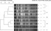
Analysis of the PFGE patterns of the genomic DNA of the 9 high-level mupirocin-resistant MRSA isolates identified 5 clusters. The results indicate that the isolates are not closely related, except for the two isolates in cluster I (Fig. 1).
Susceptibility of mupirocin-resistant MRSA to fusidic acid and retapamulin
The MIC values of fusidic acid for the low-level mupirocin-resistant MRSA isolates were ≥128 µg/ml, but the MIC and MIC90 values of fusidic acid for the high-level mupirocin-resistant isolates were ≤0.5 to ≥32 µg/ml and ≥32 µg/ml, respectively. The MIC and MIC90 values of retapamulin for both the low- and high-level mupirocin-resistant MRSA isolates were 0.5 µg/ml and 0.5 µg/ml, respectively.
Analysis of the MICs showed that all 13 low-level mupirocin-resistant isolates were resistant to fusidic acid but susceptible to retapamulin. In contrast, among the 9 high-level mupirocin-resistant isolates, 5 (55.6%) were resistant to fusidic acid, and all were susceptible to retapamulin (Table 2).
DISCUSSION
Fusidic acid and mupirocin are the most widely used topical antibiotics for the treatment of superficial skin infections caused by S. aureus and S. pyogenes. Owing to the fact that they can be purchased without a doctor's prescription in Korea, the drugs can be used indiscriminately. Misuse of topical antibiotics could lead to pathogens with increased resistance to topical antibiotics. This leads to a risk of increasing the selective survival of MRSA because it resistance to these topical antibiotics is higher than that of methicillin-sensitive S. aureus (MSSA)156.
Mupirocin, a topical antibiotic originally isolated from Pseudomonas fluorescens, inhibits bacterial protein synthesis by competitively binding to isoleucyl tRNA synthetase (encoded by IleS). It was introduced into clinical practice in 1985. Shortly after, in 1987, clinical isolates resistant to mupirocin were first reported, and the resistance rate has since then increased progressively7. In Korea, topical mupirocin has been in use since 1994 to eradicate staphylococcal infection in hospitals, and the use of mupirocin has increased at an alarming rate. Currently, the resistance rate of S. aureus to mupirocin is reported as 5% to 25.3%1234. Park et al.8 reported that 27 of 193 (14.0%) MRSA isolates in Korea were resistant to mupirocin. In our study, the prevalence of mupirocin resistance is identified to be 4.4%. The observed disparity in the resistance rate is believed to reflect the differences in the study populations, such as the study region, underlying disease, and history of antibiotic use. Mupirocin resistance in staphylococci is commonly categorized as either low-level resistance (MICs of 8~256 µg/ml) or high-level resistance (MICs >256 µg/ml). Low-level mupirocin resistance arises from point mutations in the chromosomally encoded native IleS gene, whereas high-level resistance is related to the acquisition of a plasmid containing the mupA resistance element, which possesses a modified IleS-2 gene1. Low-level mupirocin-resistant strains are considered to have no clinical significance since the concentration of mupirocin in the 2% ointment (20,000 µg/ml) exceeds the MICs for the low-level mupirocin-resistant strains. Therefore, topical mupirocin can eradicate low-level mupirocin-resistant strains. In contrast, high-level mupirocin-resistant strains that cannot be eradicated by mupirocin treatment pose a serious clinical problem. This resistance to mupirocin and other antibiotics can be transferred together by plasmids carrying various resistance genes, including mupA. Additionally, other bacteria can be a reservoir for the mupA gene. Fortunately, high-level resistant strains are observed less frequently than low-level resistant strains91011. In the present study, among 22 mupirocin-resistant isolates, 13 were low-level resistant strains and 9 were high-level resistant strains.
PFGE is one of the most prominently used methods for epidemiologic typing and also for determining the genetic relatedness of bacterial isolates. However, it requires technical expertise, long processing time, and expensive instrumental setup1213. In our present study, the PFGE patterns of chromosomal DNA were used to determine the epidemiologic molecular clonality of high-level mupirocin-resistant MRSA isolates. The results showed that all isolates, except for the two in cluster I, were unrelated. We believe that the genetically unrelated isolates independently acquired a plasmid containing the mupA gene. All of the unrelated, high-level mupirocin-resistant MRSA isolates were inhibited by retapamulin in our studies.
Fusidic acid inhibits bacterial protein synthesis by interfering with the translocation of elongation factor G from the ribosome. In the United Kingdom, 50% of the S. aureus isolates from dermatology patients were resistant to fusidic acid; on the other hand, 78% of the S. aureus isolates from atopic patients were resistant to fusidic acid14. This indicates that fusidic acid resistance is more prevalent than mupirocin resistance in S. aureus. A retrospective analysis reported that, among the 482 S. aureus isolates obtained from infected skin wounds in a Korean tertiary hospital between October 2009 and October 2011, 48.3% were MRSA, and 45.9% were resistant to fusidic acid4. Importantly, 4.8% of the S. aureus isolates were resistant to both fusidic acid and mupirocin4, demonstrating that new topical antibiotics are required to treat infections caused by mupirocin- and fusidic acid-resistant bacteria. Fusidic acid-resistant isolates are classified into two groups: low-level resistant isolates (MIC 2~32 µg/ml) and high-level resistant isolates (MIC >128 µg/ml). Low-level resistance is related to the acquisition of a plasmid containing fusB, which encodes a FusB family protein that protects drug target sites. High-level resistance results from point mutations in the chromosomally encoded native fusA gene1516. This information suggests that, while plasmid propagation could give rise to a combination of lowlevel fusidic acid resistance and high-level mupirocin resistance, chromosomal mutation could give rise to a combination of high-level fusidic acid resistance and low-level mupirocin resistance. In our current study, the rate of resistance to fusidic acid was observed to be lower in high-level mupirocin-resistant isolates compared to low-level mupirocin-resistant isolates.
Retapamulin is a derivative of pleuromutilin, which is derived from Clitopilus scyphoides, an edible mushroom. Pleuromutilin displays a unique mode of action, which involves inhibiting protein synthesis primarily by inhibiting ribosomal activity at three sites in the following manner: selectively binding to a site on the 50S subunit of the bacterial ribosome, binding to protein L3 at site P of the ribosome, and inhibiting ribosomal peptidyl transferase activity. Multi-target sites and novel binding sites that differ from those of other antibiotics minimize the development of retapamulin resistance. Currently, there are no approved CLSI or BSAC breakpoints for retapamulin; there is only an epidemiologic cutoff value recommended by EUCAST. As per the guidelines of EUCAST and Traczewski and Brown17, MICs of ≤0.5, 1, and ≥2 µg/ml can be interpreted as susceptible, intermediate, and resistant, respectively. In an in vitro study of 664 S. aureus isolates from the United Kingdom, retapamulin at concentrations of ≤0.25 mg/L inhibited 663 (99.9%) isolates including many fusidic acid-resistant and/or highly mupirocin-resistant isolates18. Candel et al.19 reported that, in Spain, retapamulin inhibited all isolates of MSSA and MRSA that were susceptible to linezolid; however, linezolid-resistant MRSA isolates were resistant to retapamulin, with MICs over 32 mg/L. A report from the United States of America in 2013 revealed that, among 155 MRSA isolates, 2.6% were resistant to retapamulin20. The test organisms included strains resistant to vancomycin, linezolid, daptomycin, and mupirocin. Thus, a small number of studies have tested the susceptibility of S. aureus to retapamulin. However, the susceptibility of Korean isolates of S. aureus to retapamulin is not known. In our study, we did not identify any mupirocin-resistant MRSA isolates that were also resistant to retapamulin.
To the best of our knowledge, this is the first report to describe the in vitro antimicrobial activity of retapamulin against clinical isolates of mupirocin-resistant MRSA. From the results of this study, we conclude that retapamulin could be an effective antibiotic for the treatment of mupirocin-resistant MRSA infections in Korea. The resistance rate to fusidic acid was lower in high-level mupirocin-resistant isolates than in low-level mupirocin-resistant isolates, suggesting the importance of additional studies with more isolates. Furthermore, the abuse of topical antibiotics should be avoided to forestall further increases in antibiotic resistance.


 XML Download