

 PDF
PDF ePub
ePub Citation
 Print
Print


Dear Editor:
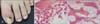
Acral angioosteoma cutis (AAOC) is a rare benign exophytic tumor characterized by bony and vascular proliferation. Clinically, it occurs on acral skin and resembles pyogenic granuloma (PG). Here, we report an unusual case of AAOC that developed on the periungual area of the great toe. A 12-year-old girl presented with a solitary tender crusted nodule on her left great toe. She had been suffering from an ingrown nail and the lesion had first appeared two months earlier after a partial nail extraction. The lesion increased in size over time. The patient was otherwise healthy. Physical examination revealed an erythematous to blackish, somewhat firm, crusted periungual nodule, measuring 5 mm in diameter, with paronychia on her left great toe (Fig. 1A). Clinically, the lesion was suspicious for PG or granulation tissue. The lesion was removed by curettage and a histopathological examination revealed a well-circumscribed polypoid tumor containing multiple bony trabeculae and small vessel proliferation in the stroma (Fig. 1B, C). Well-formed dilated capillaries that showed neither a lobular pattern nor endothelial atypia were distributed diffusely among the bony trabeculae. These findings were consistent with a diagnosis of AAOC. We followed the lesion for 1 year and observed no recurrence.
In 2006, Googe et al.1. first described AAOC as a benign tumor of unknown histogenesis that is distinctive from other cutaneous lesions showing calcification or ossification. Clinically, AAOC can be easily misdiagnosed as one of the benign acral neoplasms2345. In other similar conditions, the subungual exostosis differs from that in AAOC in that a fibrocartilaginous cap surrounds the lesion without vascular proliferation. In addition, ectopic bone formation protruding from the skeleton is typically found on radiography. Both osteoma cutis and fibro-osseous pseudotumor of the digit show ossification without vascular proliferation. Osteochondroma also has no vascular channels and is characterized by bony trabeculae covered with hyaline cartilage. PG showing metaplastic ossification is the most difficult entity to differentiate from AAOC45, but can be differentiated by its typical lobular vascular proliferation. In our case, unfortunately, radiography was not performed to examine for ectopic bone formation and a connection between the skeleton and the tumor. However, when performing curettage and biopsy, we found no connection between the tumor and underlying bony abnormalities, and the lesion was easily removed. Thus, we considered that the tumor was not an exostosis or osteochondroma.
The pathogenesis of AAOC is still unclear, but we assume that it is similar to that of other hemangiomas showing ossification. According to previous reports, vascular endothelial growth factor (VEGF) and bone morphogenetic protein (BMP) may play an important role in the development of PG with ossification2345. VEGFs and BMPs are synthesized by various cells including endothelial cells and osteoblasts in response to hypoxia, trauma, or inflammation. They then induce vascular and osteoblastic differentiation, wherein the induction is regulated through interactions with one another. We assume that these proposed functions of VEGFs and BMPs may also explain the development of AAOC.
Although there have been only two case reports of AAOC, and only limited information is available since its first description by Googe et al.1, we do not think this tumor is truly rare. Some reports of PG showing ossification did not describe lobular vascularity45. This suggests the possibility of AAOC being overlooked and misdiagnosed as PG or other acral tumors. Considering the definition of PG, we believe that some previously reported cases of ossifying PG that showed no lobular vascularity could be reclassified as AAOC. Of course more cases and investigations are needed to prove AAOC as a distinct clinicopathologic entity and not another variant of ossifying PG. AAOC should therefore be considered in the differential diagnosis of acral lesions, especially when the patient has a history of inflammation or trauma in the periungual area, as in our case.
 XML Download